


04 prism
- 2. • A prism is defined as a portion of a refracting medium bordered by two plane surfaces which are inclined at a finite angle.
- 3. Axis of the prism: A line bisecting the angle Apex: The thin edge where the intersecting surfaces meet Base: The opposite surface. Refracting/ Apical angle of the prism: The angle between the two surfaces
- 5. Prism Terminology Apex: Tip of the prism where the two refracting surfaces meet. Thinnest portion of the prism Base The bottom of the prism or the side opposite the apex or apical angle Thickest portion of the prism Orientation of an ophthalmic prism is described relative to the base
- 6. Prism Terminology Refracting angle: Angle included by the two faces of the prism (β)(α) Greater the angle, the more prism will deviate light Angle of Deviation: Amount light deviates from its original path, in degrees (ε)
- 7. Prism Action a) Prism deviates light toward the base of the prism b) Objects appear to move toward apex of the prism c) Eye must make a movement toward apex to maintain fixation on an object d) Eye moves by an amount equal to the angle of deviation
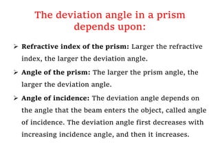
- 8. The deviation angle in a prism depends upon: Refractive index of the prism: Larger the refractive index, the larger the deviation angle. Angle of the prism: The larger the prism angle, the larger the deviation angle. Angle of incidence: The deviation angle depends on the angle that the beam enters the object, called angle of incidence. The deviation angle first decreases with increasing incidence angle, and then it increases.
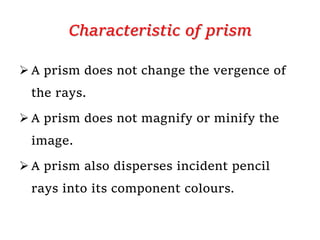
- 10. Characteristic of prism A prism does not change the vergence of the rays. A prism does not magnify or minify the image. A prism also disperses incident pencil rays into its component colours.
- 11. Image formation The object being viewed through the prism appears displaced toward the apex of the prism. Although the light rays themselves bent toward the base The image formed by a prism is erect virtual & displaced towards the apex of the prism.
- 12. There are two primary positions in which the power of a prism may be specified a) The position of minimum deviation b) The prentice position.
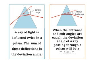
- 13. The deviation angle & Angle of minimum deviation A beam passing through an object like a prism or water drop is deflected twice: once entering, and again when exiting. The sum of these two deflections is called the deviation angle.
- 14. A ray of light is deflected twice in a prism. The sum of these deflections is the deviation angle. When the entrance and exit angles are equal, the deviation angle of a ray passing through a prism will be a minimum.
- 15. Angle of deviation is least when the angle of incidence equals the angle of emergence The angle of deviation equals half the refracting angle of the prism
- 17. The Prentice position The Prentice position is an orientation of a prism, used in optics and ophthalmology. In this position, light enters it at an angle of 90° to the first surface, so that the beam does not refract at that surface. All the deviation caused by the prism takes place at the exit surface.
- 18. The Prentice position The deviation of light in the prentice position is greater than that in the position of minimum deviation, because in the prentice position the angle of incidence does not equal the angle of emergence. Therefore the Prentice position power of any prism is greater than its power in the position of minimum deviation.
- 19. • In ophthalmology, glass prisms, e, g, trial lens prism, were classically calibrated for use in the Prentice position, • while plastic prisms, i, e, prism bars were calibrated for use in the frontal position.
- 20. For example, a 40 dioptre plastic prism held in the frontal plane will have an effective power of 41 diopters, but if it is held in the prentice position its effective power becomes 72 diopters.
- 21. Notation of prism The power of any prism can be express in various units. The Prism Dioptre (∆) A prism of one dioptre power (1∆) produces a liner apparent displacement of 1 cm, of an object O, situated at 1 m.
- 22. Notation of prism Angle of apparent deviation: Under condition of ophthalmic usage a prism of 1 prism dioptre power produces an angle of apparent deviation of ½ 0. Thus 1 prism dioptre= ½ 0 (Prism diopters in the US and degrees in Europe)
- 23. Notation of prism Centrad (𝛻): This unit differs from the prism dioptre only in that the image displacement is measured along an arc 1 m from the prism. The centrad produces a very slightly greater angle of deviation than the prism dioptre, but the difference, in practice, is negligible.
- 25. Use of prism 1) Diagnostic 2) Therapeutic 3) Instruments 4) Miscellaneous
- 26. Diagnostic use of PRISM Assessment of squint & heterophoria a) Measurement of angle objectively by prism cover test b) Measurement of angle subjectively by maddox rod c) To assess likelihood of diplopia after proposed squint surgery in adults. d) Measurement of fusional reserve e) 4 ∆D base out test
- 27. Measurement of angle objectively by prism cover test
- 28. prism cover test: Procedure The prism cover test measures the angle of deviation on near or distance fixation and in any gaze position. It combines the alternate cover test with prisms and is performed as follows: The alternate cover test is first performed to establish the direction and approximate extent of deviation.
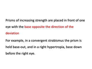
- 29. Prisms of increasing strength are placed in front of one eye with the base opposite the direction of the deviation For example, in a convergent strabismus the prism is held base-out, and in a right hypertropia, base down before the right eye.
- 30. The alternate cover test is performed continuously as stronger prisms are introduced, typically using a prism bar consisting of a column of prisms of progressive strength. The amplitude of the re-fixation movement should gradually decrease as the strength of prism approaches the extent of deviation.
- 31. The end-point is approached when no movement is seen. To ensure the maximum angle is found, the prism strength can be increased further until a movement is observed in the opposite direction (the point of reversal) and then reduced again to find the neutral value; the angle of deviation is then taken from the strength of the prism.
- 32. Measurement of angle subjectively by Maddox rod • The Maddox rod consists of a series of fused cylindrical red glass rods that convert the appearance of a white spot of light into a red streak. The optical properties of the rods cause the streak of light to be at an angle of 90° with the long axis of the rods; when the glass rods are held horizontally, the streak will be vertical and vice versa.
- 33. The rod is placed in front of the right eye (Fig: A). This dissociates the two eyes: the red streak seen by the right eye cannot be fused with the unaltered white spot of light seen by the left eye (Fig: B). The amount of dissociation (Fig: C) is measured by the superimposition of the two images using prisms. The base of the prism is placed in the position opposite to the direction of the deviation. Both vertical and horizontal deviations can be measured in this way but the test cannot differentiate a phoria from a tropia.
- 34. (A) Maddox rod test;
- 35. (B) appearance of a point of light through the Maddox rod;
- 37. • Maddox wing • The Maddox wing dissociates the eyes for near fixation (1/3 m) • and measures heterophoria.
- 38. Maddox wing The Maddox wing dissociates the eyes for near fixation (1/3 m) and measures heterophoria.
- 39. To assess likelihood of diplopia after proposed squint surgery in adults. Squint surgery in adult sometimes may cause intractable diplopia, but before surgery if we assess the squint with prism we can be aware of it to the patient.
- 40. Measurement of fusional reserve Increasingly powerful prisms are placed before one eye until fusion breaks down. This is very useful in assessing the presence of binocular vision in children below two years of age.
- 41. 4 ∆D base out test This is a delicate test for small degrees of esotropia (microtropia). A four-diopter prism placed base-out before the deviating eye causes no movement as the image remains within the suppression scotoma. When placed before the normal (fixing) eye, movement occurs.
- 42. Forms of diagnostic prisms i. Single un mounted prisms ii. Trial lens set prisms iii. Prism bars: These are bars composed of adjacent prisms of increasing power.
- 43. Therapeutic uses of prism
- 44. Therapeutic prism: Convergence insufficiency The commonest therapeutic use of prisms in the orthoptic department is in building up the fusional reserve of patients with convergence insufficiency.
- 45. Convergence insufficiency: Base out prism exercises Base out prisms can also be used to stimulate the converge reflex. The base out prism induces crossed diplopia and the patient must converge to overcome the prism strength and obtain BSV.
- 46. Convergence insufficiency: Base out prism exercises • Some practitioners give a patient a single prism and have them do gradual exercises or near tasks, while other practitioners have the patient use a prism bar and to overcome increasing prism strengths while focusing on a near target.
- 47. To relieve diplopia To relieve diplopia in certain cases of squint, these include decompanseted heterophorias, small vertical squints and some paralytic squints with diplopia in the primary position. Prisms are reserved for those patients for whom surgery is not indicated
- 48. Forms of therapeutic prism Temporary wear prisms: Used in treatment include clip- on spectacle prisms for trial wear. Eg:-Fresnel prism (pronounced fre-nell') prisms,) • To understand how a Fresnel prism works, imagine cutting off the tops of a large number of equally powered prisms and gluing them, one above the other, onto a thin piece of plastic. A Fresnel prism is only 1 mm thick. It consists of plastic sheet of parallel tiny prisms of identical refracting angle. The overall prismatic effect is the same as that of a single large prism. The sheets are lighter than a glass prism and can be stuck on to the patient’s glasses.
- 49. • Advantages of a Fresnel Prism: It is very thin and extremely lightweight. It is flexible and can be applied to an existing spectacle lens. It can be cut to any shape with scissors or a razor blade. Reduce magnification differences considerably. • Disadvantages: They are harder to clean than conventional lenses. Fresnel prisms also cause a slight loss of visual acuity caused by reflections for prisms greater than 10Δ.
- 51. Prisms in optical instruments: 1) Slit lamp bio microscope. 2) Applanation tonometer 3) keratometer
- 52. Different types of prism used in ophthalmology Porro-prism: , is a type of reflection prism used in optical instruments to alter the orientation of an image. • Porro prisms are most often used in pairs, forming a double Porro prism. A second prism, rotated 90° with respect to the first, is placed such that light will traverse both prisms. The net effect of the prism system is a beam parallel to but displaced from its original direction, with the image rotated 180°. As before, the handedness of the image is unchanged.
- 53. • Double Porro prism systems are used in small optical telescopes to re-orient an inverted image (an arrangement is known as an image erection system), and especially in many binoculars where they both erect the image and provide a longer, folded distance between the objective lenses and the eyepieces. • Commonly, the two components of the double Porro system are cemented together, and the prisms may be truncated to save weight and size.
- 54. Single Porro prism. Double Porro prism
- 55. • Dove prism: • A Dove prism is a type of reflective prism which is used to invert an image. Dove prisms are shaped from a truncated right-angle prism. A beam of light entering one of the sloped faces of the prism undergoes total internal reflection from the inside of the longest (bottom) face and emerges from the opposite sloped face. Images passing through the prism are flipped, and because only one reflection takes place, the image is inverted but not laterally transposed.
- 56. Dove prism: A Dove prism is a type of reflective prism which is used to invert an image. Dove prisms are shaped from a truncated right-angle prism. A beam of light entering one of the sloped faces of the prism undergoes total internal reflection from the inside of the longest (bottom) face and emerges from the opposite sloped face. Images passing through the prism are flipped, and because only one reflection takes place, the image is inverted but not laterally transposed.
- 57. The Wollaston prism • A Wollaston prism is an optical device, invented by William Hyde Wollaston, that manipulates polarized light. It separates light into two separate linearly polarized outgoing beams with orthogonal polarization. The two beams will be polarized according to the optical axis of the two right angle prisms.
- 58. The Wollaston prism • The Wollaston prism consists of two orthogonal prisms of birefringent material— typically a uniaxial material such as calcite. These prisms are cemented together on their base (traditionally with Canada balsam) to form two right triangle prisms with perpendicular optic axes.
- 59. The Wollaston prism • Outgoing light beams diverge from the prism as ordinary and extraordinary rays due to the differences in the indexes of refraction, with the angle of divergence determined by the prisms' wedge angle and the wavelength of the light. Commercial prisms are available with divergence angles from less than 1° to about 45°. • Use in: Keratometer