MANAGMENT OF GASTRIC
CARCINOMA
MODERATOR : Dr. HAILU,ASSISTANT PROF. General and upper GI
surgery
By- Tsion Tilahun (GSRIII)
JULY 2020
OUTLINE
2
• Introduction
• Epidemiology
• Clinical presentation
• Diagnostic workups and staging
• Treatment
Anatomy
12/27/2024 3
Anatomy
• Innervation:
– Parasympathetic via the
vagus.
• Left anterior and right
posterior.
– Sympathetic via the
celiac plexus.
12/27/2024 4
Lymphatic drainage
12/27/2024 5
Zone 1 , draining the pyloric portion of the greater curve.
Zone 2 , draining from the upper half of the greater
curve.
Zone 3 , drains the proximal two-thirds of the stomach
and the upper lesser curve, and surrounds the left
gastric artery.
Zone 4, drains the distal portion of the lesser curve and
pylorus
All four drain into the celiac group of nodes and into the
thoracic duct.
12/27/2024 6
Zone 1
Zone 2
Zone 3
Zone 4
Lymphatic drainage of the stomach & LND
Stations draining the
stomach as to the
JRSGC
Stations 3-6 are
commonly removed
with D1 gastrectomy
Stations 1, 2, & 7 -12
are commonly
removed with D2
gastrectomy
Epidemiology
 4th MC Ca in the world
 the 2nd leading cause of Ca death
 M:F = 3:2
 It’s a disease of the elders with peak incidence in the
7th decade of life
 More common in low socioeconomic class citizens
 It is more common & has a higher mortality in African
Americans, Asian Americans, & Hispanics compared
with whites)
Cont..
 Incidence <10 (US) Vs 40 /100000 (highest in Asia,
Latin America & Caribbean)
 It has decreased by 2/3 in the past 50 yrs. mostly in
intestinal histology , the distal Ca
 Its the MC cancer in Japan as a result screening in
Japan was started in the 1970s since then the MR has
decreased by 50%
 Globally there is proximal migration of tumors seen
so the distribution currently is closer to 40% distal,
30% middle, & 30% proximal
Ethiopia
Epid..cont.
1. PRIMARY MALIGNANT NEOPLASMS OF THE STOMACH
 The three most common neoplasms are:
• Adenocarcinoma (95%)
• Lymphoma (4%)
• Malignant GIST (1%)
 Other rare malignancies:
• Carcinoid
• Angiosarcoma
• Carcino-sarcoma
• Squamous cell carcinoma
2. Secondary involvement
• Due to heamatogenous metastasis from other sites (e.g., melanoma or breast)
• More commonly, malignant tumors from adjacent organs invade the stomach
by direct extension (e.g., colon or pancreas ) or by peritoneal seeding (e.g.,
ovary)
3
Risk factors
Nutritional
Low fruit and vegetable
Salted meat/fish
High nitrate
High complex carbohydrate
Environmental
Poor food preparation
Lack of refrigeration
contaminated drinking
water
Smoking
Medical
H-pylori
Gastric atrophy
Pernicious anemia
Polyps
Menetrier disease
EBV
Prior gastric surgery
Other
Low socio-economic
Male gender
12
13
Diet
High salted meat/fish and pickled foods
Damage stomach mucosa and increases the
susceptibility to carcinogenesis
High nitrate N-nitroso(carcinogen)
Intake of green, leafy vegetables and citrus fruits,
which contain antioxidants such as ascorbate and
beta-carotene is protective
Smoking
Increases risk by 60% in men and 20%in women
12/27/2024 14
Genetics
commonly affected genes are P53,COX 2 and CDH1
gene
Hereditary diffuse gastric cancer
 Inactivates e-cadherin (cell adhesion molecule)
2 of more gastric cancer cases in the 1st
/2nd
degree relatives
with age<50
80% increased risk
 Lynch syndrome(HNPCC) – intestinal type and 10% higher risk
 Familial adenomatous polyposis - 10% increased risk
 Li-fraumeni syndrome – P53 gene mutation
Risk Factors
15
Helicobacter pylori
Classified as definite carcinogen
Associated with 6x increases risk in developing
adenocarcinoma
Triggers inflammation at the corpus mucosa that
results in atrophy and intestinal metaplasia
Alterations depend on both the presence of bacterial
proteins and the host immune response
The cagA strain causes more mucosal inflammation
and thus a higher risk of gastric cancer
16
Pernicious anemia
Destruction of parietal and chief cells by
autoimmune reaction
Achlorhydria that accompanies this condition
favors bacterial proliferation, which generates
carcinogenic nitrosamines from nitrates
Blood group A
20% incidence
Mostly associated with diffuse gastric cancer
17
Epstein–Barr virus (EBV)
Detected in 2-16% of gastric adenocarcinoma
Role in gastric carcinogenesis is not yet established
Higher frequency in tumors of the proximal and Male
predominance
Previous gastric surgery
Occur after ten of surgery
Overall risk is 3-10%
Pathogenesis related to bile reflux , achlorhydria and
atrophic gastritis
Clinical features
 Vague discomfort and/or indigestion
 Epigastric pain that is constant, non-radiating, and
unrelieved by food ingestion
 Diffuse mural disease may present with early satiety due to
decreased distensiability so weight loss , anorexia and other
constitutional SmS
 Proximal tumors may present with dysphagia where as
antral tumors as GOO
 May Presents as either acute UGIB with frank heamatemesis
(5-15%) or most commonly as occult GIB with some form of
anemia (40%) with melena (ulcerated lesions)
Clinical..
P/E
 is unremarkable i.e. cachexia , pallor , Palpable
abdominal mass , HSM , jaundice, or ascites as an
evidence of metastasis
• left supraclavicular node(Virchow’s)
• Periumbilical node (sister marry joseph node)
• Left axillary node(irish)
• Peritoneal spread can present with an enlarged ovary
(Krukenberg's tumor or a mass in the cul-de-sac on
rectal examination (Blumer's shelf.
Clinical..
• Paraneoplastic manifestations
• Dermatologic findings like diffuse seborrheic keratoses
(sign of Leser-Trelat) or acanthosis nigricans which is
characterized by velvety and darkly pigmented patches on
skin folds.
• Neither finding is specific for gastric cancer.
• Other paraneoplastic abnormalities that can occur in
gastric cancer include:-
- microangiopathic hemolytic anemia
-membranous nephropathy
-hypercoagulable states (Trousseau's syndrome).
Polyarteritis nodosa has been reported as the single
manifestation of an early and surgically curable gastric cancer
Diagnostic workups and staging
• CBC , chemistry panel, LFTs, Serum-Albumin and
coagulation studies, OBT
• Two main modalities for gastric cancer screening
upper endoscopy (Gold standard)
contrast radiography
• false-negative barium studies can occur in as many
as 50 percent of cases
• barium study may be superior to upper endoscopy
is in patients with linitis plastica
• barium study may be superior to upper
endoscopy is in patients with linitis plastica
 Tumor markers like CEA, CA-125, CA-19-9, B-
HCG
 serum pepsinogen, Serum trefoil factor 3
(TFF3), microRNAs
SCREENING
Controversial
• Universal screening:-
Japan, for individuals older than 50 years with conventional double-contrast barium radiograph with
photofluorography every year or upper endoscopy every two to three years .
In Korea, upper endoscopy is recommended every two years for individuals aged 40 to 75 years
• Selective screening of high-risk subgroups
individuals at increased risk for gastric cancer include those with the following
Gastric adenomas
Pernicious anemia
Gastric intestinal metaplasia
Familial adenomatous polyposis
Lynch syndrome
Peutz-Jeghers syndrome
Juvenile polyposis syndrome
STAGING EVALUATION
Complete clinical or preoperative staging of patients with gastric cancer
includes:
• Physical examination, including evaluation of appropriate nodal areas
(especially left supraclavicular nodes) as well as the abdomen and rectal
examination.
• CT scans of abdomen and pelvis, chest
CT is not very accurate for assessing the depth of tumor invasion of the
stomach wall or regional nodal
• Endoscopic ultrasound (EUS) may provide more accurate staging evaluation
of the tumor and nodal stage(>5mm) than CT and also allows for
preoperative biopsies.(80% accurate)
• Staging laparoscopy and peritoneal cytology :advantage of directly
visualizing the liver surface, peritoneum, and local lymph nodes, and
permitting biopsy of any suspicious lesions
2. Gastric Ca Management 2020.pptx2. Gastric Ca Management 2020.pptx
12/27/2024 28
T1 T2 T3 T4a T4b
N0
Stage Ia Stage Ib Stage IIa Stage IIb Stage IIIb
N1
Stage Ib Stage IIa Stage IIb Stage IIIa Stage IIIb
N2
Stage IIa Stage IIb Stage IIIa Stage IIIb Stage IIIc
N3
Stage IIb Stage IIIa Stage IIIb Stage IIIc Stage IIIc
M1
Stage IV Stage IV Stage IV Stage IV Stage IV
Treatment Treat ….
Core point of treatment
• Extent of surgical resection resection
• Extent of lymph node dissection
• Chemo radio therapy
Surgery
• The goal of curative surgery is complete resection of all
tumor with a grossly negative margin of at least 5 cm as
well with an adequate lymphadenectomy performed (R0
resection)
Indicators of unresectabiliy
• distant metastases
• invasion of a major vascular structure, such as the aorta ,hepatic
artery ,celiac axis/proximal splenic artery
• Lymph nodes behind or inferior to the pancreas, aorto-caval region,
into the mediastinum, or in the porta hepatis
• Distal splenic artery involvement is not an indicator of unresectability
Surg….
Distal radical subtotal gastrectomy is standard
operation
Reconstruction is by billroth II gastrojejunotomy or
Roux –en- gastrojejunostomy .
Total gasrectomy with Roux-en-Y
esophagojejunostomy may be required
for R0 resection and proximal gastric
adenoma,large midgastric lesions or
infiltrative disease (eg, linitis plastica)
Techniques
1.Begin at pylorus with high
ligation of R gastroepiploic
artery
2 along the greather curvature
3 spare LGEA and short gastric
vesels (not for sub total)
4 posterior antrum separted from
anterior pancreas and base of
the transverse mesocolon by
division of fine connective
tissue attachements.
Cont..
5.Gastrohepatic ligament is
incised , leser curvature is
dissected
6. Dissection is extended
toward the celiac axis
7. Proximal duodenum is
divided (care for common
bile duct and hepatic artery
• Proximal loop of jejunum is
chosen and apposed to
stomach.
• Jejunum can be delivered
through incision in the
transverse mesocolon or
anterior to transverse
mesocolon (malignance)
Endoscopic Resection
General guidelines for endoscopic resection of
early gastric cancer ;(T1a)
1. Tumor limited to the mucosa,
2. No lymphovascular invasion,
3. Tumor smaller than 2 cm,
4. No ulceration, and
5. Well or moderately differentiated
histopathology
Extent of lymph node dissection
More than 15 respected lymph node is
required
• D1 lymphadenectomy -
dissection of the
perigastric lymph nodes
only(3-6)
• D2 lymphadenectomy
removal of nodes along the
hepatic, left gastric, celiac
and splenic arteries & in the
splenic hilum (stations 1-
12).
• D3 dissection is a D2+ per
aortic .
D1 vs D2 Lymphadenectomy
2. Gastric Ca Management 2020.pptx2. Gastric Ca Management 2020.pptx
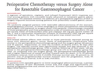
Chemotherapy and Chemoradiotherapy
• Neoadjuvant/perioperative chemotherapy
• Neoadjuvant chemoradiotherapy
2. Gastric Ca Management 2020.pptx2. Gastric Ca Management 2020.pptx
POSTTREATMENT SURVEILLANCE
• History and physical examination every three to
six months for one to three years, then every six
months for years 4 and 5, then annually
• Complete blood count (CBC) and chemistry
profile, as clinically indicated
• Radiologic imaging or endoscopy, as clinically
indicated
• Monitor for vitamin B12 deficiency in surgically
treated patients and treat as indicated
Gastric GIST
• Originally thought to be a type of smooth muscle
sarcoma,
• they are now known to be a distinct tumor
derived from the interstitial cells of Cajal, an
intestinal pacemaker cell.
• They are most commonly found in the stomach
(40% to 60%), small intestine (30%), and colon
(15%).
57
Cont.
• They vary considerably in their presentation and clinical
course, ranging from small benign tumors to massive lesions
with necrosis, hemorrhage, and wide metastases.
• can manifest at any age, peak age >50 years.
• an equal male-to-female ratio or a slight male predominance
• Most GISTs manifest symptomatically, typically with bleeding
or vague abdominal pain or discomfort.
• Many patients remain asymptomatic, and their tumors are
discovered incidentally at the time of other surgery or,
increasingly, during imaging performed for other indications
GIST cont…
• They are frequently identified by
immunohistochemical staining for the c-kit proto-
oncogene (CD117), which is overexpressed in
95% of these tumors, and for CD34, which is
positive in 60% to 70% of GISTs.
• Investigations are same with gastric
adenocarcinoma
58
Suggested guideline for assessing malignant potential of GIST of
different sizes and mitotic rate
1. Benign (no tumor-related mortality)
-No larger than 2 cm, no more than 5 mitoses/50 HPF
2.Probably benign (<3% with progressive disease)
- >2 cm but ≤5 cm; no more than 5 mitoses/50 HPF
3.Uncertain or low malignant potential
-No larger than 2 cm; >5 mitoses/50 HPF
4.Low to moderate malignant potential (12%-15% tumor-related mortality)
->10 cm; no more than 5 mitoses/HPF
• >2 cm but ≤5 cm; >5 mitoses/50 HPF
5.High malignant potential (49%-86% tumor-related mortality)
• ->5 cm but ≤10 cm; >5 mitoses/50 HPF
• >10 cm; >5 mitoses/50 HPF
59
TREATMENT
• The mainstay of treatment is complete surgical resection
• Tumors greater than 2 cm in diameter should be resected
• Tumors that are less than 2 cm and have high-risk features on endoscopy and
EUS, such as irregular borders, ulceration, and heterogeneity, should be
resected
• tumors without such features can be observed with repeat endoscopy and EUS
at 6- to 12-month intervals
• Depending on tumor size, options of resection include:
 wide local excision
 Enucleation
 sleeve gastrectomy

total gastrectomy, with or without en bloc resection of adjacent organs.
• No lymph node dissection is required as lymph node metastases are rare.
60
Adjuvant Therapy for GIST
• tyrosine kinase inhibitor imatinib (Gleevec) is used as adjuvant ,
neoadjuvant and palliative therapy.
• In patients with metastatic or unresectable disease, imatinib (400
mg daily) showed an overall 2-year survival of 70% compared with
25% for patients on traditional chemotherapy.
• In the adjuvant setting, tumors 3 cm or larger who underwent
complete resection and were treated with imatinib for 1 year had a
recurrence rate of 8% compared with 20% for untreated patients
• Minimum of 3 years as adjuvant following complete resection .
 Indications for tyrosine kinase inhibitor
 moderate to high risk for recurrence or metastasis(>3cm or
>5mitosis/hpf) after resection of resectable ds
 For unresectable tumour as neoadjuvant, optimal duration of tx
uncertain.
 For metastatic disease and patients with resected primary disease at
moderate risk of recurrence, indefinite treatment with imatinib
.
61
• Five-year survival following resection for
GIST is about 50%.
• Most patients with low-grade lesions
are cured (80% 5-year survival), but
most patients with high-grade lesions
are not (30% 5-year survival).
62
Reference
1. SCHWARTZ’S PRINCIPLES OF SURGERY , 11th
edition
2. Maingot’s ABDOMINAL OPERATIONS 13th
edition
3. Shackelford’s SURGERYof the ALIMENTARY TRACT, 8th
edition
4. Uptodate 21.6
5. Zollenger atlas of surgical operation 9th
edition.
7. Kobayashi
T, Kimura T. [Long-term outcome of preoperative chemothe
rapy with 5'-deoxy-5-fluorouridine (5'-DFUR) for gastric can
cer].
8. Gan To Kagaku Ryoho 2000; 27:1521.
Nio Y, Koike M, Omori H, et al. A randomized consent desig
n trial of
neoadjuvant chemotherapy with tegafur plus uracil
69

More Related Content

PPT
gastriccancer
PPT
gastriccancer types classified and manage
PPT
gastriccancer.ppt
PPT
gastriccancer.ppt
PPT
gastriccancer.ppt
PDF
gastriccancer-160627133725.pdf
PPTX
Gastric Cancer ( stomach tumor )
PPT
carcinoma of stomach
gastriccancer
gastriccancer types classified and manage
gastriccancer.ppt
gastriccancer.ppt
gastriccancer.ppt
gastriccancer-160627133725.pdf
Gastric Cancer ( stomach tumor )
carcinoma of stomach

Similar to 2. Gastric Ca Management 2020.pptx2. Gastric Ca Management 2020.pptx (20)

PPT
Cancergastritis200810
PPTX
Gastric cancer seminar
PPTX
4. Gastric Cancer.pptx666666666666666666666666666
PPTX
4. Gastric Cancer.pptx7777777777777777777777
PPTX
GASTRIC CANCER.pptx
PPTX
GASTRIC CANCER.pptx
PPTX
GASTRIC CARCINOMA- EPIGASTRIC LUMPS- Abdominal Lumps.pptx
PPTX
gastriccancer-160428190410.pptx
PPTX
22. Gastric Cancer powerpoint presentation
PPTX
Carcinoma of Stomach - presentation by residents
PPT
Gastric Carcinoma
PPTX
Gastric carcinoma
PPT
Gastric cancer
PPTX
gastric cancer
PPT
Git 4th GC18.
PPT
Gastric cancer
PPTX
clinical approach to gastric cancer.pptx
PPTX
Malignant Neoplasms of Stomach.pptx
PPTX
Carcinoma of stomach
PPTX
Stomach cancer
Cancergastritis200810
Gastric cancer seminar
4. Gastric Cancer.pptx666666666666666666666666666
4. Gastric Cancer.pptx7777777777777777777777
GASTRIC CANCER.pptx
GASTRIC CANCER.pptx
GASTRIC CARCINOMA- EPIGASTRIC LUMPS- Abdominal Lumps.pptx
gastriccancer-160428190410.pptx
22. Gastric Cancer powerpoint presentation
Carcinoma of Stomach - presentation by residents
Gastric Carcinoma
Gastric carcinoma
Gastric cancer
gastric cancer
Git 4th GC18.
Gastric cancer
clinical approach to gastric cancer.pptx
Malignant Neoplasms of Stomach.pptx
Carcinoma of stomach
Stomach cancer
Ad

More from Aidrus Abdi (16)

PPTX
Case presentation PUD ahmed.pptxpptxpptx
PPTX
Case presentation, incicional hernia.pptx
PPTX
Diaphragmatic injury.pptxDiaphragmatic injury.pptx
PPTX
Approach to a patient with CNS tumor.pptx
PPTX
Antibiotic use in surgery.pptxpptxpptxpptx
PPTX
Case presentation Colovesical fistula.pptx
PPTX
5-min talk on Meckel diverticulum.pptx5-min talk on Meckel diverticulum.pptx
PPTX
5-min talk on GB perforation.pptx5-min talk on GB perforation.pptx
PPTX
Air leak.pptxAir leak.pptxAir leak.pptxAir leak.pptx
PPTX
Anorectal Malformation soll.pptxAnorectal Malformation soll.pptx
PPTX
septic A Seminar.pptx septic A Seminar.pptx
PPTX
AEDH.pptxAEDH.pptxAEDH.pptxAEDH.pptxAEDH.pptx
PPTX
csf basics.pptxcsf basics.pptxcsf basics.pptx
PPTX
Thyroidectomy minilik seminar.pptTechniques of Thyroidectomy.pptTechniques of...
PPTX
Thyroidectomy .pptxTechniques of Thyroidectomy.ppt
PPT
Techniques of Thyroidectomy.pptTechniques of Thyroidectomy.ppt
Case presentation PUD ahmed.pptxpptxpptx
Case presentation, incicional hernia.pptx
Diaphragmatic injury.pptxDiaphragmatic injury.pptx
Approach to a patient with CNS tumor.pptx
Antibiotic use in surgery.pptxpptxpptxpptx
Case presentation Colovesical fistula.pptx
5-min talk on Meckel diverticulum.pptx5-min talk on Meckel diverticulum.pptx
5-min talk on GB perforation.pptx5-min talk on GB perforation.pptx
Air leak.pptxAir leak.pptxAir leak.pptxAir leak.pptx
Anorectal Malformation soll.pptxAnorectal Malformation soll.pptx
septic A Seminar.pptx septic A Seminar.pptx
AEDH.pptxAEDH.pptxAEDH.pptxAEDH.pptxAEDH.pptx
csf basics.pptxcsf basics.pptxcsf basics.pptx
Thyroidectomy minilik seminar.pptTechniques of Thyroidectomy.pptTechniques of...
Thyroidectomy .pptxTechniques of Thyroidectomy.ppt
Techniques of Thyroidectomy.pptTechniques of Thyroidectomy.ppt
Ad

Recently uploaded (20)

PPTX
Nutrition needs in a Surgical Patient.pptx
PPTX
RESEARCH APPROACH & DESIGN.pptx presented by preeti kulshrestha
PDF
heliotherapy- types and advantages procedure
PDF
Nematodes - by Sanjan PV 20-52.pdf based on all aspects
PPTX
PSYCHIATRIC SEQUALAE OF HEAD INJURY.pptx
PDF
communicable diseases for healthcare - Part 1.pdf
PPTX
Surgical anatomy, physiology and procedures of esophagus.pptx
PPTX
Computed Tomography: Hardware and Instrumentation
PDF
Seizures and epilepsy (neurological disorder)- AMBOSS.pdf
PPTX
Local Anesthesia Local Anesthesia Local Anesthesia
PPTX
Phamacology Presentation (Anti cance drugs).pptx
DOCX
ORGAN SYSTEM DISORDERS Zoology Class Ass
PDF
The Role of Medicinal Plants in Alleviating Symptoms of Diabetes-Related Com...
PPTX
GAIT IN HUMAN AMD PATHOLOGICAL GAIT ...............
PDF
Tackling Intensified Climatic Civil and Meteorological Aviation Weather Chall...
PDF
neonatology-for-nurses.pdfggghjjkkkkkkjhhg
PPTX
Emergencies in Anaesthesia by Dr SAMI.pptx
PPTX
Geriatrics_(0).pptxxvvbbbbbbbnnnnnnnnnnk
PDF
Diabetes mellitus - AMBOSS.pdf
PDF
Cranial nerve palsies (I-XII) - AMBOSS.pdf
Nutrition needs in a Surgical Patient.pptx
RESEARCH APPROACH & DESIGN.pptx presented by preeti kulshrestha
heliotherapy- types and advantages procedure
Nematodes - by Sanjan PV 20-52.pdf based on all aspects
PSYCHIATRIC SEQUALAE OF HEAD INJURY.pptx
communicable diseases for healthcare - Part 1.pdf
Surgical anatomy, physiology and procedures of esophagus.pptx
Computed Tomography: Hardware and Instrumentation
Seizures and epilepsy (neurological disorder)- AMBOSS.pdf
Local Anesthesia Local Anesthesia Local Anesthesia
Phamacology Presentation (Anti cance drugs).pptx
ORGAN SYSTEM DISORDERS Zoology Class Ass
The Role of Medicinal Plants in Alleviating Symptoms of Diabetes-Related Com...
GAIT IN HUMAN AMD PATHOLOGICAL GAIT ...............
Tackling Intensified Climatic Civil and Meteorological Aviation Weather Chall...
neonatology-for-nurses.pdfggghjjkkkkkkjhhg
Emergencies in Anaesthesia by Dr SAMI.pptx
Geriatrics_(0).pptxxvvbbbbbbbnnnnnnnnnnk
Diabetes mellitus - AMBOSS.pdf
Cranial nerve palsies (I-XII) - AMBOSS.pdf

2. Gastric Ca Management 2020.pptx2. Gastric Ca Management 2020.pptx

  • 1. MANAGMENT OF GASTRIC CARCINOMA MODERATOR : Dr. HAILU,ASSISTANT PROF. General and upper GI surgery By- Tsion Tilahun (GSRIII) JULY 2020
  • 2. OUTLINE 2 • Introduction • Epidemiology • Clinical presentation • Diagnostic workups and staging • Treatment
  • 4. Anatomy • Innervation: – Parasympathetic via the vagus. • Left anterior and right posterior. – Sympathetic via the celiac plexus. 12/27/2024 4
  • 5. Lymphatic drainage 12/27/2024 5 Zone 1 , draining the pyloric portion of the greater curve. Zone 2 , draining from the upper half of the greater curve. Zone 3 , drains the proximal two-thirds of the stomach and the upper lesser curve, and surrounds the left gastric artery. Zone 4, drains the distal portion of the lesser curve and pylorus All four drain into the celiac group of nodes and into the thoracic duct.
  • 6. 12/27/2024 6 Zone 1 Zone 2 Zone 3 Zone 4
  • 7. Lymphatic drainage of the stomach & LND Stations draining the stomach as to the JRSGC Stations 3-6 are commonly removed with D1 gastrectomy Stations 1, 2, & 7 -12 are commonly removed with D2 gastrectomy
  • 8. Epidemiology  4th MC Ca in the world  the 2nd leading cause of Ca death  M:F = 3:2  It’s a disease of the elders with peak incidence in the 7th decade of life  More common in low socioeconomic class citizens  It is more common & has a higher mortality in African Americans, Asian Americans, & Hispanics compared with whites)
  • 9. Cont..  Incidence <10 (US) Vs 40 /100000 (highest in Asia, Latin America & Caribbean)  It has decreased by 2/3 in the past 50 yrs. mostly in intestinal histology , the distal Ca  Its the MC cancer in Japan as a result screening in Japan was started in the 1970s since then the MR has decreased by 50%  Globally there is proximal migration of tumors seen so the distribution currently is closer to 40% distal, 30% middle, & 30% proximal
  • 11. Epid..cont. 1. PRIMARY MALIGNANT NEOPLASMS OF THE STOMACH  The three most common neoplasms are: • Adenocarcinoma (95%) • Lymphoma (4%) • Malignant GIST (1%)  Other rare malignancies: • Carcinoid • Angiosarcoma • Carcino-sarcoma • Squamous cell carcinoma 2. Secondary involvement • Due to heamatogenous metastasis from other sites (e.g., melanoma or breast) • More commonly, malignant tumors from adjacent organs invade the stomach by direct extension (e.g., colon or pancreas ) or by peritoneal seeding (e.g., ovary) 3
  • 12. Risk factors Nutritional Low fruit and vegetable Salted meat/fish High nitrate High complex carbohydrate Environmental Poor food preparation Lack of refrigeration contaminated drinking water Smoking Medical H-pylori Gastric atrophy Pernicious anemia Polyps Menetrier disease EBV Prior gastric surgery Other Low socio-economic Male gender 12
  • 13. 13 Diet High salted meat/fish and pickled foods Damage stomach mucosa and increases the susceptibility to carcinogenesis High nitrate N-nitroso(carcinogen) Intake of green, leafy vegetables and citrus fruits, which contain antioxidants such as ascorbate and beta-carotene is protective Smoking Increases risk by 60% in men and 20%in women
  • 14. 12/27/2024 14 Genetics commonly affected genes are P53,COX 2 and CDH1 gene Hereditary diffuse gastric cancer  Inactivates e-cadherin (cell adhesion molecule) 2 of more gastric cancer cases in the 1st /2nd degree relatives with age<50 80% increased risk  Lynch syndrome(HNPCC) – intestinal type and 10% higher risk  Familial adenomatous polyposis - 10% increased risk  Li-fraumeni syndrome – P53 gene mutation
  • 15. Risk Factors 15 Helicobacter pylori Classified as definite carcinogen Associated with 6x increases risk in developing adenocarcinoma Triggers inflammation at the corpus mucosa that results in atrophy and intestinal metaplasia Alterations depend on both the presence of bacterial proteins and the host immune response The cagA strain causes more mucosal inflammation and thus a higher risk of gastric cancer
  • 16. 16 Pernicious anemia Destruction of parietal and chief cells by autoimmune reaction Achlorhydria that accompanies this condition favors bacterial proliferation, which generates carcinogenic nitrosamines from nitrates Blood group A 20% incidence Mostly associated with diffuse gastric cancer
  • 17. 17 Epstein–Barr virus (EBV) Detected in 2-16% of gastric adenocarcinoma Role in gastric carcinogenesis is not yet established Higher frequency in tumors of the proximal and Male predominance Previous gastric surgery Occur after ten of surgery Overall risk is 3-10% Pathogenesis related to bile reflux , achlorhydria and atrophic gastritis
  • 18. Clinical features  Vague discomfort and/or indigestion  Epigastric pain that is constant, non-radiating, and unrelieved by food ingestion  Diffuse mural disease may present with early satiety due to decreased distensiability so weight loss , anorexia and other constitutional SmS  Proximal tumors may present with dysphagia where as antral tumors as GOO  May Presents as either acute UGIB with frank heamatemesis (5-15%) or most commonly as occult GIB with some form of anemia (40%) with melena (ulcerated lesions)
  • 19. Clinical.. P/E  is unremarkable i.e. cachexia , pallor , Palpable abdominal mass , HSM , jaundice, or ascites as an evidence of metastasis • left supraclavicular node(Virchow’s) • Periumbilical node (sister marry joseph node) • Left axillary node(irish) • Peritoneal spread can present with an enlarged ovary (Krukenberg's tumor or a mass in the cul-de-sac on rectal examination (Blumer's shelf.
  • 20. Clinical.. • Paraneoplastic manifestations • Dermatologic findings like diffuse seborrheic keratoses (sign of Leser-Trelat) or acanthosis nigricans which is characterized by velvety and darkly pigmented patches on skin folds. • Neither finding is specific for gastric cancer. • Other paraneoplastic abnormalities that can occur in gastric cancer include:- - microangiopathic hemolytic anemia -membranous nephropathy -hypercoagulable states (Trousseau's syndrome). Polyarteritis nodosa has been reported as the single manifestation of an early and surgically curable gastric cancer
  • 21. Diagnostic workups and staging • CBC , chemistry panel, LFTs, Serum-Albumin and coagulation studies, OBT • Two main modalities for gastric cancer screening upper endoscopy (Gold standard) contrast radiography • false-negative barium studies can occur in as many as 50 percent of cases • barium study may be superior to upper endoscopy is in patients with linitis plastica
  • 22. • barium study may be superior to upper endoscopy is in patients with linitis plastica
  • 23.  Tumor markers like CEA, CA-125, CA-19-9, B- HCG  serum pepsinogen, Serum trefoil factor 3 (TFF3), microRNAs
  • 24. SCREENING Controversial • Universal screening:- Japan, for individuals older than 50 years with conventional double-contrast barium radiograph with photofluorography every year or upper endoscopy every two to three years . In Korea, upper endoscopy is recommended every two years for individuals aged 40 to 75 years • Selective screening of high-risk subgroups individuals at increased risk for gastric cancer include those with the following Gastric adenomas Pernicious anemia Gastric intestinal metaplasia Familial adenomatous polyposis Lynch syndrome Peutz-Jeghers syndrome Juvenile polyposis syndrome
  • 25. STAGING EVALUATION Complete clinical or preoperative staging of patients with gastric cancer includes: • Physical examination, including evaluation of appropriate nodal areas (especially left supraclavicular nodes) as well as the abdomen and rectal examination. • CT scans of abdomen and pelvis, chest CT is not very accurate for assessing the depth of tumor invasion of the stomach wall or regional nodal • Endoscopic ultrasound (EUS) may provide more accurate staging evaluation of the tumor and nodal stage(>5mm) than CT and also allows for preoperative biopsies.(80% accurate) • Staging laparoscopy and peritoneal cytology :advantage of directly visualizing the liver surface, peritoneum, and local lymph nodes, and permitting biopsy of any suspicious lesions
  • 28. 12/27/2024 28 T1 T2 T3 T4a T4b N0 Stage Ia Stage Ib Stage IIa Stage IIb Stage IIIb N1 Stage Ib Stage IIa Stage IIb Stage IIIa Stage IIIb N2 Stage IIa Stage IIb Stage IIIa Stage IIIb Stage IIIc N3 Stage IIb Stage IIIa Stage IIIb Stage IIIc Stage IIIc M1 Stage IV Stage IV Stage IV Stage IV Stage IV
  • 29. Treatment Treat …. Core point of treatment • Extent of surgical resection resection • Extent of lymph node dissection • Chemo radio therapy
  • 30. Surgery • The goal of curative surgery is complete resection of all tumor with a grossly negative margin of at least 5 cm as well with an adequate lymphadenectomy performed (R0 resection) Indicators of unresectabiliy • distant metastases • invasion of a major vascular structure, such as the aorta ,hepatic artery ,celiac axis/proximal splenic artery • Lymph nodes behind or inferior to the pancreas, aorto-caval region, into the mediastinum, or in the porta hepatis • Distal splenic artery involvement is not an indicator of unresectability
  • 31. Surg…. Distal radical subtotal gastrectomy is standard operation Reconstruction is by billroth II gastrojejunotomy or Roux –en- gastrojejunostomy . Total gasrectomy with Roux-en-Y esophagojejunostomy may be required for R0 resection and proximal gastric adenoma,large midgastric lesions or infiltrative disease (eg, linitis plastica)
  • 32. Techniques 1.Begin at pylorus with high ligation of R gastroepiploic artery 2 along the greather curvature 3 spare LGEA and short gastric vesels (not for sub total) 4 posterior antrum separted from anterior pancreas and base of the transverse mesocolon by division of fine connective tissue attachements.
  • 33. Cont.. 5.Gastrohepatic ligament is incised , leser curvature is dissected 6. Dissection is extended toward the celiac axis 7. Proximal duodenum is divided (care for common bile duct and hepatic artery
  • 34. • Proximal loop of jejunum is chosen and apposed to stomach. • Jejunum can be delivered through incision in the transverse mesocolon or anterior to transverse mesocolon (malignance)
  • 35. Endoscopic Resection General guidelines for endoscopic resection of early gastric cancer ;(T1a) 1. Tumor limited to the mucosa, 2. No lymphovascular invasion, 3. Tumor smaller than 2 cm, 4. No ulceration, and 5. Well or moderately differentiated histopathology
  • 36. Extent of lymph node dissection More than 15 respected lymph node is required • D1 lymphadenectomy - dissection of the perigastric lymph nodes only(3-6) • D2 lymphadenectomy removal of nodes along the hepatic, left gastric, celiac and splenic arteries & in the splenic hilum (stations 1- 12). • D3 dissection is a D2+ per aortic .
  • 37. D1 vs D2 Lymphadenectomy
  • 43. Chemotherapy and Chemoradiotherapy • Neoadjuvant/perioperative chemotherapy • Neoadjuvant chemoradiotherapy
  • 46. POSTTREATMENT SURVEILLANCE • History and physical examination every three to six months for one to three years, then every six months for years 4 and 5, then annually • Complete blood count (CBC) and chemistry profile, as clinically indicated • Radiologic imaging or endoscopy, as clinically indicated • Monitor for vitamin B12 deficiency in surgically treated patients and treat as indicated
  • 47. Gastric GIST • Originally thought to be a type of smooth muscle sarcoma, • they are now known to be a distinct tumor derived from the interstitial cells of Cajal, an intestinal pacemaker cell. • They are most commonly found in the stomach (40% to 60%), small intestine (30%), and colon (15%). 57
  • 48. Cont. • They vary considerably in their presentation and clinical course, ranging from small benign tumors to massive lesions with necrosis, hemorrhage, and wide metastases. • can manifest at any age, peak age >50 years. • an equal male-to-female ratio or a slight male predominance • Most GISTs manifest symptomatically, typically with bleeding or vague abdominal pain or discomfort. • Many patients remain asymptomatic, and their tumors are discovered incidentally at the time of other surgery or, increasingly, during imaging performed for other indications
  • 49. GIST cont… • They are frequently identified by immunohistochemical staining for the c-kit proto- oncogene (CD117), which is overexpressed in 95% of these tumors, and for CD34, which is positive in 60% to 70% of GISTs. • Investigations are same with gastric adenocarcinoma 58
  • 50. Suggested guideline for assessing malignant potential of GIST of different sizes and mitotic rate 1. Benign (no tumor-related mortality) -No larger than 2 cm, no more than 5 mitoses/50 HPF 2.Probably benign (<3% with progressive disease) - >2 cm but ≤5 cm; no more than 5 mitoses/50 HPF 3.Uncertain or low malignant potential -No larger than 2 cm; >5 mitoses/50 HPF 4.Low to moderate malignant potential (12%-15% tumor-related mortality) ->10 cm; no more than 5 mitoses/HPF • >2 cm but ≤5 cm; >5 mitoses/50 HPF 5.High malignant potential (49%-86% tumor-related mortality) • ->5 cm but ≤10 cm; >5 mitoses/50 HPF • >10 cm; >5 mitoses/50 HPF 59
  • 51. TREATMENT • The mainstay of treatment is complete surgical resection • Tumors greater than 2 cm in diameter should be resected • Tumors that are less than 2 cm and have high-risk features on endoscopy and EUS, such as irregular borders, ulceration, and heterogeneity, should be resected • tumors without such features can be observed with repeat endoscopy and EUS at 6- to 12-month intervals • Depending on tumor size, options of resection include:  wide local excision  Enucleation  sleeve gastrectomy  total gastrectomy, with or without en bloc resection of adjacent organs. • No lymph node dissection is required as lymph node metastases are rare. 60
  • 52. Adjuvant Therapy for GIST • tyrosine kinase inhibitor imatinib (Gleevec) is used as adjuvant , neoadjuvant and palliative therapy. • In patients with metastatic or unresectable disease, imatinib (400 mg daily) showed an overall 2-year survival of 70% compared with 25% for patients on traditional chemotherapy. • In the adjuvant setting, tumors 3 cm or larger who underwent complete resection and were treated with imatinib for 1 year had a recurrence rate of 8% compared with 20% for untreated patients • Minimum of 3 years as adjuvant following complete resection .  Indications for tyrosine kinase inhibitor  moderate to high risk for recurrence or metastasis(>3cm or >5mitosis/hpf) after resection of resectable ds  For unresectable tumour as neoadjuvant, optimal duration of tx uncertain.  For metastatic disease and patients with resected primary disease at moderate risk of recurrence, indefinite treatment with imatinib . 61
  • 53. • Five-year survival following resection for GIST is about 50%. • Most patients with low-grade lesions are cured (80% 5-year survival), but most patients with high-grade lesions are not (30% 5-year survival). 62
  • 54. Reference 1. SCHWARTZ’S PRINCIPLES OF SURGERY , 11th edition 2. Maingot’s ABDOMINAL OPERATIONS 13th edition 3. Shackelford’s SURGERYof the ALIMENTARY TRACT, 8th edition 4. Uptodate 21.6 5. Zollenger atlas of surgical operation 9th edition. 7. Kobayashi T, Kimura T. [Long-term outcome of preoperative chemothe rapy with 5'-deoxy-5-fluorouridine (5'-DFUR) for gastric can cer]. 8. Gan To Kagaku Ryoho 2000; 27:1521. Nio Y, Koike M, Omori H, et al. A randomized consent desig n trial of neoadjuvant chemotherapy with tegafur plus uracil 69

Editor's Notes

  • #7: level N1 (i.e., stations 3 to 6), level N2 (i.e., stations 1, 2, 7, 8, and 11), and level N3 (i.e., stations 9, 10, and 12) nodes.
  • #8: Gastric cancer is the 14th most common cancer and cause of cancer death in the USA, with an estimated 22,000 new cases/year and more than 10,000 deaths
  • #9: Although there has been an increase in proximal tumors in Japan, most are distal gastric cancers
  • #11: Benign tumors : -Polyp -Leiomyoma -Inflammatory lesions -Heterotopic pancreas
  • #13: Dietary nitrates have been impugned as a possible cause of gastric cancer. Gastric bacteria (more common in the achlorhydric stomach of patients with atrophic gastritis, a risk factor for gastric cancer) convert nitrate into nitrite, a proven carcinogen.
  • #19: Unfortunately most patients present in later stages of disease, with evidence of metastatic or locally advanced tumor
  • #23: erum pepsinogen — A low serum pepsinogen I concentration and a low serum pepsinogen I/II ratio are suggestive of the presence of atrophic gastritis, a risk factor for gastric cancer. Serum trefoil factor 3 (TFF3) is a small stable protein expressed in the goblet cells of the small and large intestine and in gastric intestinal metaplasia. In one study, the sensitivity and specificity of serum TFF3 for detection of gastric cancer were both 81 percent compared with 45 and 88 percent, respectively, At least three microRNAs (miRNAs), miRNA-421, miRNA 18a, and miR-106a, are highly expressed in gastric cancers and are detectable in peripheral blood and gastric aspirates [13-15]. Assays for multiple miRNAs may further
  • #47: They are rarely associated with familial syndromes such as GISTparaganglioma syndrome (Carney triad), neurofibromatosis and von Hippel-Lindau disease, but most develop de novo.
  • #51: Recurrence rates are approximately 40%, and most patients who experience recurrence demonstrate metastasis to the liver, with one third having only isolated local recurrence. Recurrence can occur 20 years later, and long-term follow-up is warranted. Long-term disease-free survival is approximately 50%, with 20% to 80% of patients dying of their disease. Although there are no dichotomous criteria that are able to define benign versus malignant lesions histologically, the most important risk factors for malignancy are tumor size larger than 10 cm and more than five mitoses/50 high-power fields (HPF). Based on a long-term follow-up study of 1700 patients with gastric GISTs, guidelines for assessing malignant potential based on the combination of these two factors have been developed
  • #52: However, as a result of current trials, for patients with metastatic disease and patients with resected primary disease at moderate risk of recurrence, indefinite treatment with imatinib has been approved by the U.S. Food and Drug Administration. These findings led to the development of effective systemic therapies in the form of small molecule tyrosine kinase inhibitors (TKIs), of which the prototype is imatinib . These agents block signaling via KIT and PDGFRA by binding to the adenosine triphosphate-binding pocket required for phosphorylation and activation of the receptor. The end result is inhibition of tumor proliferation. We recommend adjuvant treatment with a tyrosine kinase inhibitor ( imatinib 400 mg daily) for a minimum of three years rather than one year in patients who have a completely resected primary high-risk GIST The optimal duration of neoadjuvant imatinib is uncertain, and we individualize this decision based upon drug tolerance, tumor location and extent, and the urgency of surgical treatment.