SlideShare a Scribd company logo
A Comprehensive Review
of Cell Isolation Methods
Isolation of one or multiple cell types from a heterogeneous
population is an integral part of modern biological research
and routine clinical diagnosis and treatment. Purification of
specific cells is essential for basic cell biology research,
cellular enumeration in specific pathologies and cell-based
regenerative therapies. The central principle of separating any
cell type from a population is to utilize one or more
properties that are unique to that cell type.
Basic Principle of Cell
Isolation
A cell isolation procedure can either be a positive selection or a
negative selection – the former aims at isolating the target cell
type from the entire population, usually with specific antibodies
while the latter strategy involves the depletion of all cell types
of the population resulting in only the target cells remaining.
Due to the use of specific antibodies targeting
a particular cell type, positive selection yields a
higher purity of the desired population.
Furthermore, a cell population isolated through
positive selection can be sequentially purified
through several cycles of the procedure, a
benefit that negative selective cannot provide.
Positive Selection Negative Selection
It is more complicated to design an antibody
cocktail to deplete all the non-target cells making
negative selection less efficient vis-à-vis purity.
Positively selected cells carry antibodies and other
labeling agents that may interfere with downstream
culture and assays – if that is a concern, it is
preferable to use a negative selection method.
VS
Cell Characteristics As The Basis For
Cell Separation
The physical properties of size and
density are commonly used for the
bulk recovery of cells; either by
sedimentation, filtration or density
gradient centrifugation.
Cell size and density
Specific binding of surface antigens
to either antibodies or aptamers can
selectively capture cells of the specific
surface phenotype. The captured cells
are subsequently detected with the
help of measurable probes.
Surface markers
This feature determines the extent of
attachment of cells to plastic and
other polymer surfaces and can be
used to separate adherent cells from
suspension/free-floating cells.
Surface charge & adhesion
Different cell types can be distinguished
by shape, histological staining, media-
selective growth, redox potential and
other visual and behavioral properties
which can then be harnessed to isolate
those cells.
Cell morphology & physiology
Adherent Cells
These cells require a suitable surface to attach to thrive.
This attachment occurs through the interaction between
the cell adhesion molecules (CAMs) on the cell surface and
the corresponding ligands on the culture surface.
These cells naturally do not require an attachment surface
and occur in suspension in the body, usually in fluids like
blood and lymph. Examples include lymphocytes,
granulocytes, and other immune cells.
Cancer cells that have lost contact inhibition and
anchorage dependency. In serum-free conditions without
any anchorage facilities, cancer cells can form spheroids
and amorphous aggregates.
Suspension Cells
Cancer Cells
Cell Isolation Through Adhesion
Properties
Adherent Culture Suspension Culture
Adherent cultures – example: macrophages,
fibroblasts, mesenchymal cells
Macrophages are routinely isolated from bone
marrow or peripheral blood. Right after the isolation
of mononuclear cells, they can be seeded on coated
polystyrene plates along-with serum and
monocyte/macrophage differentiation cytokine
cocktail. After 5-7 days, the cells differentiate and
form an adherent monolayer while the unwanted
cells remain in suspension and are discarded.
Suspension cultures – examples: stem cells,
embryoid bodies, tumorspheres.
Cells naturally growing in suspension and cells
that have lost anchorage dependency can be
separated from the adherent cells by culturing in
ultra-low attachment plates in the absence of
serum. The desired cells either grow in a single-
cell suspension or aggregate to form floating
spheroids. The adherent cells die out without the
support of an attachment surface.
Advantages and Limitations of Adhesion-Based Isolation
Adhesion-based methods are technically simple, reproducible and mostly cost-effective which yield a good crop of the cells. The
limitation is that the purity of the recovered cells is low and there is always a risk of cross-contamination with other cells as well as
with bacteria.
Cell Density/Size-Based Separation
Separation techniques relying on the size and density of cells are
frequently used to separate specific cell types from bulk peripheral blood
(PB) and bone marrow (BM). The most commonly used methods in this
category include density gradient centrifugation and filtration.
They are also called ‘bulk sorting’ methods since they help isolate
relatively large cell populations in a short duration. Although bulk sorting
methods are an essential part of several clinical and biotechnology
procedures because of high yields, the purity and homogeneity of the
harvested cells are quite low compared to other cell separation
procedures.
Density Gradient Centrifugation
Filtration
The biological sample is centrifuged in a
suitable gradient medium at the
appropriate speed till the different cell
types are fractionated into different layers
or phases depending on their respective
densities.
Filtration-based cell separation is a size-
based method wherein cells smaller than
the pore size of the specific filtration
device pass through and larger cells are
trapped.
Advantages and Limitations
The procedure is technically simple and cost-effective.
It can be scaled up or down as per requirement with minimal
adjustments.
The yield of cells obtained, especially for blood samples, is
high.
The purity of the different cell fractions obtained is low,
especially when fractionating blood.
The procedure is time-consuming and low throughput.
Simple, easy to perform and reproducible.
Filters containing the captured cells can be directly used in
downstream assays.
Poor specificity and purity – rare cells are frequently lost, and
false positives are common.
The surface phenotype of some cancer cells is lost during the
process, which affects downstream assays.
A specific cell type can be selected over unwanted cell populations by culturing them in a medium that
provides some selective advantage to the desired cell type. In routine cell culture, a particular cell type can
be enriched by adding specific growth factors and cytokines – typical examples include enriching
populations of a specific lineage and differentiation stage.
Methods Based on
Cell Physiology and Morphology
This strategy can be best explained by the HAT medium selection often used
in hybridoma technology. HAT stands for hypoxanthine-aminopterin-
thymidine: aminopterin blocks de-novo DNA synthesis and hypoxanthine
and thymidine provide the raw materials for the alternative ‘salvage pathway’.
The key enzyme needed for the salvage pathway is HGPRT – hypoxanthine
guanine phosphoribosyltransferase and only cells expressing the HGPRT
gene can survive in the HAT medium.
Metabolic/Biosynthetic Enzymes
If the transfected cells express an antibiotic resistance
gene, the specific antibiotic is added to the medium at a
concentration that is lethal to the cells which do not
express the resistance gene. Common antibiotics used
for mammalian cell selection include bleomycin,
puromycin and hygromycin.
Antibiotic Resistance
The Principle of Selective Growth
The time and labor required
for the expansion and
maintenance of the relevant
clones
The emergence of
spontaneous resistant clones
that do not carry the gene of
interest
The high costs of some
reagents and specialized
media
Reproducibility Relative ease of performance Adequate cell yield
The Advantages and Limitations of Selective Media Isolation
Advantages
Limitations
Laser capture microdissection (LCM) was developed to isolate pure cell
populations from heterogeneous tissues on the basis of their natural
morphology or specific histological/immunological staining.
Laser Capture Microdissection
A variety of source materials can be utilized for LCM technology:
examples include live cells from cell cultures, formalin-fixed paraffin
embedded tissues, frozen tissues, fresh tissues, metaphase spreads,
and blood smears. Tissue sections (frozen or paraffin embedded) are
the most commonly used samples and for optimal capture and
analysis.
The Advantages of LCM
Depending on the
laser voltage and
tissue structure, as
many as several
thousands of cells can
be collected in a short
period
A wide range of
tissues can be
subjected to LCM
LCM microscope and
platforms are easy to
operate and can be
easily synergized with
other assays
LCM collected cells
can be successfully
used for downstream
assays requiring
functionally active
nucleic acids and
proteins.
Due to the high
precision laser beams,
the damage to
adjacent tissues is
minimal, and one
tissue section can be
probed several times
For removal of selected cells post laser targeting;
the tissue sections cannot be coverslipped.
Without mounting medium and coverslip, the
refractive index of the tissue is altered.
Most LCM platforms employ lasers whose
minimal spot size is 7.5µm, and this is not
precise enough to isolate single cells.
Another technical challenge that results due to
tissue drying (especially frozen tissues) is the
lack of adherence of the dissected tissue onto
the film or inverted cap.
The Limitations of LCM
Website: www.creative-bioarray.com
E-mail: info@creative-bioarray.com
Cell Surface Marker-
Based Techniques
Cellular surfaces are populated with a multitude of ligands – proteins,
carbohydrates, and glycoproteins - which perform various biological
functions and give every cell a unique surface phenotype.
Antibody-mediated cell detection and isolation techniques became
commonplace with the discovery of the cluster of differentiation (CD)
markers – surface receptors usually involved in cell signaling and
adhesion.
The unique CD profiles of different cell types are used to define and
separate different populations within a tissue, isolate rare cells like
adult stem cells, cancer stem cells, etc. and differentiate between
control and treated cells depending on the experimental design.
Fluorescence-Activated Cell Sorting (FACS)
The Fluorescence Activated
Cell Sorting (FACS) was
invented by Bonner and
Herzenberg for sorting
viable cells with the help of
fluorescent probes.
The populations stained
with different fluorophore-
tagged antibodies can be
separated by the different
fluorescent signals that
they generate – in terms of
wavelength and intensities.
FACS is used exclusively for
the positive selection and
isolation of cells.
FACS is a highly sensitive and high
throughput procedure for isolating
cells from heterogeneous
populations.
It is the ideal method of
simultaneously sorting multiple
populations based on just their
immuno-phenotype.
FACS is a highly versatile technology
that can separate cells based not
only on surface markers but also cell
size and granularity, cell cycle status,
intracellular cytokine expression,
metabolic status, etc.
The Advantages of FACS
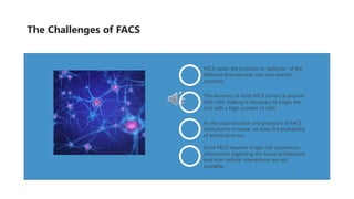
FACS raises the problem of ‘spillover’ of the
different fluorophores into non-specific
channels.
The recovery of most FACS sorters is around
50%-70% making it necessary to begin the
sort with a high number of cells.
As the sophistication and precision of FACS
instruments increase, so does the probability
of technical errors.
Since FACS requires single-cell suspension,
information regarding the tissue architecture
and inter-cellular interactions are not
available.
The Challenges of FACS
Immuno-magnetic separation of cells is based on the
deflection of cells in a magnetic gradient field.
Based on the type of magnetic particles used,
immunomagnetic separation are mainly of two kinds –
Dynabeads®-based and MACS™ technologies. Dynabeads® can be coupled with antibodies
or other ligands to targeting of specific cells
or immunoprecipitation reactions.
The target – or non-target cells depending on
the selection strategy – are labeled with 50nm
magnetic microbeads conjugated antibodies
and then subjected to a magnetic field.
Dynabeads®
MACS
Immuno-magnetic
techniques can be
used for both positive
and negative selection
of cells.
Magnetic Separation
MACS is a high throughput,
selective and rapid method for
isolating target populations or
removing undesired cells.
Advantages
Advantages
MACS can be scaled up or down
depending on the desired yield
and the downstream applications.
Advantages
It can be extended to a wide range
of cells and over various platforms
including microfluidic devices.
Advantages
The MACS columns help isolate
rare cells with high specificity.
The Advantages of Magnetic Separation
In case sorting with Dynabeads®, the latter need to be eluted
out owing to their large sizes as it may affect downstream
assays.
Separating cells using magnet separators is not very efficient
as draining out the unbound cells.
For delicate cells, binding of the magnetic particles may cause
mechanical shear. MACS columns should be used instead of
separators to minimize the shear.
The Limitations of Magnetic Separation
Aptamers are single-stranded oligonucleotides
– DNA or RNA – that can bind to highly specific
targets based on structural conformation.
Aptamers were first generated using a
procedure called SELEX – Systemic Evolution of
Ligands by Exponential enrichment – which
involves the sequential binding of the
oligonucleotides to target molecules.
Aptamer Technology
Direct
binding
Target
induced
structural
switch
Sandwich
binding
Target
induced
dissociation
It is the simplest assay
where aptamers labeled
with a single moiety of a
fluorophore or a
luminophore directly bind
to the target and the
signal can be easily read
out with a suitable
detector.
In the absence of the target,
the fluorophore-labeled
DNA aptamer forms a
partial duplex with another
(non-specific) aptamer
labeled with a quencher.
When the target is
introduced, the aptamer
binds to the target, thereby
releasing the quencher
from the fluorophore and
triggering the latter’s signal.
The specific unlabeled
aptamers immobilized on
a solid phase first capture
the target cells, which is
followed by the addition
of the biotinylated version
of the same aptamers. The
signal is generated
through further binding of
streptavidin-conjugated
HRP to the biotin
molecules as in ELISA.
DNA aptamers coiled with
gold nanoparticles (AuNPs)
are used for this assay. As
soon as the aptamers see
the target, they uncoil and
bind to the target cells
and release the AuNPs.
The latter are precipitated
in a salt solution bringing
about a color change.
Aptamer Technology
The preparation of
aptamers is completely
in vitro and thus
obviates the use of live
animals.
The repertoire of
specific aptamers that
can be synthesized for
targeting any cell is
virtually limitless.
The chemical synthesis
of aptamers and the
scale-out PCRs are easy
to perform and cost-
effective.
The aptamer assays are
highly reproducible, and
variability is generally
very low between
different batches.
Advantages of FACS
Due to the vast number of aptamers generated, there
is always the risk of non-specific binding. Therefore
several clones need to be tested for specificity which
can be time-consuming.
The safety and bio-compatibility concerns of aptamers
have not been addressed so far, therefore it not yet
approved for clinical applications.
The cell yield is very low which does not make it an
ideal method for the isolation of rare cells.
Certain cell isolation techniques make use of more than one cell characteristic (as classified above) and are therefore
the combination of at least two different techniques.
The objective is to either combine the advantages of both techniques or overcome the limitations of one of them.
The combination is usually that of an immuno-label-based technique with a label-free technique utilizing a cell property
like density, size, morphology, etc.
Combination Isolation Techniques
USA
Address: SUITE 115, 17 Ramsey Road, Shirley, NY
11967, USA
Tel: 1-631-626-9181
Fax: 1-631-614-7828
info@creative-bioarray.com
Europe
Tel: 44-208-123-7131
info@creative-bioarray.com
Email: info@creative-bioarray.com
Contact Us

More Related Content

PPTX
Cell Sorting Techniques
PPT
cell isolation and cell isolation methods
PPT
cell isolation and its method for all types of cell
PDF
Mastering the Challenges of Primary Cell Culture: Unlocking True In Vivo Insi...
DOCX
Characterization strategies for primary cell culture term paper
PPTX
Cancer: Underlying pathophysiological mechanisms and novel treatment approach...
PDF
Cancer: Underlying pathophysiological mechanisms and novel treatment approach...
PPTX
Cell synchronization
Cell Sorting Techniques
cell isolation and cell isolation methods
cell isolation and its method for all types of cell
Mastering the Challenges of Primary Cell Culture: Unlocking True In Vivo Insi...
Characterization strategies for primary cell culture term paper
Cancer: Underlying pathophysiological mechanisms and novel treatment approach...
Cell synchronization

Similar to A Comprehensive Review of Cell Isolation Methods (20)

PDF
Primary cell culture guide
PPTX
Animal cell culture studies in Bpharm III sem.pptx
PPTX
CHARACTERISATION OF CELLS LINES
PDF
An Overview Of Cell Culture Extended-dikompresi.pdf
PDF
Animal cell culture (Basics)
PPT
Basics of animal cells culture
PPTX
Mouse Mesenchymal Stem cells: Advanced pre-screening tools for Toxicological ...
PPTX
Protoplast culture
PPTX
Animal cells basic concepts
PPTX
Laboratory diagnosis of neoplasm
PDF
Advancements in Cell Culture Research: Custom Cell Isolation
PPTX
Cell proliferation assay services
PPT
12. Cell synchronization and Immortalization
PPTX
Screening of anticancer drugs
PPTX
Applications of Flow Cytometry | Cell Analysis
PDF
Virus Isolation II.pdf
PPTX
Animal cell culture
PPTX
Alternative animal experimentation technique
PDF
Flow Cytometry: Guide to Understanding Applications and Benefits | The Lifesc...
PPT
1. Hallmarks of cancer.ppt
Primary cell culture guide
Animal cell culture studies in Bpharm III sem.pptx
CHARACTERISATION OF CELLS LINES
An Overview Of Cell Culture Extended-dikompresi.pdf
Animal cell culture (Basics)
Basics of animal cells culture
Mouse Mesenchymal Stem cells: Advanced pre-screening tools for Toxicological ...
Protoplast culture
Animal cells basic concepts
Laboratory diagnosis of neoplasm
Advancements in Cell Culture Research: Custom Cell Isolation
Cell proliferation assay services
12. Cell synchronization and Immortalization
Screening of anticancer drugs
Applications of Flow Cytometry | Cell Analysis
Virus Isolation II.pdf
Animal cell culture
Alternative animal experimentation technique
Flow Cytometry: Guide to Understanding Applications and Benefits | The Lifesc...
1. Hallmarks of cancer.ppt
Ad

More from Creative-Bioarray (20)

PPTX
Enrichment, Isolation and Characterization of CTCs
PPTX
A Comprehensive Review of Stem Cell Research
PPTX
A Brief Guide to Tissue Fixation and Its Importance in Pathology Analysis
PPTX
A Comprehensive Review of Cell Culture Media
PPTX
Multiplexing Immunohistochemistry: A Powerful Tool for Comprehensive Tissue A...
PPTX
Patch Clamp Assay - A detailed step-by-step description of the standard patch...
PDF
Kinase Screening
PDF
RNA ISH ASSAY
PPTX
Immunohistochemistry
PPTX
Flow cytometry
PPTX
Caco-2 Permeability Assay
PPTX
Comparison of caco 2 with other cell-based models for intestinal permeability...
PPTX
Caco 2 cell permeability assay
PPTX
Technologies to study kinases
PDF
Differentiation of ipsc
PDF
Cell Viability
PDF
Spotlight on ipsc reprogramming
PPTX
Flow cytometry
PPTX
Histology services
PPTX
Atp binding cassette (abc) transporter
Enrichment, Isolation and Characterization of CTCs
A Comprehensive Review of Stem Cell Research
A Brief Guide to Tissue Fixation and Its Importance in Pathology Analysis
A Comprehensive Review of Cell Culture Media
Multiplexing Immunohistochemistry: A Powerful Tool for Comprehensive Tissue A...
Patch Clamp Assay - A detailed step-by-step description of the standard patch...
Kinase Screening
RNA ISH ASSAY
Immunohistochemistry
Flow cytometry
Caco-2 Permeability Assay
Comparison of caco 2 with other cell-based models for intestinal permeability...
Caco 2 cell permeability assay
Technologies to study kinases
Differentiation of ipsc
Cell Viability
Spotlight on ipsc reprogramming
Flow cytometry
Histology services
Atp binding cassette (abc) transporter
Ad

Recently uploaded (20)

PDF
Optimiser vos workloads AI/ML sur Amazon EC2 et AWS Graviton
PDF
Mobile App Security Testing_ A Comprehensive Guide.pdf
PDF
Diabetes mellitus diagnosis method based random forest with bat algorithm
PDF
Dropbox Q2 2025 Financial Results & Investor Presentation
PPTX
KOM of Painting work and Equipment Insulation REV00 update 25-dec.pptx
PDF
Chapter 3 Spatial Domain Image Processing.pdf
PDF
Review of recent advances in non-invasive hemoglobin estimation
PDF
Encapsulation theory and applications.pdf
PPTX
20250228 LYD VKU AI Blended-Learning.pptx
PDF
A comparative analysis of optical character recognition models for extracting...
PDF
gpt5_lecture_notes_comprehensive_20250812015547.pdf
PDF
7 ChatGPT Prompts to Help You Define Your Ideal Customer Profile.pdf
PDF
Build a system with the filesystem maintained by OSTree @ COSCUP 2025
PDF
cuic standard and advanced reporting.pdf
PDF
Machine learning based COVID-19 study performance prediction
PDF
Empathic Computing: Creating Shared Understanding
DOCX
The AUB Centre for AI in Media Proposal.docx
PPTX
Digital-Transformation-Roadmap-for-Companies.pptx
PPTX
A Presentation on Artificial Intelligence
PDF
Agricultural_Statistics_at_a_Glance_2022_0.pdf
Optimiser vos workloads AI/ML sur Amazon EC2 et AWS Graviton
Mobile App Security Testing_ A Comprehensive Guide.pdf
Diabetes mellitus diagnosis method based random forest with bat algorithm
Dropbox Q2 2025 Financial Results & Investor Presentation
KOM of Painting work and Equipment Insulation REV00 update 25-dec.pptx
Chapter 3 Spatial Domain Image Processing.pdf
Review of recent advances in non-invasive hemoglobin estimation
Encapsulation theory and applications.pdf
20250228 LYD VKU AI Blended-Learning.pptx
A comparative analysis of optical character recognition models for extracting...
gpt5_lecture_notes_comprehensive_20250812015547.pdf
7 ChatGPT Prompts to Help You Define Your Ideal Customer Profile.pdf
Build a system with the filesystem maintained by OSTree @ COSCUP 2025
cuic standard and advanced reporting.pdf
Machine learning based COVID-19 study performance prediction
Empathic Computing: Creating Shared Understanding
The AUB Centre for AI in Media Proposal.docx
Digital-Transformation-Roadmap-for-Companies.pptx
A Presentation on Artificial Intelligence
Agricultural_Statistics_at_a_Glance_2022_0.pdf

A Comprehensive Review of Cell Isolation Methods

  • 1. A Comprehensive Review of Cell Isolation Methods Isolation of one or multiple cell types from a heterogeneous population is an integral part of modern biological research and routine clinical diagnosis and treatment. Purification of specific cells is essential for basic cell biology research, cellular enumeration in specific pathologies and cell-based regenerative therapies. The central principle of separating any cell type from a population is to utilize one or more properties that are unique to that cell type.
  • 2. Basic Principle of Cell Isolation A cell isolation procedure can either be a positive selection or a negative selection – the former aims at isolating the target cell type from the entire population, usually with specific antibodies while the latter strategy involves the depletion of all cell types of the population resulting in only the target cells remaining.
  • 3. Due to the use of specific antibodies targeting a particular cell type, positive selection yields a higher purity of the desired population. Furthermore, a cell population isolated through positive selection can be sequentially purified through several cycles of the procedure, a benefit that negative selective cannot provide. Positive Selection Negative Selection It is more complicated to design an antibody cocktail to deplete all the non-target cells making negative selection less efficient vis-à-vis purity. Positively selected cells carry antibodies and other labeling agents that may interfere with downstream culture and assays – if that is a concern, it is preferable to use a negative selection method. VS
  • 4. Cell Characteristics As The Basis For Cell Separation The physical properties of size and density are commonly used for the bulk recovery of cells; either by sedimentation, filtration or density gradient centrifugation. Cell size and density Specific binding of surface antigens to either antibodies or aptamers can selectively capture cells of the specific surface phenotype. The captured cells are subsequently detected with the help of measurable probes. Surface markers This feature determines the extent of attachment of cells to plastic and other polymer surfaces and can be used to separate adherent cells from suspension/free-floating cells. Surface charge & adhesion Different cell types can be distinguished by shape, histological staining, media- selective growth, redox potential and other visual and behavioral properties which can then be harnessed to isolate those cells. Cell morphology & physiology
  • 5. Adherent Cells These cells require a suitable surface to attach to thrive. This attachment occurs through the interaction between the cell adhesion molecules (CAMs) on the cell surface and the corresponding ligands on the culture surface. These cells naturally do not require an attachment surface and occur in suspension in the body, usually in fluids like blood and lymph. Examples include lymphocytes, granulocytes, and other immune cells. Cancer cells that have lost contact inhibition and anchorage dependency. In serum-free conditions without any anchorage facilities, cancer cells can form spheroids and amorphous aggregates. Suspension Cells Cancer Cells
  • 6. Cell Isolation Through Adhesion Properties Adherent Culture Suspension Culture Adherent cultures – example: macrophages, fibroblasts, mesenchymal cells Macrophages are routinely isolated from bone marrow or peripheral blood. Right after the isolation of mononuclear cells, they can be seeded on coated polystyrene plates along-with serum and monocyte/macrophage differentiation cytokine cocktail. After 5-7 days, the cells differentiate and form an adherent monolayer while the unwanted cells remain in suspension and are discarded. Suspension cultures – examples: stem cells, embryoid bodies, tumorspheres. Cells naturally growing in suspension and cells that have lost anchorage dependency can be separated from the adherent cells by culturing in ultra-low attachment plates in the absence of serum. The desired cells either grow in a single- cell suspension or aggregate to form floating spheroids. The adherent cells die out without the support of an attachment surface.
  • 7. Advantages and Limitations of Adhesion-Based Isolation Adhesion-based methods are technically simple, reproducible and mostly cost-effective which yield a good crop of the cells. The limitation is that the purity of the recovered cells is low and there is always a risk of cross-contamination with other cells as well as with bacteria.
  • 8. Cell Density/Size-Based Separation Separation techniques relying on the size and density of cells are frequently used to separate specific cell types from bulk peripheral blood (PB) and bone marrow (BM). The most commonly used methods in this category include density gradient centrifugation and filtration. They are also called ‘bulk sorting’ methods since they help isolate relatively large cell populations in a short duration. Although bulk sorting methods are an essential part of several clinical and biotechnology procedures because of high yields, the purity and homogeneity of the harvested cells are quite low compared to other cell separation procedures.
  • 9. Density Gradient Centrifugation Filtration The biological sample is centrifuged in a suitable gradient medium at the appropriate speed till the different cell types are fractionated into different layers or phases depending on their respective densities. Filtration-based cell separation is a size- based method wherein cells smaller than the pore size of the specific filtration device pass through and larger cells are trapped. Advantages and Limitations Advantages and Limitations The procedure is technically simple and cost-effective. It can be scaled up or down as per requirement with minimal adjustments. The yield of cells obtained, especially for blood samples, is high. The purity of the different cell fractions obtained is low, especially when fractionating blood. The procedure is time-consuming and low throughput. Simple, easy to perform and reproducible. Filters containing the captured cells can be directly used in downstream assays. Poor specificity and purity – rare cells are frequently lost, and false positives are common. The surface phenotype of some cancer cells is lost during the process, which affects downstream assays.
  • 10. A specific cell type can be selected over unwanted cell populations by culturing them in a medium that provides some selective advantage to the desired cell type. In routine cell culture, a particular cell type can be enriched by adding specific growth factors and cytokines – typical examples include enriching populations of a specific lineage and differentiation stage. Methods Based on Cell Physiology and Morphology
  • 11. This strategy can be best explained by the HAT medium selection often used in hybridoma technology. HAT stands for hypoxanthine-aminopterin- thymidine: aminopterin blocks de-novo DNA synthesis and hypoxanthine and thymidine provide the raw materials for the alternative ‘salvage pathway’. The key enzyme needed for the salvage pathway is HGPRT – hypoxanthine guanine phosphoribosyltransferase and only cells expressing the HGPRT gene can survive in the HAT medium. Metabolic/Biosynthetic Enzymes If the transfected cells express an antibiotic resistance gene, the specific antibiotic is added to the medium at a concentration that is lethal to the cells which do not express the resistance gene. Common antibiotics used for mammalian cell selection include bleomycin, puromycin and hygromycin. Antibiotic Resistance The Principle of Selective Growth
  • 12. The time and labor required for the expansion and maintenance of the relevant clones The emergence of spontaneous resistant clones that do not carry the gene of interest The high costs of some reagents and specialized media Reproducibility Relative ease of performance Adequate cell yield The Advantages and Limitations of Selective Media Isolation Advantages Limitations
  • 13. Laser capture microdissection (LCM) was developed to isolate pure cell populations from heterogeneous tissues on the basis of their natural morphology or specific histological/immunological staining. Laser Capture Microdissection A variety of source materials can be utilized for LCM technology: examples include live cells from cell cultures, formalin-fixed paraffin embedded tissues, frozen tissues, fresh tissues, metaphase spreads, and blood smears. Tissue sections (frozen or paraffin embedded) are the most commonly used samples and for optimal capture and analysis.
  • 14. The Advantages of LCM Depending on the laser voltage and tissue structure, as many as several thousands of cells can be collected in a short period A wide range of tissues can be subjected to LCM LCM microscope and platforms are easy to operate and can be easily synergized with other assays LCM collected cells can be successfully used for downstream assays requiring functionally active nucleic acids and proteins. Due to the high precision laser beams, the damage to adjacent tissues is minimal, and one tissue section can be probed several times
  • 15. For removal of selected cells post laser targeting; the tissue sections cannot be coverslipped. Without mounting medium and coverslip, the refractive index of the tissue is altered. Most LCM platforms employ lasers whose minimal spot size is 7.5µm, and this is not precise enough to isolate single cells. Another technical challenge that results due to tissue drying (especially frozen tissues) is the lack of adherence of the dissected tissue onto the film or inverted cap. The Limitations of LCM
  • 16. Website: www.creative-bioarray.com E-mail: info@creative-bioarray.com Cell Surface Marker- Based Techniques Cellular surfaces are populated with a multitude of ligands – proteins, carbohydrates, and glycoproteins - which perform various biological functions and give every cell a unique surface phenotype. Antibody-mediated cell detection and isolation techniques became commonplace with the discovery of the cluster of differentiation (CD) markers – surface receptors usually involved in cell signaling and adhesion. The unique CD profiles of different cell types are used to define and separate different populations within a tissue, isolate rare cells like adult stem cells, cancer stem cells, etc. and differentiate between control and treated cells depending on the experimental design.
  • 17. Fluorescence-Activated Cell Sorting (FACS) The Fluorescence Activated Cell Sorting (FACS) was invented by Bonner and Herzenberg for sorting viable cells with the help of fluorescent probes. The populations stained with different fluorophore- tagged antibodies can be separated by the different fluorescent signals that they generate – in terms of wavelength and intensities. FACS is used exclusively for the positive selection and isolation of cells.
  • 18. FACS is a highly sensitive and high throughput procedure for isolating cells from heterogeneous populations. It is the ideal method of simultaneously sorting multiple populations based on just their immuno-phenotype. FACS is a highly versatile technology that can separate cells based not only on surface markers but also cell size and granularity, cell cycle status, intracellular cytokine expression, metabolic status, etc. The Advantages of FACS
  • 19. FACS raises the problem of ‘spillover’ of the different fluorophores into non-specific channels. The recovery of most FACS sorters is around 50%-70% making it necessary to begin the sort with a high number of cells. As the sophistication and precision of FACS instruments increase, so does the probability of technical errors. Since FACS requires single-cell suspension, information regarding the tissue architecture and inter-cellular interactions are not available. The Challenges of FACS
  • 20. Immuno-magnetic separation of cells is based on the deflection of cells in a magnetic gradient field. Based on the type of magnetic particles used, immunomagnetic separation are mainly of two kinds – Dynabeads®-based and MACS™ technologies. Dynabeads® can be coupled with antibodies or other ligands to targeting of specific cells or immunoprecipitation reactions. The target – or non-target cells depending on the selection strategy – are labeled with 50nm magnetic microbeads conjugated antibodies and then subjected to a magnetic field. Dynabeads® MACS Immuno-magnetic techniques can be used for both positive and negative selection of cells. Magnetic Separation
  • 21. MACS is a high throughput, selective and rapid method for isolating target populations or removing undesired cells. Advantages Advantages MACS can be scaled up or down depending on the desired yield and the downstream applications. Advantages It can be extended to a wide range of cells and over various platforms including microfluidic devices. Advantages The MACS columns help isolate rare cells with high specificity. The Advantages of Magnetic Separation
  • 22. In case sorting with Dynabeads®, the latter need to be eluted out owing to their large sizes as it may affect downstream assays. Separating cells using magnet separators is not very efficient as draining out the unbound cells. For delicate cells, binding of the magnetic particles may cause mechanical shear. MACS columns should be used instead of separators to minimize the shear. The Limitations of Magnetic Separation
  • 23. Aptamers are single-stranded oligonucleotides – DNA or RNA – that can bind to highly specific targets based on structural conformation. Aptamers were first generated using a procedure called SELEX – Systemic Evolution of Ligands by Exponential enrichment – which involves the sequential binding of the oligonucleotides to target molecules. Aptamer Technology
  • 24. Direct binding Target induced structural switch Sandwich binding Target induced dissociation It is the simplest assay where aptamers labeled with a single moiety of a fluorophore or a luminophore directly bind to the target and the signal can be easily read out with a suitable detector. In the absence of the target, the fluorophore-labeled DNA aptamer forms a partial duplex with another (non-specific) aptamer labeled with a quencher. When the target is introduced, the aptamer binds to the target, thereby releasing the quencher from the fluorophore and triggering the latter’s signal. The specific unlabeled aptamers immobilized on a solid phase first capture the target cells, which is followed by the addition of the biotinylated version of the same aptamers. The signal is generated through further binding of streptavidin-conjugated HRP to the biotin molecules as in ELISA. DNA aptamers coiled with gold nanoparticles (AuNPs) are used for this assay. As soon as the aptamers see the target, they uncoil and bind to the target cells and release the AuNPs. The latter are precipitated in a salt solution bringing about a color change. Aptamer Technology
  • 25. The preparation of aptamers is completely in vitro and thus obviates the use of live animals. The repertoire of specific aptamers that can be synthesized for targeting any cell is virtually limitless. The chemical synthesis of aptamers and the scale-out PCRs are easy to perform and cost- effective. The aptamer assays are highly reproducible, and variability is generally very low between different batches.
  • 26. Advantages of FACS Due to the vast number of aptamers generated, there is always the risk of non-specific binding. Therefore several clones need to be tested for specificity which can be time-consuming. The safety and bio-compatibility concerns of aptamers have not been addressed so far, therefore it not yet approved for clinical applications. The cell yield is very low which does not make it an ideal method for the isolation of rare cells.
  • 27. Certain cell isolation techniques make use of more than one cell characteristic (as classified above) and are therefore the combination of at least two different techniques. The objective is to either combine the advantages of both techniques or overcome the limitations of one of them. The combination is usually that of an immuno-label-based technique with a label-free technique utilizing a cell property like density, size, morphology, etc. Combination Isolation Techniques
  • 28. USA Address: SUITE 115, 17 Ramsey Road, Shirley, NY 11967, USA Tel: 1-631-626-9181 Fax: 1-631-614-7828 info@creative-bioarray.com Europe Tel: 44-208-123-7131 info@creative-bioarray.com Email: info@creative-bioarray.com Email: info@creative-bioarray.com Contact Us