SlideShare a Scribd company logo
A p p r o a c h t o
G ra n u l o m a t o u s
Ly m p h A d e n i t i s
( G L A )
PA R T H I V M E H TA
A H M E D A B A D
Acknowledgements
TEAM Pulmocare
Granulomatous
Lymph Adenitis
(GLA)
Granulomatous Lymph Adenitis
(GLA)
• Granulomatous Lymph Adenitis is a
reactive phenomenon of lymph nodes
that displays a distinctive pattern of
chronic inflammation and histiocytes.
• It is caused by a heterogeneous
collection of diseases, including
reactive, infectious and malignant
etiologies.
GLA – Parthiv Mehta 20180805 MEHSANA
Granulomatous Lymph Adenitis
(GLA)
•Histologically, granulomatous
lymphadenitis (i.e. granulomas)
be divided into 3 distinct
–Based on the type of inflammation
present (acute v/s chronic), and
–whether or not necrosis
accompanies the inflammation
GLA – Parthiv Mehta 20180805 MEHSANA
Granulomatous Lymph Adenitis
(GLA)
GLA – Parthiv Mehta 20180805 MEHSANA
Approach
Approach
GLA – Parthiv Mehta 20180805 MEHSANA
Granulomatous Lymph Adenitis
(GLA)
GLA – Parthiv Mehta 20180805 MEHSANA
Granulomatous Lymph Adenitis
(GLA)
• The common causes of GLA are
• (A) Infective agents like mycobacteria, fungi,
parasites, etc. and
• (B) Non-infective etiologies like Sarcoidosis,
Foreign bodies, Wegener’s granulomatosis,
Crohn’s disease, etc. In addition,
• (C) Certain neoplasms are also known to be
associated with a granulomatous response in the
parenchyma e.g. Hodgkin’s disease
• Approach to Differential diagnosis and
management demand a skillful interpretation of
clinical findings and histology.
GLA – Parthiv Mehta 20180805 MEHSANA
Approach
GLA – Parthiv Mehta 20180805 MEHSANA
GLA - Etiology
• Acute v/s Chronic
• Caseating v/s Non-caseating
• Infective v/s Non-infective
• Neoplastic v/s Non-neoplastic
Granulomatous Lymph Adenitis
(GLA)
Noninfectious GLA
1) Sarcoidosis lymphadenitis 2) Sarcoid-like lymphadenitis 3)Berylliosis
Infectious GLA
A. Suppurative
1) Tularemia lymphadenitis 2) Cat scratch lymphadenitis
3) Yersinia lymphadenitis 4) Lymphogranuloma venereum
5) Fungal infection
B. Non-suppurative
1) Tuberculous Lymphadenitis 2) Atypical Mycobacterial infection
3) BCG-Lymphadenitis 4) Toxoplasma Lymphadenitis
5) Leprosy 6) Syphilis
7) Brucellosis 8) Fungal infection (Cryptococcus,
Histoplasma, Coccidioidomycosis,
Pneumocystis)
GLA – Parthiv Mehta 20180805 MEHSANA
Approach
GLA – Parthiv Mehta 20180805 MEHSANA
Approach 01
HISTORY
&
CLINICAL
EXAMIATION
E X T R E M E LY I M P O R TA N T, A N D C A N , I N
FA C T,
O F F E R C R I T I C A L D I A G N O S T I C C L U E S
History and Clinical Examination
• Factors that can assist in identifying the
etiology of lymphadenopathy include
– Age of patient,
– Duration of lymphadenopathy,
– Exposures,
– Associated symptoms, and
– Location (localized vs. generalized).
– Time course of enlargement,
– Tenderness to palpation,
– Recent infections, recent immunizations, and
medications.
GLA – Parthiv Mehta 20180805 MEHSANA
History and Clinical Examination
• History of exposure to infection or
infectious agents is a particularly helpful
clue.
• History of animal exposure helps to
suspect.
• Has the patient travelled and possibly
been exposed to area specific infectious
agents?
• Mycoses (fungal infections) are
endemic in many parts of the world,
Koch’s is most likely in family contacts.
GLA – Parthiv Mehta 20180805 MEHSANA
History and Clinical Examination
• Some granulomatous diseases have a particular
predilection for affecting certain body sites.
Eosinophilic Granuloma –––– Bone
Wegener’s Granulomatosis –– Face
Giant Cell Granuloma –––––– Oral Cavity
Granuloma Annulare ––––––– Skin
Various Infectious Agents –––– CNS
GLA – Parthiv Mehta 20180805 MEHSANA
Clue - 1 : Site of Lymph nodes
GLA – Parthiv Mehta 20180805 MEHSANA
Clue - 1 : Site of Lymph nodes
GLA – Parthiv Mehta 20180805 MEHSANA
Clue - 1 : Site of Lymph nodes
GLA – Parthiv Mehta 20180805 MEHSANA
Clue - 1 : Site of Lymph nodes
GLA – Parthiv Mehta 20180805 MEHSANA
Clue - 2: Character and size of
Lymph nodes
•Warmth, overlying erythema, tenderness, mobility,
fluctuance and consistency.
•A painless, hard, irregular mass or a firm, rubbery lesion
that is immobile or fixed may represent a malignancy,
although in general, qualitative characteristics are unable
to reliably predict malignancy.
•Shotty lymphadenopathy is the presence of multiple small
lymph nodes that feel like “buck shots” under the skin.
This usually implies reactive lymphadenopathy from viral
infection.
•Painful or tender lymphadenopathy is nonspecific and
may represent possible inflammation caused by
infection, but it can also be the result of hemorrhage
Approach 01
History & Clinical Examination
• Q.1 – Is that a Lymph node?
–Enlarged parotid
–Thyroglossal cyst
–Abscess
–Branchial cyst
GLA – Parthiv Mehta 20180805 MEHSANA
Approach 01
History & Clinical Examination
• Q. 2 Is it Acute of Chronic?
• Q.3 Localized or generalized??
• Q.4 Are there associated systemic /
localizing symptoms / signs???
• Q.5 What is the character?
–Tender / nontender
–Mobile & Separate / Fixed & matted
–Firm & rubbery / Painless & stone hard
GLA – Parthiv Mehta 20180805 MEHSANA
Approach 01
History & Clinical Examination
• Q.6. Are there usual epidemiological
clues?
–Exposure to TB
–Sexual exposure
–Travel
–Exposure to bird droppings
–Exposure to cats
GLA – Parthiv Mehta 20180805 MEHSANA
Approach 02
INVESTIGATIONS
J U D I C I O U S U S E C A N A D D T O
C O N F I D E N C E , C O S T E F F E C T I V E N E S S A N D
L E A D T O P R O G N O S T I C A T I O N
Investigation - Approach
• If history and physical examination findings
suggest a benign or self-limited process,
reassurance can be provided and follow-up
arranged if lymphadenopathy persists.
• Findings suggestive of infectious or
autoimmune etiologies may require specific
testing and treatment as indicated.
• If malignancy is considered unlikely based on
history and physical examination, localized
lymphadenopathy can be observed for four
weeks.
• Generalized lymphadenopathy should prompt
routine laboratory testing and testing for
Laboratory Diagnosis
• Usually, routine laboratory tests are
nondiagnostic; cultures of blood, skin lesions or
lymph nodes are frequently negative.
• If there is suspicion of an infectious agent,
there are specific serologic studies that can
confirm the diagnosis.
• Polymerase chain reaction (PCR) and
antibody titre also are very helpful.
• Direct and indirect fluorescence antibody
tests are also available and have good
sensitivities.
• FNAC and/or Biopsy are a must to establish
cause GLA – Parthiv Mehta 20180805 MEHSANA
Clues and Initial Testing
for GLA
Historical clues
Suggested
diagnoses
Initial testing
Fever, night sweats,
weight loss,
Node located in
supraclavicular,
popliteal, or iliac region,
Bruising, Splenomegaly
Leukemia,
Lymphoma,
Solid tumor
Metastasis
CBC, ESR
Imaging - USG or CT may
be considered
Biopsy- Nodal or Bone
Marrow
Fever, chills, malaise,
sore throat, nausea,
vomiting, diarrhea;
No other red flag
symptoms
Bacterial or viral
pharyngitis,
Hepatitis,
Influenza,
Mononucleosis,
Tuberculosis (if
exposed),
Rubella
Limited illnesses may not
require any additional
testing; (depending on
clinical assessment,
consider CBC, LFT,
cultures, and disease-
specific serologies as
needed)
Clues and Initial Testing
for GLA
Historical clues Suggested diagnoses Initial testing
High-risk sexual
behavior
Chancroid,
HIV infection,
Lympho-granuloma
Venereum,
Syphilis
HIV-1/HIV-2
immunoassay, Culture of
lesions,
Nucleic acid amplification
for chlamydia,
Migration inhibitory
factor
Test
GLA – Parthiv Mehta 20180805 MEHSANA
Clues and Initial Testing
for GLA
Historical clues
Suggested
diagnoses
Initial testing
Animal or food contact
Cats Cat-scratch disease
(Bartonella)
Serology and PCR
Toxoplasmosis Serology
Rabbits, or sheep or
cattle wool, hair, or
hides
Anthrax Per CDC guidelines
Brucellosis Serology and PCR
Tularemia Blood culture and
serology
Undercooked meat Anthrax Per CDC guidelines
Brucellosis Serology and PCR
Toxoplasmosis Serology
Clues and Initial Testing
for GLA
Historical clues
Suggested
diagnoses
Initial testing
Recent travel,
insect bites
Diagnoses based on
endemic region
Serology and testing
as indicated by
suspected exposure
Arthralgias, rash,
joint stiffness,
fever, chills,
muscle weakness
Rheumatoid
arthritis,
Sjögren syndrome,
dermatomyositis,
systemic lupus
erythematosus
Antinuclear antibody,
anti-doubled-
stranded DNA, ESR,
CBC,
Rheumatoid factor,
Creatine kinase,
electromyography, or
muscle biopsy as
indicated
GLA – Parthiv Mehta 20180805 MEHSANA
Histopathological Diagnosis
• Direct approach
–Needle
–True cut – Core Needle Biopsy
–Excision
GLA – Parthiv Mehta 20180805 MEHSANA
FNAC in Diagnosis of GLA
• Easy to perform
• Trained Physician / Pathologist /
Radiologist can do it
• Diagnostic yield approximately 60%
–More so in Lymphoma / Malignancy
–Cervical lymph nodes
Fine needle aspiration cytology (FNAC) in the diagnosis of granulomatous lymphadenitis
UMJ. 2006 Jan; 75(1): 59–64
Evaluation of fine needle aspiration cytology of lymph nodes in KMC, Teaching hospital.
KUMJ. 2009 Apr-Jun;7(26):139-42.
GLA – Parthiv Mehta 20180805 MEHSANA
FNAC in Diagnosis of GLA
Choice of node
• Largest node
• Avoid inguinal & axillary
• Supra clavicular-highest diagnostic yield
• Complications
GLA – Parthiv Mehta 20180805 MEHSANA
Excision Biopsy in Diagnosis of
GLA
• Incisional
–External approach
• Laparoscopic
–Thoracoscopic
–Mediastinoscopic
–Laparoscopic
Larger tissue.. All possible investigations
GLA – Parthiv Mehta 20180805 MEHSANA
Histological Diagnosis of GLA
• The histologic identification of granulomatous
inflammation is a helpful predictor of diagnostic
etiology.
• Histologic patterns (foreign-body, necrotizing,
non-necrotizing, suppurative, and a diffuse
histiocytic reaction) can narrow the clinical
differential diagnosis.
• A definitive diagnosis can often be made with the
aid of ancillary testing (special stains and or
molecular diagnostics).
• An open clinical dialogue between the clinician
and the pathologist allows for appropriate use
of ancillary laboratory techniques and
ultimately accurate and effective patient care.GLA – Parthiv Mehta 20180805 MEHSANA
Histological Diagnosis of GLA
• 23% of diagnoses could not identify
the specific etiology via H&E stain at
the time of biopsy.
• Etiology identification improved to
90.8% with clinical features,
radiographic findings, and improved
laboratory methodologies, including
molecular techniques, culture, IHC
profiles, and serologic values.
S. Mukhopadhyay, et al. Pulmonary necrotizing granulomas of unknown cause: Chest, 144 (3) (2013), pp.
813-824
GLA – Parthiv Mehta 20180805 MEHSANA
Radiodiagnosis
• USG - Surface
–Must
–Defines structure
–Helps identify site
–Precision
GLA – Parthiv Mehta 20180805 MEHSANA
Radiodiagnosis
• USG - EBUS
–Must
–Defines structure
–Helps identify site
–Precision
GLA – Parthiv Mehta 20180805 MEHSANA
Radiodiagnosis
• X-ray Chest
–Hilar Adenopathy
–Progress / Resolution
–Thoracic structures
–Associated Pulmonary Infiltrates
–Associated Pleural Pathology
GLA – Parthiv Mehta 20180805 MEHSANA
Radiodiagnosis
CT Scan
• Lymph nodes
• Probable structure
• Vascularity
• Associated thoracic pathology
• Likely approach
• Likely diagnosis clue - PET CT
GLA – Parthiv Mehta 20180805 MEHSANA
Approach 02
Investigations
1.CBC with differential….
–Atypical lymphocytosis
–Eosinophilia
–Pancytopenia
2. Biochemistry
–Serum Uric Acid
–Serum Kidney, Liver functions
GLA – Parthiv Mehta 20180805 MEHSANA
Approach 02
Investigations
Localized Adenopathy
1. Throat culture
2. Urethral/cervical swabs
3. Blood culture
4. USG / CT Scan
5. Biopsy
6. Bone Marrow biopsy
GLA – Parthiv Mehta 20180805 MEHSANA
Approach 02
Investigations
Generalised Adenopathy
1. Heterophile test (For IM due to EBV)
2. VDRL
3. Antibody titer of viruses, fungi,
toxoplasmosis
4. Anti nuclear antibodies
5. Rheumatoid factor
GLA – Parthiv Mehta 20180805 MEHSANA
Approach 02
Investigations
Hilar Adenopathy
1.Mantoux test
2.Chest X-RAY, CT
3.ACE determination
4.Bronchoscopy - EBUS
5.Mediastinoscopy
GLA – Parthiv Mehta 20180805 MEHSANA
Approach 03
DECISION
MAKING
and
GUIDANCE
W E L L I N F O R M E D P H Y S I C I A N I S T H E B E S T
P E R S O N T O H E L P
Decision Making
Decision Making
GLA – Parthiv Mehta 20180805 MEHSANA
Decision Making - 01
Decision Making - 02
Decision Making - 03
Guidance - Initial Management
• Patients worry about the cause of their abnormal
lymph nodes.
• To adequately address their fears, the physician
should ask the patient about his or her concerns
and respond to questions about specific diagnoses.
• When biopsy is deferred, the physician should
explain to the patient the rationale for waiting.
• Patients should be cautioned to remain alert for
the reappearance of the nodes because
lymphomatous nodes have been known to
temporarily regress.
GLA – Parthiv Mehta 20180805 MEHSANA
Approach
Final Comment
L E T U S M A K E L I F E S I M P L E
Tb or no TB?
Tb or no TB?
GLA – Parthiv Mehta 20180805 MEHSANA
Tb or no TB?
• In countries with high incidence of
TB, TB is considered firstly in differential
diagnosis of granulomatous diseases.
• Detailed analysis and physical
examinations should be done in
differential diagnosis of granulomatous
diseases, and
TB must be excluded.
GLA – Parthiv Mehta 20180805 MEHSANA
Tb or not TB?
• Organisms are easily detectable by
proper staining, immuno-histochemical
and molecular biological methods such
as PCR
• Accurate pathological and
microbiological diagnosis can lead to
precise treatment.
MDR / XDR TB must be excluded.GLA – Parthiv Mehta 20180805 MEHSANA
Let us make it simple..
Clinical Recommendation
Evidence
rating
USG should be used as the initial imaging
modality for children up to 14 years presenting
with a neck mass with or without fever.
C
CT Scan should be used as the initial imaging
modality for children older than 14 years and
adults presenting with solitary or multiple neck
masses.
C
Corticosteroids should be avoided until a
definitive diagnosis of lymphadenopathy is made
because they could potentially mask or delay
histologic diagnosis of leukemia or lymphoma.
C
FNAC may be used to differentiate malignant
from reactive lymphadenopathy.
C
AJFP. 2016 Dec 1;94(11):896-903.GLA – Parthiv Mehta 20180805 MEHSANA
Let us make it simple…
• In most patients, lymphadenopathy has a
readily diagnosable infectious cause.
• A diagnosis of less obvious causes can often
be made after considering the patient's age,
the duration of the lymphadenopathy and
whether localizing signs or symptoms,
constitutional signs or epidemiologic clues are
present.
• When the cause of the lymphadenopathy
remains unexplained, a three- to four-week
observation period is appropriate when the
clinical setting indicates a high probability of
benign disease.GLA – Parthiv Mehta 20180805 MEHSANA
Let us make it simple…
GLA
Arise, Awake..
Stop Not Till the Goal is Reached..

More Related Content

PPTX
Chronic Lymphocytic Leukemia
PPT
Non-Hodgkin’s Lymphoma (NHL).ppt
PPTX
MALToma
PPT
Acute myeloid leukemia
PPTX
Pre malignant lesions of skin
PPTX
leukemoid reaction and leukemia
PPTX
Ulcers of the intestine
Chronic Lymphocytic Leukemia
Non-Hodgkin’s Lymphoma (NHL).ppt
MALToma
Acute myeloid leukemia
Pre malignant lesions of skin
leukemoid reaction and leukemia
Ulcers of the intestine

What's hot (20)

PPTX
Generalized lymphadenopathy
PPT
non-hodgkin’s-lymphoma
PPTX
Myelodysplastic Syndromes ppt
PPTX
Paraneoplastic syndromes
PPTX
Non hodgkin lymphoma
PPTX
Ulcerative lesion 4 6-2016
PPTX
Cutaneous pseudolymphoma
PPTX
Paraneoplastic syndromes
PPTX
Myelodysplastic syndrome
PDF
PPTX
Myelodysplastic syndrome according to WHO 2016
PPTX
Adnexal tumours of the skin and familial syndromes.
PPTX
Immunofluorescence in dermatopathology
PPTX
Chronic myeloid Leukemia
PPTX
Chronic lymphocytic leukemia
PPTX
Giant cell lesions of bone
PPT
Thrombophilia
PPT
Trop spl syndr
PPTX
Chronic leukemias
Generalized lymphadenopathy
non-hodgkin’s-lymphoma
Myelodysplastic Syndromes ppt
Paraneoplastic syndromes
Non hodgkin lymphoma
Ulcerative lesion 4 6-2016
Cutaneous pseudolymphoma
Paraneoplastic syndromes
Myelodysplastic syndrome
Myelodysplastic syndrome according to WHO 2016
Adnexal tumours of the skin and familial syndromes.
Immunofluorescence in dermatopathology
Chronic myeloid Leukemia
Chronic lymphocytic leukemia
Giant cell lesions of bone
Thrombophilia
Trop spl syndr
Chronic leukemias
Ad

Similar to Approach to lymphadenitis pmm (20)

PPT
32577_Lymphadenopathy.ppt
PPT
Lymphadenopathy.ppt
PPTX
Approach to lymphnode pathology
PPT
Management of Lymphadenopathy
PPTX
Diagnosis and examination of swollen lymph nodes ppt.pptx
PPTX
Generalised Lymphadenopathy medicine .pptx
PPT
lymphadenopathy_and_malignancy.ppt
PPTX
Lymphadenopathy
PPTX
Journal club - Dr NM 28.2.25. pptx
PPTX
Eosinophils in lymph node
PPTX
Lymphoma clinical feature & dx-Dr.Rajesh
PPTX
Non neoplastic lymphadenopathy
PDF
38th Publication- JFMPC- 3rd Name.pdf
PPTX
Sutton 9 limfoma
PPTX
FlashPath - Sarcoidosis
PDF
Lymphadenopathy in general
PPTX
Generalized lymphadenopathy
PPTX
14. Hodgkins Lymphoma powerpoint presentation.pptx
PPT
Non hodgkin lymphoma
PPTX
Non- Hodgkins & Hodgkins lymphoma ppt presentation
32577_Lymphadenopathy.ppt
Lymphadenopathy.ppt
Approach to lymphnode pathology
Management of Lymphadenopathy
Diagnosis and examination of swollen lymph nodes ppt.pptx
Generalised Lymphadenopathy medicine .pptx
lymphadenopathy_and_malignancy.ppt
Lymphadenopathy
Journal club - Dr NM 28.2.25. pptx
Eosinophils in lymph node
Lymphoma clinical feature & dx-Dr.Rajesh
Non neoplastic lymphadenopathy
38th Publication- JFMPC- 3rd Name.pdf
Sutton 9 limfoma
FlashPath - Sarcoidosis
Lymphadenopathy in general
Generalized lymphadenopathy
14. Hodgkins Lymphoma powerpoint presentation.pptx
Non hodgkin lymphoma
Non- Hodgkins & Hodgkins lymphoma ppt presentation
Ad

More from Parthiv Mehta (20)

PPTX
Musical Intervention in ICU - Parthiv Mehta
PDF
COPD easy understanding 2020_march
PPTX
Pre operative pulmonary evaluation 2019
PPTX
Parthiv Mehta Rational Use of Antibiotics 20180722
PPTX
Nac in copd india experience_pmm_2018
PPTX
Reactive airway obstruction in children detection management_2018_pmm
PPTX
Bronchitis afpa rdmc_06_dr meghanaben mehta_20180422
PPT
Treatment of tb afpa rdmc_06_dr. amit thaker_20180422
PPTX
Oxygen theraphy afpa rdmc_06_dr. r.k.mehta_20180422
PPTX
Inhalation devices afpa rdmc_06_dr. dhiren mehta_20180422
PPTX
Pulmonary fibrosis afpa rdmc_06_dr. amit shah_20180422
PPTX
Acute exacerbation of bronchial asthma dr. mukesh bhatt afpa_rdmc_06_20180422
PPTX
Breathlessness afpa rdmc_06_dr. abhay dixit_20182204
PPT
Tb overview dr parthiv mehta-afpa_rdmc_05_20171210
PPTX
Surgery in thoracic diseases afpa rdmc_06_20182204
PPTX
Respiratory mcq rdmc_2018
PPTX
Pneumonia in pregnancy april2018 pmm_aogs
PPTX
Ali to ards in pregnancy parthiv mehta_2018_aogs
PPTX
Pneumonia in opd to icu 2018 pmm
PPTX
Traditional medicines in respiratory system 2018 pmm
Musical Intervention in ICU - Parthiv Mehta
COPD easy understanding 2020_march
Pre operative pulmonary evaluation 2019
Parthiv Mehta Rational Use of Antibiotics 20180722
Nac in copd india experience_pmm_2018
Reactive airway obstruction in children detection management_2018_pmm
Bronchitis afpa rdmc_06_dr meghanaben mehta_20180422
Treatment of tb afpa rdmc_06_dr. amit thaker_20180422
Oxygen theraphy afpa rdmc_06_dr. r.k.mehta_20180422
Inhalation devices afpa rdmc_06_dr. dhiren mehta_20180422
Pulmonary fibrosis afpa rdmc_06_dr. amit shah_20180422
Acute exacerbation of bronchial asthma dr. mukesh bhatt afpa_rdmc_06_20180422
Breathlessness afpa rdmc_06_dr. abhay dixit_20182204
Tb overview dr parthiv mehta-afpa_rdmc_05_20171210
Surgery in thoracic diseases afpa rdmc_06_20182204
Respiratory mcq rdmc_2018
Pneumonia in pregnancy april2018 pmm_aogs
Ali to ards in pregnancy parthiv mehta_2018_aogs
Pneumonia in opd to icu 2018 pmm
Traditional medicines in respiratory system 2018 pmm

Recently uploaded (20)

PPTX
surgery guide for USMLE step 2-part 1.pptx
PPTX
Slider: TOC sampling methods for cleaning validation
DOC
Adobe Premiere Pro CC Crack With Serial Key Full Free Download 2025
PDF
Deadly Stampede at Yaounde’s Olembe Stadium Forensic.pdf
PPTX
15.MENINGITIS AND ENCEPHALITIS-elias.pptx
PPT
Breast Cancer management for medicsl student.ppt
PPT
CHAPTER FIVE. '' Association in epidemiological studies and potential errors
PPT
OPIOID ANALGESICS AND THEIR IMPLICATIONS
PPTX
Electromyography (EMG) in Physiotherapy: Principles, Procedure & Clinical App...
PPTX
Pathophysiology And Clinical Features Of Peripheral Nervous System .pptx
PPTX
Uterus anatomy embryology, and clinical aspects
PPTX
CME 2 Acute Chest Pain preentation for education
PPTX
DENTAL CARIES FOR DENTISTRY STUDENT.pptx
PPTX
Imaging of parasitic D. Case Discussions.pptx
PPTX
CEREBROVASCULAR DISORDER.POWERPOINT PRESENTATIONx
PPT
ASRH Presentation for students and teachers 2770633.ppt
PPTX
Fundamentals of human energy transfer .pptx
PPT
genitourinary-cancers_1.ppt Nursing care of clients with GU cancer
PPTX
NEET PG 2025 Pharmacology Recall | Real Exam Questions from 3rd August with D...
PPTX
1 General Principles of Radiotherapy.pptx
surgery guide for USMLE step 2-part 1.pptx
Slider: TOC sampling methods for cleaning validation
Adobe Premiere Pro CC Crack With Serial Key Full Free Download 2025
Deadly Stampede at Yaounde’s Olembe Stadium Forensic.pdf
15.MENINGITIS AND ENCEPHALITIS-elias.pptx
Breast Cancer management for medicsl student.ppt
CHAPTER FIVE. '' Association in epidemiological studies and potential errors
OPIOID ANALGESICS AND THEIR IMPLICATIONS
Electromyography (EMG) in Physiotherapy: Principles, Procedure & Clinical App...
Pathophysiology And Clinical Features Of Peripheral Nervous System .pptx
Uterus anatomy embryology, and clinical aspects
CME 2 Acute Chest Pain preentation for education
DENTAL CARIES FOR DENTISTRY STUDENT.pptx
Imaging of parasitic D. Case Discussions.pptx
CEREBROVASCULAR DISORDER.POWERPOINT PRESENTATIONx
ASRH Presentation for students and teachers 2770633.ppt
Fundamentals of human energy transfer .pptx
genitourinary-cancers_1.ppt Nursing care of clients with GU cancer
NEET PG 2025 Pharmacology Recall | Real Exam Questions from 3rd August with D...
1 General Principles of Radiotherapy.pptx

Approach to lymphadenitis pmm

  • 1. A p p r o a c h t o G ra n u l o m a t o u s Ly m p h A d e n i t i s ( G L A ) PA R T H I V M E H TA A H M E D A B A D
  • 4. Granulomatous Lymph Adenitis (GLA) • Granulomatous Lymph Adenitis is a reactive phenomenon of lymph nodes that displays a distinctive pattern of chronic inflammation and histiocytes. • It is caused by a heterogeneous collection of diseases, including reactive, infectious and malignant etiologies. GLA – Parthiv Mehta 20180805 MEHSANA
  • 5. Granulomatous Lymph Adenitis (GLA) •Histologically, granulomatous lymphadenitis (i.e. granulomas) be divided into 3 distinct –Based on the type of inflammation present (acute v/s chronic), and –whether or not necrosis accompanies the inflammation GLA – Parthiv Mehta 20180805 MEHSANA
  • 6. Granulomatous Lymph Adenitis (GLA) GLA – Parthiv Mehta 20180805 MEHSANA
  • 8. Approach GLA – Parthiv Mehta 20180805 MEHSANA
  • 9. Granulomatous Lymph Adenitis (GLA) GLA – Parthiv Mehta 20180805 MEHSANA
  • 10. Granulomatous Lymph Adenitis (GLA) • The common causes of GLA are • (A) Infective agents like mycobacteria, fungi, parasites, etc. and • (B) Non-infective etiologies like Sarcoidosis, Foreign bodies, Wegener’s granulomatosis, Crohn’s disease, etc. In addition, • (C) Certain neoplasms are also known to be associated with a granulomatous response in the parenchyma e.g. Hodgkin’s disease • Approach to Differential diagnosis and management demand a skillful interpretation of clinical findings and histology. GLA – Parthiv Mehta 20180805 MEHSANA
  • 11. Approach GLA – Parthiv Mehta 20180805 MEHSANA
  • 12. GLA - Etiology • Acute v/s Chronic • Caseating v/s Non-caseating • Infective v/s Non-infective • Neoplastic v/s Non-neoplastic
  • 13. Granulomatous Lymph Adenitis (GLA) Noninfectious GLA 1) Sarcoidosis lymphadenitis 2) Sarcoid-like lymphadenitis 3)Berylliosis Infectious GLA A. Suppurative 1) Tularemia lymphadenitis 2) Cat scratch lymphadenitis 3) Yersinia lymphadenitis 4) Lymphogranuloma venereum 5) Fungal infection B. Non-suppurative 1) Tuberculous Lymphadenitis 2) Atypical Mycobacterial infection 3) BCG-Lymphadenitis 4) Toxoplasma Lymphadenitis 5) Leprosy 6) Syphilis 7) Brucellosis 8) Fungal infection (Cryptococcus, Histoplasma, Coccidioidomycosis, Pneumocystis) GLA – Parthiv Mehta 20180805 MEHSANA
  • 14. Approach GLA – Parthiv Mehta 20180805 MEHSANA
  • 15. Approach 01 HISTORY & CLINICAL EXAMIATION E X T R E M E LY I M P O R TA N T, A N D C A N , I N FA C T, O F F E R C R I T I C A L D I A G N O S T I C C L U E S
  • 16. History and Clinical Examination • Factors that can assist in identifying the etiology of lymphadenopathy include – Age of patient, – Duration of lymphadenopathy, – Exposures, – Associated symptoms, and – Location (localized vs. generalized). – Time course of enlargement, – Tenderness to palpation, – Recent infections, recent immunizations, and medications. GLA – Parthiv Mehta 20180805 MEHSANA
  • 17. History and Clinical Examination • History of exposure to infection or infectious agents is a particularly helpful clue. • History of animal exposure helps to suspect. • Has the patient travelled and possibly been exposed to area specific infectious agents? • Mycoses (fungal infections) are endemic in many parts of the world, Koch’s is most likely in family contacts. GLA – Parthiv Mehta 20180805 MEHSANA
  • 18. History and Clinical Examination • Some granulomatous diseases have a particular predilection for affecting certain body sites. Eosinophilic Granuloma –––– Bone Wegener’s Granulomatosis –– Face Giant Cell Granuloma –––––– Oral Cavity Granuloma Annulare ––––––– Skin Various Infectious Agents –––– CNS GLA – Parthiv Mehta 20180805 MEHSANA
  • 19. Clue - 1 : Site of Lymph nodes GLA – Parthiv Mehta 20180805 MEHSANA
  • 20. Clue - 1 : Site of Lymph nodes GLA – Parthiv Mehta 20180805 MEHSANA
  • 21. Clue - 1 : Site of Lymph nodes GLA – Parthiv Mehta 20180805 MEHSANA
  • 22. Clue - 1 : Site of Lymph nodes GLA – Parthiv Mehta 20180805 MEHSANA
  • 23. Clue - 2: Character and size of Lymph nodes •Warmth, overlying erythema, tenderness, mobility, fluctuance and consistency. •A painless, hard, irregular mass or a firm, rubbery lesion that is immobile or fixed may represent a malignancy, although in general, qualitative characteristics are unable to reliably predict malignancy. •Shotty lymphadenopathy is the presence of multiple small lymph nodes that feel like “buck shots” under the skin. This usually implies reactive lymphadenopathy from viral infection. •Painful or tender lymphadenopathy is nonspecific and may represent possible inflammation caused by infection, but it can also be the result of hemorrhage
  • 24. Approach 01 History & Clinical Examination • Q.1 – Is that a Lymph node? –Enlarged parotid –Thyroglossal cyst –Abscess –Branchial cyst GLA – Parthiv Mehta 20180805 MEHSANA
  • 25. Approach 01 History & Clinical Examination • Q. 2 Is it Acute of Chronic? • Q.3 Localized or generalized?? • Q.4 Are there associated systemic / localizing symptoms / signs??? • Q.5 What is the character? –Tender / nontender –Mobile & Separate / Fixed & matted –Firm & rubbery / Painless & stone hard GLA – Parthiv Mehta 20180805 MEHSANA
  • 26. Approach 01 History & Clinical Examination • Q.6. Are there usual epidemiological clues? –Exposure to TB –Sexual exposure –Travel –Exposure to bird droppings –Exposure to cats GLA – Parthiv Mehta 20180805 MEHSANA
  • 27. Approach 02 INVESTIGATIONS J U D I C I O U S U S E C A N A D D T O C O N F I D E N C E , C O S T E F F E C T I V E N E S S A N D L E A D T O P R O G N O S T I C A T I O N
  • 28. Investigation - Approach • If history and physical examination findings suggest a benign or self-limited process, reassurance can be provided and follow-up arranged if lymphadenopathy persists. • Findings suggestive of infectious or autoimmune etiologies may require specific testing and treatment as indicated. • If malignancy is considered unlikely based on history and physical examination, localized lymphadenopathy can be observed for four weeks. • Generalized lymphadenopathy should prompt routine laboratory testing and testing for
  • 29. Laboratory Diagnosis • Usually, routine laboratory tests are nondiagnostic; cultures of blood, skin lesions or lymph nodes are frequently negative. • If there is suspicion of an infectious agent, there are specific serologic studies that can confirm the diagnosis. • Polymerase chain reaction (PCR) and antibody titre also are very helpful. • Direct and indirect fluorescence antibody tests are also available and have good sensitivities. • FNAC and/or Biopsy are a must to establish cause GLA – Parthiv Mehta 20180805 MEHSANA
  • 30. Clues and Initial Testing for GLA Historical clues Suggested diagnoses Initial testing Fever, night sweats, weight loss, Node located in supraclavicular, popliteal, or iliac region, Bruising, Splenomegaly Leukemia, Lymphoma, Solid tumor Metastasis CBC, ESR Imaging - USG or CT may be considered Biopsy- Nodal or Bone Marrow Fever, chills, malaise, sore throat, nausea, vomiting, diarrhea; No other red flag symptoms Bacterial or viral pharyngitis, Hepatitis, Influenza, Mononucleosis, Tuberculosis (if exposed), Rubella Limited illnesses may not require any additional testing; (depending on clinical assessment, consider CBC, LFT, cultures, and disease- specific serologies as needed)
  • 31. Clues and Initial Testing for GLA Historical clues Suggested diagnoses Initial testing High-risk sexual behavior Chancroid, HIV infection, Lympho-granuloma Venereum, Syphilis HIV-1/HIV-2 immunoassay, Culture of lesions, Nucleic acid amplification for chlamydia, Migration inhibitory factor Test GLA – Parthiv Mehta 20180805 MEHSANA
  • 32. Clues and Initial Testing for GLA Historical clues Suggested diagnoses Initial testing Animal or food contact Cats Cat-scratch disease (Bartonella) Serology and PCR Toxoplasmosis Serology Rabbits, or sheep or cattle wool, hair, or hides Anthrax Per CDC guidelines Brucellosis Serology and PCR Tularemia Blood culture and serology Undercooked meat Anthrax Per CDC guidelines Brucellosis Serology and PCR Toxoplasmosis Serology
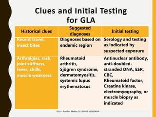
  • 33. Clues and Initial Testing for GLA Historical clues Suggested diagnoses Initial testing Recent travel, insect bites Diagnoses based on endemic region Serology and testing as indicated by suspected exposure Arthralgias, rash, joint stiffness, fever, chills, muscle weakness Rheumatoid arthritis, Sjögren syndrome, dermatomyositis, systemic lupus erythematosus Antinuclear antibody, anti-doubled- stranded DNA, ESR, CBC, Rheumatoid factor, Creatine kinase, electromyography, or muscle biopsy as indicated GLA – Parthiv Mehta 20180805 MEHSANA
  • 34. Histopathological Diagnosis • Direct approach –Needle –True cut – Core Needle Biopsy –Excision GLA – Parthiv Mehta 20180805 MEHSANA
  • 35. FNAC in Diagnosis of GLA • Easy to perform • Trained Physician / Pathologist / Radiologist can do it • Diagnostic yield approximately 60% –More so in Lymphoma / Malignancy –Cervical lymph nodes Fine needle aspiration cytology (FNAC) in the diagnosis of granulomatous lymphadenitis UMJ. 2006 Jan; 75(1): 59–64 Evaluation of fine needle aspiration cytology of lymph nodes in KMC, Teaching hospital. KUMJ. 2009 Apr-Jun;7(26):139-42. GLA – Parthiv Mehta 20180805 MEHSANA
  • 36. FNAC in Diagnosis of GLA Choice of node • Largest node • Avoid inguinal & axillary • Supra clavicular-highest diagnostic yield • Complications GLA – Parthiv Mehta 20180805 MEHSANA
  • 37. Excision Biopsy in Diagnosis of GLA • Incisional –External approach • Laparoscopic –Thoracoscopic –Mediastinoscopic –Laparoscopic Larger tissue.. All possible investigations GLA – Parthiv Mehta 20180805 MEHSANA
  • 38. Histological Diagnosis of GLA • The histologic identification of granulomatous inflammation is a helpful predictor of diagnostic etiology. • Histologic patterns (foreign-body, necrotizing, non-necrotizing, suppurative, and a diffuse histiocytic reaction) can narrow the clinical differential diagnosis. • A definitive diagnosis can often be made with the aid of ancillary testing (special stains and or molecular diagnostics). • An open clinical dialogue between the clinician and the pathologist allows for appropriate use of ancillary laboratory techniques and ultimately accurate and effective patient care.GLA – Parthiv Mehta 20180805 MEHSANA
  • 39. Histological Diagnosis of GLA • 23% of diagnoses could not identify the specific etiology via H&E stain at the time of biopsy. • Etiology identification improved to 90.8% with clinical features, radiographic findings, and improved laboratory methodologies, including molecular techniques, culture, IHC profiles, and serologic values. S. Mukhopadhyay, et al. Pulmonary necrotizing granulomas of unknown cause: Chest, 144 (3) (2013), pp. 813-824 GLA – Parthiv Mehta 20180805 MEHSANA
  • 40. Radiodiagnosis • USG - Surface –Must –Defines structure –Helps identify site –Precision GLA – Parthiv Mehta 20180805 MEHSANA
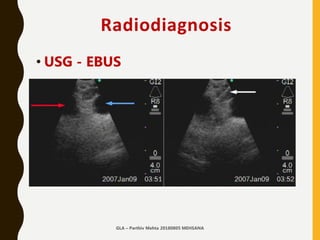
  • 41. Radiodiagnosis • USG - EBUS –Must –Defines structure –Helps identify site –Precision GLA – Parthiv Mehta 20180805 MEHSANA
  • 42. Radiodiagnosis • X-ray Chest –Hilar Adenopathy –Progress / Resolution –Thoracic structures –Associated Pulmonary Infiltrates –Associated Pleural Pathology GLA – Parthiv Mehta 20180805 MEHSANA
  • 43. Radiodiagnosis CT Scan • Lymph nodes • Probable structure • Vascularity • Associated thoracic pathology • Likely approach • Likely diagnosis clue - PET CT GLA – Parthiv Mehta 20180805 MEHSANA
  • 44. Approach 02 Investigations 1.CBC with differential…. –Atypical lymphocytosis –Eosinophilia –Pancytopenia 2. Biochemistry –Serum Uric Acid –Serum Kidney, Liver functions GLA – Parthiv Mehta 20180805 MEHSANA
  • 45. Approach 02 Investigations Localized Adenopathy 1. Throat culture 2. Urethral/cervical swabs 3. Blood culture 4. USG / CT Scan 5. Biopsy 6. Bone Marrow biopsy GLA – Parthiv Mehta 20180805 MEHSANA
  • 46. Approach 02 Investigations Generalised Adenopathy 1. Heterophile test (For IM due to EBV) 2. VDRL 3. Antibody titer of viruses, fungi, toxoplasmosis 4. Anti nuclear antibodies 5. Rheumatoid factor GLA – Parthiv Mehta 20180805 MEHSANA
  • 47. Approach 02 Investigations Hilar Adenopathy 1.Mantoux test 2.Chest X-RAY, CT 3.ACE determination 4.Bronchoscopy - EBUS 5.Mediastinoscopy GLA – Parthiv Mehta 20180805 MEHSANA
  • 48. Approach 03 DECISION MAKING and GUIDANCE W E L L I N F O R M E D P H Y S I C I A N I S T H E B E S T P E R S O N T O H E L P
  • 50. Decision Making GLA – Parthiv Mehta 20180805 MEHSANA
  • 54. Guidance - Initial Management • Patients worry about the cause of their abnormal lymph nodes. • To adequately address their fears, the physician should ask the patient about his or her concerns and respond to questions about specific diagnoses. • When biopsy is deferred, the physician should explain to the patient the rationale for waiting. • Patients should be cautioned to remain alert for the reappearance of the nodes because lymphomatous nodes have been known to temporarily regress. GLA – Parthiv Mehta 20180805 MEHSANA
  • 55. Approach Final Comment L E T U S M A K E L I F E S I M P L E
  • 56. Tb or no TB?
  • 57. Tb or no TB? GLA – Parthiv Mehta 20180805 MEHSANA
  • 58. Tb or no TB? • In countries with high incidence of TB, TB is considered firstly in differential diagnosis of granulomatous diseases. • Detailed analysis and physical examinations should be done in differential diagnosis of granulomatous diseases, and TB must be excluded. GLA – Parthiv Mehta 20180805 MEHSANA
  • 59. Tb or not TB? • Organisms are easily detectable by proper staining, immuno-histochemical and molecular biological methods such as PCR • Accurate pathological and microbiological diagnosis can lead to precise treatment. MDR / XDR TB must be excluded.GLA – Parthiv Mehta 20180805 MEHSANA
  • 60. Let us make it simple.. Clinical Recommendation Evidence rating USG should be used as the initial imaging modality for children up to 14 years presenting with a neck mass with or without fever. C CT Scan should be used as the initial imaging modality for children older than 14 years and adults presenting with solitary or multiple neck masses. C Corticosteroids should be avoided until a definitive diagnosis of lymphadenopathy is made because they could potentially mask or delay histologic diagnosis of leukemia or lymphoma. C FNAC may be used to differentiate malignant from reactive lymphadenopathy. C AJFP. 2016 Dec 1;94(11):896-903.GLA – Parthiv Mehta 20180805 MEHSANA
  • 61. Let us make it simple… • In most patients, lymphadenopathy has a readily diagnosable infectious cause. • A diagnosis of less obvious causes can often be made after considering the patient's age, the duration of the lymphadenopathy and whether localizing signs or symptoms, constitutional signs or epidemiologic clues are present. • When the cause of the lymphadenopathy remains unexplained, a three- to four-week observation period is appropriate when the clinical setting indicates a high probability of benign disease.GLA – Parthiv Mehta 20180805 MEHSANA
  • 62. Let us make it simple… GLA
  • 63. Arise, Awake.. Stop Not Till the Goal is Reached..