SlideShare a Scribd company logo
AWAKE INTUBATION
PREPARATION AND BLOCKS
ZIKRULLAH
BACKGROUND
• First awake intubation was performed by Dr.
Macewend in 1880 without topical or regional
blocks , sedatives, analgesics.
When do we need Awake Intubation??
• History of the patient and Airway assessment is
suggestive of a
-Difficult mask ventilation.
-Difficult conventional Laryngoscopy and
Intubation.
• In such circumstances, awake intubation becomes
the most prudent and secure option to maintain the
patency of Airway. 3
DIFFICULT AIRWAY
GENERAL ANESTHESIA
+/- PARALYSIS
RECOGNIZED
PROPER
PREPARATION
ASA DIFFICULT AIRWAY ALGORITHM
UNRECOGNIZED
AWAKE
INTUBATION
CHOICES
SUCCEED
FAIL
SURGICAL
AIRWAY
MASK
VENTILATION
NO
YES
EMERGENCY
PATHWAY
NON -EMERGENCY
PATHWAY
LMA
COMBITUBE
TTJV
INTUBATION
CHOICES
INTUBATION
CHOICES
SURGICAL
AIRWAY
SUCCEED
FAIL
CONFIRM
ANESTHESIA
WITH MASK
VENTILATION
AWAKEN
SURGICAL
AIRWAY
EXTUBATE
OVER JET
STYLET
REGIONAL
ANESTHESIA
CANCEL
CASE
REGROUP
Intubation choices include use of different
laryngoscope blades, LMA as an intubation
conduit (with or without fiberoptic guidance),
fiberoptic intubation, intubating stylet or tube
changer, light wand, retrograde intubation,
and blind oral or nasal intubation.
*
*
*
AWAKEN
WHY AWAKE INTUBATION IN Pt. WITH DA ???
1. The natural airway is preserved
2. Spontaneous breathing is maintained
3. A patient who is awake and well topicalized is
easier to intubate
4. The patient can still protect his or her airway
from aspiration.
5. Patients are able to monitor their own
neurologic symptoms (for example, the patient
with potential cervical pathology).
INDICATIONS FOR AWAKE INTUBATION
1. Previous history of difficult intubation.
2. Anticipated difficult airway (assessed on physical
examination) .
3. Trauma to the following:
Face
Upper airway
Cervical spine
4. Anticipated difficult mask ventilation
5. Severe risk of aspiration
6. Respiratory failure
7. Severe hemodynamic instability
6
Elements of Awake Intubation(AI)
Element Underlying Concept or Action
Explanation Patients understand Safety
Desiccation Dry the Airway
Dilatation Prepare the Nose
Tropicalization Obtund Reflexes
Sedation Maintain the patient’s airway
control
Procrastination AI Cannot and Should not be
RUSHED
THE PREOPERATIVE VISIT
A. REVIEWING OLD CHARTS
Focus on four important features:
• Degree of difficulty of the endotracheal intubation
• Positioning of the patient during laryngoscopy
• Equipment used
• Whether the technique that was used previously is
familiar
B. PREPARATION OF THE PATIENT
• A detailed explanation of the technique is
provided, and questions are answered.
• Reason should be explained to the patient,
emphasizing that this is done for the patient's
safety.
• The patient is informed of what he or she can do
to assist in a smooth intubation.
PREMEDICATION
• Alleviate anxiety
• Provide a clear and dry airway
• Protect against the risk of aspiration
• Enable adequate topicalization of the airway
A. SEDATIVES/HYPNOTICS
• 1. BENZODIAZEPINES
• Excellent anxiolytics, amnestics, sedatives, and
hypnotics.
• MIDAZOLAM
• Dosages : 0.07 -0.15 mg/kg i.m.
• 0.01 – 0.1 mg/kg i.v.
• decrease the dose (∼30%) if narcotics or other central
nervous system depressants are administered
concomitantly.
• 2. OPIOIDS
• Sedatives
• Antitussive properties depress laryngeal reflexes
further
• Alter the respiratory pattern to a slower and
deeper rhythm.
• FENTANYL: 1 to 2 μg/kg i.v.
• onset in about 2 to 3 minutes
• Transmucosally, the onset of effect is 5 to 15
minutes, with peak analgesia within 20 to 30
minutes
• SUFENTANIL: 1 μg/kg i.v. onset in 1 to 3 minutes
• 3. INTRAVENOUS HYPNOTICS
• DEXMEDETOMIDINE:
• Highly specific α2-adrenoreceptor agonist
• Sedative, analgesic, without a change in ventilatory status
• Dose :1 μg/kg IV over 10 minutes
• :infusion of 0.2 to 0.7 μg/kg/hr
• PROPOFOL
• Sedating dosage : 0.25 mg/kg IV
• Excellent sedative with a smooth plane achieved
• Good acceptance by patients
• Amnesia, adequate anxiolysis, and beneficial antiemetic
properties.
B. ASPIRATION PROPHYLAXIS
• patients with full stomach (e.g., trauma victims) or be obese with DA
• H2-RECEPTOR ANTAGONISTS
• Cimetidine and Ranitidine
• Selective and competitive antagonists that block secretion ofH+
• Ranitidine : 0.25-1.0 mg/kg i.v
• Cimetidine : 5-10 mg /kg PO
• METOCLOPRAMIDE
• Dopamine antagonist
• Doses are 0.15 to 0.3 mg/kg IV.
• For complete aspiration prophylaxis, a combination of
nonparticulate antacid, H2-receptor blocking agent, and
metoclopramide may be used.
• C. ANTISIALOGOGUES
• Secretions obscure the view
• Prevents the local anesthetics from reaching intended areas,
• Administered approximately 1.5 hours prior to the
application of local anesthetics
• Atropine:
• Dosage : 0.01-0.02 mg/kg i.v.
• : 0.02 mg/kg i.m.
• Glycopyrrolate :
• Dose: 0.005-0.01 mg/kg
• Onset in 1 to 2 minutes
• 20 min. before intubation
D. Nasal mucosal vasoconstrictors
• Patient requires awake nasal intubation,
adequate anesthesia of this area, along with
vasoconstriction, is essential
• Agents commonly used are 4% cocaine and 2%
lidocaine with 1% phenylephrine
• Adequate anesthesia and vasoconstriction can be
achieved in 10 to 15 minutes
• Nasal decongestants 0.025% to 0.05%
oxymetazoline hydrochloride nasal solution
sprayed twice in each nostril
PREOPERATIVE PREPARATIONS
• 1. STAFF
• One additional individual to serve as an
assistant in DA management.
• Preferred whenever possible is a second
anesthesiologist who can assist in the
monitoring and ventilation of the patient
• 2. MONITORS
• During AI, the routine use of ECG, noninvasive
BP monitor, pulse oximetry, capnography, and
a precordial stethoscope is required as part of
standard basic intraoperative monitoring
• 3. SUPPLEMENTAL OXYGEN
• Supplemental oxygen (O2) should be considered
during the entire process of DA management
• In addition to the standard methods there are
nonconventional methods for increasing the
fractional concentration of o2in inspired gas
(fio2):
• suction port of the FOB,
• Atomizer during topicalization, or
• Elective transtracheal jet ventilation (TTJV)
4 . AIRWAY EQUIPMENT
• Suggested Contents of the Portable Unit for Difficult Airway
Management :
• 1. Rigid laryngoscope blades
• 2. Endotracheal tubes of assorted sizes.
• 3. Endotracheal tube guides.
• 4. Fiberoptic intubation equipment.
• 5. Retrograde intubation equipment.
• 6. At least one device for emergency nonsurgical airway
ventilation.
• 7. Equipment suitable for emergency surgical airway
access
• 8. An exhaled CO2 detector.
• 9. Pulse oximetry unit.
• 10. Portable O2 tank.
TOPICALIZATION
• A. LIDOCAINE
• Available in various preparations including
aqueous (1%, 2%, and 4%) and viscous (1%)
solutions, ointment (1%), and aerosol preparation
• Xylocaine 10% metered-dose oral spray (astra)
delivers 10 mg per spray and rapidly anesthetizes
the upper airway.
• B. BENZOCAINE
• Benzocaine is available for use as 10%, 15%, and
20% solutions
• C. COCAINE
• Local anesthesia and vasoconstriction when applied
topically
• Available as 4% and 10% solutions, for topical application.
• The 10% solution is not used because of a very high
incidence of toxic effects.
• The maximum dose should not exceed 200 mg or 1 mg/kg.
• Cocaine should be used with caution in patients with
known hypersensitivity, coronary artery disease,
hypertension, pseudocholinesterase deficiency,
preeclampsia or hyperthyroidism as well as in children,
elderly patients, and patients receiving monoamine oxidase
inhibitors
• E. Application Techniques
• 1. ATOMIZERS
• Tetracaine : 0.3% to 0.5% with epinephrine
1:200,000 maximum 4 to 7 mL,
• Lidocaine :4% maximum 10 mL,
• placed in an atomizer , connected to the oxygen
tank (flow, 8 to 10 L/min), and sprayed in the
oropharynx for 10-second periods with 20-
second rest intervals for about 20 minutes
Awake intubation
• 2. NEBULIZERS
• The ultrasonic nebulizer utilizes 5 mL of 4%
lidocaine to be nebulized with O2 (6 to 8 L/min).
• The size of the droplet depends on the flow of O2
and the type of nebulizer.
• With O2 flow less than 6 L/min, droplet sizes of 30
to 60 μm can be achieved, coating the mucosa up
to the trachea.
• Other less commonly used techniques for topical
anesthesia include lozenges (amethocaine
lozenges 60 mg) and gargle with 4% lidocaine gel.
• NERVE BLOCKS
SENSORY NERVE SUPPLY 0F AIRWAY
• A. NASAL CAVITY AND NASOPHARYNX
• The majority of the innervation is derived
from two sources: the sphenopalatine
ganglion and the anterior ethmoidal nerve.
• Located in the pterygopalatine fossa
posterior to the middle turbinate.
• The greater and lesser palatine nerves,
provide sensory innervation to the nasal
turbinates and to two thirds of the posterior
nasal septum
SPHENOPALATINE NERVE BLOCK:
NASAL APPROACH
• Long cotton-tipped applicators soaked in either
4% cocaine or 4% lidocaine with epinephrine
1:200,000 applied over the mucosal surface
overlying the ganglion
• The applicator should be angled at approximately
45-degree angle to the hard palate until the upper
posterior wall of the nasopharynx (sphenoid bone)
is reached.
• The applicators are then left in place for
approximately 5 to 10 minutes
Awake intubation
• ANTERIOR ETHMOIDAL NERVE BLOCK
• The anterior ethmoidal nerve is blocked by the
insertion of a long cotton-tipped applicator, soaked
in either 4% cocaine or 4% lidocaine with
epinephrine 1:200,000, that is placed parallel to the
dorsal surface of the nose until it meets the anterior
surface of the cribriform plate .
• The applicator is held in position for 5 to 10 minutes.
B. OROPHARYNX
• The somatic and visceral afferent fibers of the
oropharynx are supplied by a plexus derived from
the vagus, facial, and glossopharyngeal nerves
(GPN).
• GPN supplies sensory innervation to the posterior
third of the tongue (lingual branch), vallecula,
anterior surface of the epiglottis, posterior and
lateral walls of the pharynx, and the tonsillar
pillars.
• 2. GLOSSOPHARYNGEAL NERVE BLOCK
• The oropharynx is topicalized
• patient is placed in the sitting position and asked
to protrude the tongue anteriorly
• displaces the tongue medially, forming a gutter or
trough
• The gutter ends in a cul-de-sac formed by the
base of the palatoglossal arch
• A 25-gauge spinal needle is inserted at the base
of the cul-de- sac and advanced 0.25 to 0.5 cm
• Two milliliters of 1% to 2% lidocaine is injected,
and the procedure is repeated on the other side
• COMPLICATIONS: headache,pharyngeal
abcess,hematoma,paralysis of pharyngeal
muscles with airway obstruction
. LARYNX
• The SLN supplies sensory innervation to the
base of the tongue, vallecula, epiglottis,
aryepiglottic folds, arytenoids, and down to
but excluding the vocal cords.
• The SLN originates as a branch of the vagus
nerve, lying deep to the carotid artery. It then
travels anteriorly and at the level of the cornu
of the hyoid bone branches
• Position And Landmarks
• The patient is placed in the supine position, head
slightly extended.
• The cornu of the hyoid bone lies beneath the angle of
the mandible and anterior to the carotid artery
• The superior cornu of the thyroid cartilage can be
recognized by palpating the thyroid notch and tracing
the upper edge of the thyroid cartilage posteriorly.
EXTERNAL APPROACH:
• The patient is placed supine with the neck extended.
• The hyoid bone is displaced laterally toward the side
to be blocked,
• 25-gauge, 2.5-cm needle is walked off the greater
cornu of the hyoid bone inferiorly and advanced 2 to
3 mm. As the needle passes through the thyrohyoid
membrane, a slight loss of resistance is felt, and 3 mL
of 1% lignocaine solution is injected superficial and
deep to this structure. The block is then repeated on
the opposite side.
36
Awake intubation
INTERNAL APPROACH: PYRIFORM FOSSA
• Patient is asked to open the mouth widely, and the
tongue is grasped using a guaze pad or tongue
blade.
• A right angle forcep (e.g., Jackson-Krause) is
covered with anesthetic-soaked guaze and is slid
over the lateral posterior curvature of tongue and
down into the pyriform sinuses bilaterally.
• Cotton swabs are held in place for 5 minutes.
38
39
D. TRACHEA AND VOCAL CORDS
Translaryngeal block
Patient selection
– A translaryngeal block results in anesthesia of the
trachea below the vocal cords.
– However, injection of local anesthetic usually
stimulates the cough reflex, and this block should
be avoided in patients in whom coughing is
undesirable.
Drug
– 3-4 ml of 4% lidocaine
40
– Patient lies supine with neck slightly extended
– Cricothyroid memb. is identified.
Position
– A 22 gauge or smaller needle is inserted into
midline until air can be freely aspirated .
– 3ml of LA is injected rapidly
– Needle should be removed rapidly since the pt
usually coughs vigorously.
– This helps in spreading the LA under the vocal
cord thus causing rapid anaesthesia of trachea
&carina.
Awake intubation
44
Potential problem
– Coughing
– Bleeding from thyroid gland & its blood vessel.
– Tracheal injury.
– Subcutaneous emphysema.
SPRAY AS YOU GO TECHNIQUE
• Noninvasive and involves injecting local anesthetics
through the suction port of the FOB.
• It requires attaching a triple stopcock to the proximal
portion of the suction port in order to connect
oxygen tubing to flow at 2 to 4 L/min.
• Under direct vision through the bronchoscope,
targeted areas are sprayed with aliquots of 0.2 to 1.0
mL of 2% to 4% lidocaine. The physician then waits
30 to 60 seconds before advancing to deeper
structures and repeating the maneuver.
46
LABAT TECHNIQUE
• Method of anesthetizing the vocal cords and trachea.
• This technique requires a laryngeal mirror, a head
lamp, and a Labat needle.
• Sitting position, mouth wide open, the tongue is
pulled outward.
47
• In a right hand–dominant physician the left hand
holds the laryngeal mirror over the oropharynx in
order to identify the vocal cords. The right hand then
holds the Labat needle and syringe and drips local
anesthetic (4 mL of 2% lidocaine) over the vocal
cords.
The most difficult part of Awake
intubation is – the decision to do it.
THANKS

More Related Content

PPTX
Airway local blocks
PPTX
Awake fibrioptic Intubation in difficult airway
PPT
Congenital heart disease,anesthetic management
PPTX
Anaesthesia and disorders of adrenal cortex
PPT
Fiberoptic intubation
PPTX
Neuromuscular Monitoring
PPTX
Blind oral and nasal intubation
PPTX
Monitoring depth of anesthesia
Airway local blocks
Awake fibrioptic Intubation in difficult airway
Congenital heart disease,anesthetic management
Anaesthesia and disorders of adrenal cortex
Fiberoptic intubation
Neuromuscular Monitoring
Blind oral and nasal intubation
Monitoring depth of anesthesia

What's hot (20)

PPTX
Baska mask
PPTX
Breathing circuit's
PDF
Awake intubation distribution
PPTX
Caudal anesthesia
PPTX
Intra operative hypoxia and hypercarbia
PDF
caudal anesthesia.pdf
PPT
Anesthesia management for pituitary tumor
PPT
Anaesthesia for spine surgery
PDF
Management of intraoperative bronchospasm
PPTX
Interscalene & supraclavicular nerve blocks
PPT
Complete fiberoptic
PPTX
Scalp block and New GCS (GCS-P)
PPTX
Low flow anaesthesia
PPTX
Rapid sequence spinal anesthesia (RSS).pptx
PPTX
ANESTHESIA MANAGEMENT OF CLEFT LIP & PALATE
PPTX
Intro to Hypoxic pulmonary vasoconstriction
PPT
Delayed recovery from anaesthesia.ppt
PDF
Desflurane
PPTX
Tonsillectomy - anaesthetic consideration
PPTX
Physics In Anaesthesia
Baska mask
Breathing circuit's
Awake intubation distribution
Caudal anesthesia
Intra operative hypoxia and hypercarbia
caudal anesthesia.pdf
Anesthesia management for pituitary tumor
Anaesthesia for spine surgery
Management of intraoperative bronchospasm
Interscalene & supraclavicular nerve blocks
Complete fiberoptic
Scalp block and New GCS (GCS-P)
Low flow anaesthesia
Rapid sequence spinal anesthesia (RSS).pptx
ANESTHESIA MANAGEMENT OF CLEFT LIP & PALATE
Intro to Hypoxic pulmonary vasoconstriction
Delayed recovery from anaesthesia.ppt
Desflurane
Tonsillectomy - anaesthetic consideration
Physics In Anaesthesia

Similar to Awake intubation (20)

PPTX
11. oxygen therapy 2.pptx
PPTX
2 Airway Management.pptx
PPTX
Airway Management.pptx
PPT
Bronchoscopy, Diagnostic technique
PPTX
Microlaryngeal surgery Microlaryngeal surgery Microlaryngeal surgery
PPTX
Prof. Romanowski Talk 10-13-23.pptx
PPTX
Extubation presentation
PDF
Emergency Care - Airway Management Principles
PPTX
Oxygen therapy
PPTX
laryngoscopy thoracoscopy thoracocentesis pulmonary angiograpy.pptx
PPTX
Airway Management and importance .jhu.pptx
PPTX
Oxygen administration procedure, indication, complications
PPTX
OXYGEN ADMINISTRATION IN HOSPITAL G61.pptx
PPTX
AIRWAY AND VENTILATORY MANAGEMENT OF TRAUMA PATIENTS 1 -.pptx
PPTX
AIRWAY MANAGEMENT.pptx In ent ,tracheostomy
PPTX
Tracheal extubation
PPT
Difficult airway management for nursing staff
PPTX
Oxygen administration procedure indication complications
PPTX
Clinical procedures
PPTX
Nasopharyngeal Airway.pptx
11. oxygen therapy 2.pptx
2 Airway Management.pptx
Airway Management.pptx
Bronchoscopy, Diagnostic technique
Microlaryngeal surgery Microlaryngeal surgery Microlaryngeal surgery
Prof. Romanowski Talk 10-13-23.pptx
Extubation presentation
Emergency Care - Airway Management Principles
Oxygen therapy
laryngoscopy thoracoscopy thoracocentesis pulmonary angiograpy.pptx
Airway Management and importance .jhu.pptx
Oxygen administration procedure, indication, complications
OXYGEN ADMINISTRATION IN HOSPITAL G61.pptx
AIRWAY AND VENTILATORY MANAGEMENT OF TRAUMA PATIENTS 1 -.pptx
AIRWAY MANAGEMENT.pptx In ent ,tracheostomy
Tracheal extubation
Difficult airway management for nursing staff
Oxygen administration procedure indication complications
Clinical procedures
Nasopharyngeal Airway.pptx

More from ZIKRULLAH MALLICK (20)

PPT
Antiemetics in medicine and anaesthesia.ppt
PPTX
BURN and its related anaesthesia complication
PPTX
fiberoptic bronchoscopy - airway securing FOI
PPTX
Bain’s circuit (Mapelson D) by Zikrullah mallick
PPTX
ANTEPARTUM HEMMORRHAGE - pregnancy complication
PPTX
anesthetic effect in IOP surgery and its drugs action
PPT
Anesthetic Considerations of Physiological Changes During Preg.ppt
PPTX
ANESTHETIC CONSIDERATION IN SMOKERS.pptx
PPTX
ANESTHESIA FOR MRI AND CT SCANs suite room
PPTX
Anatomy & nerve supply of birth canal .pptx
PPTX
ANAESTHETIC CONSIDERATION ON TRACHEOESOHAGEAL FISTULA .pptx
PPTX
age related changes in cvs and respiratory system.pptx
PPTX
a case of abdominal aorta aneurysm-- AAA
PPTX
Acid-Base-Equilibrium-Clinical-Concepts-and-Acid - Copy.pptx
PPT
Physiological functions of liver - and liver function test
PPTX
Journal club covid vaccine neurological complications
PPTX
DIABETES MELLITUS- Preop, Intraoperative management and considerations
PPTX
PPTX
Digoxin- GLYCOSIDE
PPTX
Diclofenac
Antiemetics in medicine and anaesthesia.ppt
BURN and its related anaesthesia complication
fiberoptic bronchoscopy - airway securing FOI
Bain’s circuit (Mapelson D) by Zikrullah mallick
ANTEPARTUM HEMMORRHAGE - pregnancy complication
anesthetic effect in IOP surgery and its drugs action
Anesthetic Considerations of Physiological Changes During Preg.ppt
ANESTHETIC CONSIDERATION IN SMOKERS.pptx
ANESTHESIA FOR MRI AND CT SCANs suite room
Anatomy & nerve supply of birth canal .pptx
ANAESTHETIC CONSIDERATION ON TRACHEOESOHAGEAL FISTULA .pptx
age related changes in cvs and respiratory system.pptx
a case of abdominal aorta aneurysm-- AAA
Acid-Base-Equilibrium-Clinical-Concepts-and-Acid - Copy.pptx
Physiological functions of liver - and liver function test
Journal club covid vaccine neurological complications
DIABETES MELLITUS- Preop, Intraoperative management and considerations
Digoxin- GLYCOSIDE
Diclofenac

Recently uploaded (20)

PPTX
CEREBROVASCULAR DISORDER.POWERPOINT PRESENTATIONx
PPTX
neonatal infection(7392992y282939y5.pptx
PDF
Deadly Stampede at Yaounde’s Olembe Stadium Forensic.pdf
PPTX
Imaging of parasitic D. Case Discussions.pptx
PPTX
History and examination of abdomen, & pelvis .pptx
PPT
Management of Acute Kidney Injury at LAUTECH
PPT
Breast Cancer management for medicsl student.ppt
PPTX
Acid Base Disorders educational power point.pptx
PPTX
JUVENILE NASOPHARYNGEAL ANGIOFIBROMA.pptx
PDF
Intl J Gynecology Obste - 2021 - Melamed - FIGO International Federation o...
PDF
Handout_ NURS 220 Topic 10-Abnormal Pregnancy.pdf
PPTX
post stroke aphasia rehabilitation physician
PPT
Copy-Histopathology Practical by CMDA ESUTH CHAPTER(0) - Copy.ppt
PDF
NEET PG 2025 | 200 High-Yield Recall Topics Across All Subjects
PPTX
Respiratory drugs, drugs acting on the respi system
PPT
genitourinary-cancers_1.ppt Nursing care of clients with GU cancer
PPTX
Slider: TOC sampling methods for cleaning validation
PDF
Human Health And Disease hggyutgghg .pdf
PPTX
Chapter-1-The-Human-Body-Orientation-Edited-55-slides.pptx
DOCX
RUHS II MBBS Microbiology Paper-II with Answer Key | 6th August 2025 (New Sch...
CEREBROVASCULAR DISORDER.POWERPOINT PRESENTATIONx
neonatal infection(7392992y282939y5.pptx
Deadly Stampede at Yaounde’s Olembe Stadium Forensic.pdf
Imaging of parasitic D. Case Discussions.pptx
History and examination of abdomen, & pelvis .pptx
Management of Acute Kidney Injury at LAUTECH
Breast Cancer management for medicsl student.ppt
Acid Base Disorders educational power point.pptx
JUVENILE NASOPHARYNGEAL ANGIOFIBROMA.pptx
Intl J Gynecology Obste - 2021 - Melamed - FIGO International Federation o...
Handout_ NURS 220 Topic 10-Abnormal Pregnancy.pdf
post stroke aphasia rehabilitation physician
Copy-Histopathology Practical by CMDA ESUTH CHAPTER(0) - Copy.ppt
NEET PG 2025 | 200 High-Yield Recall Topics Across All Subjects
Respiratory drugs, drugs acting on the respi system
genitourinary-cancers_1.ppt Nursing care of clients with GU cancer
Slider: TOC sampling methods for cleaning validation
Human Health And Disease hggyutgghg .pdf
Chapter-1-The-Human-Body-Orientation-Edited-55-slides.pptx
RUHS II MBBS Microbiology Paper-II with Answer Key | 6th August 2025 (New Sch...

Awake intubation

  • 2. BACKGROUND • First awake intubation was performed by Dr. Macewend in 1880 without topical or regional blocks , sedatives, analgesics.
  • 3. When do we need Awake Intubation?? • History of the patient and Airway assessment is suggestive of a -Difficult mask ventilation. -Difficult conventional Laryngoscopy and Intubation. • In such circumstances, awake intubation becomes the most prudent and secure option to maintain the patency of Airway. 3
  • 4. DIFFICULT AIRWAY GENERAL ANESTHESIA +/- PARALYSIS RECOGNIZED PROPER PREPARATION ASA DIFFICULT AIRWAY ALGORITHM UNRECOGNIZED AWAKE INTUBATION CHOICES SUCCEED FAIL SURGICAL AIRWAY MASK VENTILATION NO YES EMERGENCY PATHWAY NON -EMERGENCY PATHWAY LMA COMBITUBE TTJV INTUBATION CHOICES INTUBATION CHOICES SURGICAL AIRWAY SUCCEED FAIL CONFIRM ANESTHESIA WITH MASK VENTILATION AWAKEN SURGICAL AIRWAY EXTUBATE OVER JET STYLET REGIONAL ANESTHESIA CANCEL CASE REGROUP Intubation choices include use of different laryngoscope blades, LMA as an intubation conduit (with or without fiberoptic guidance), fiberoptic intubation, intubating stylet or tube changer, light wand, retrograde intubation, and blind oral or nasal intubation. * * * AWAKEN DIFFICULT AIRWAY GENERAL ANESTHESIA +/- PARALYSIS RECOGNIZED PROPER PREPARATION ASA DIFFICULT AIRWAY ALGORITHM UNRECOGNIZED AWAKE INTUBATION CHOICES SUCCEED FAIL SURGICAL AIRWAY MASK VENTILATION NO YES EMERGENCY PATHWAY NON -EMERGENCY PATHWAY LMA COMBITUBE TTJV INTUBATION CHOICES INTUBATION CHOICES SURGICAL AIRWAY SUCCEED FAIL CONFIRM ANESTHESIA WITH MASK VENTILATION AWAKEN SURGICAL AIRWAY EXTUBATE OVER JET STYLET REGIONAL ANESTHESIA CANCEL CASE REGROUP Intubation choices include use of different laryngoscope blades, LMA as an intubation conduit (with or without fiberoptic guidance), fiberoptic intubation, intubating stylet or tube changer, light wand, retrograde intubation, and blind oral or nasal intubation. * * * AWAKEN
  • 5. WHY AWAKE INTUBATION IN Pt. WITH DA ??? 1. The natural airway is preserved 2. Spontaneous breathing is maintained 3. A patient who is awake and well topicalized is easier to intubate 4. The patient can still protect his or her airway from aspiration. 5. Patients are able to monitor their own neurologic symptoms (for example, the patient with potential cervical pathology).
  • 6. INDICATIONS FOR AWAKE INTUBATION 1. Previous history of difficult intubation. 2. Anticipated difficult airway (assessed on physical examination) . 3. Trauma to the following: Face Upper airway Cervical spine 4. Anticipated difficult mask ventilation 5. Severe risk of aspiration 6. Respiratory failure 7. Severe hemodynamic instability 6
  • 7. Elements of Awake Intubation(AI) Element Underlying Concept or Action Explanation Patients understand Safety Desiccation Dry the Airway Dilatation Prepare the Nose Tropicalization Obtund Reflexes Sedation Maintain the patient’s airway control Procrastination AI Cannot and Should not be RUSHED
  • 8. THE PREOPERATIVE VISIT A. REVIEWING OLD CHARTS Focus on four important features: • Degree of difficulty of the endotracheal intubation • Positioning of the patient during laryngoscopy • Equipment used • Whether the technique that was used previously is familiar
  • 9. B. PREPARATION OF THE PATIENT • A detailed explanation of the technique is provided, and questions are answered. • Reason should be explained to the patient, emphasizing that this is done for the patient's safety. • The patient is informed of what he or she can do to assist in a smooth intubation.
  • 10. PREMEDICATION • Alleviate anxiety • Provide a clear and dry airway • Protect against the risk of aspiration • Enable adequate topicalization of the airway
  • 11. A. SEDATIVES/HYPNOTICS • 1. BENZODIAZEPINES • Excellent anxiolytics, amnestics, sedatives, and hypnotics. • MIDAZOLAM • Dosages : 0.07 -0.15 mg/kg i.m. • 0.01 – 0.1 mg/kg i.v. • decrease the dose (∼30%) if narcotics or other central nervous system depressants are administered concomitantly.
  • 12. • 2. OPIOIDS • Sedatives • Antitussive properties depress laryngeal reflexes further • Alter the respiratory pattern to a slower and deeper rhythm. • FENTANYL: 1 to 2 μg/kg i.v. • onset in about 2 to 3 minutes • Transmucosally, the onset of effect is 5 to 15 minutes, with peak analgesia within 20 to 30 minutes • SUFENTANIL: 1 μg/kg i.v. onset in 1 to 3 minutes
  • 13. • 3. INTRAVENOUS HYPNOTICS • DEXMEDETOMIDINE: • Highly specific α2-adrenoreceptor agonist • Sedative, analgesic, without a change in ventilatory status • Dose :1 μg/kg IV over 10 minutes • :infusion of 0.2 to 0.7 μg/kg/hr • PROPOFOL • Sedating dosage : 0.25 mg/kg IV • Excellent sedative with a smooth plane achieved • Good acceptance by patients • Amnesia, adequate anxiolysis, and beneficial antiemetic properties.
  • 14. B. ASPIRATION PROPHYLAXIS • patients with full stomach (e.g., trauma victims) or be obese with DA • H2-RECEPTOR ANTAGONISTS • Cimetidine and Ranitidine • Selective and competitive antagonists that block secretion ofH+ • Ranitidine : 0.25-1.0 mg/kg i.v • Cimetidine : 5-10 mg /kg PO • METOCLOPRAMIDE • Dopamine antagonist • Doses are 0.15 to 0.3 mg/kg IV. • For complete aspiration prophylaxis, a combination of nonparticulate antacid, H2-receptor blocking agent, and metoclopramide may be used.
  • 15. • C. ANTISIALOGOGUES • Secretions obscure the view • Prevents the local anesthetics from reaching intended areas, • Administered approximately 1.5 hours prior to the application of local anesthetics • Atropine: • Dosage : 0.01-0.02 mg/kg i.v. • : 0.02 mg/kg i.m. • Glycopyrrolate : • Dose: 0.005-0.01 mg/kg • Onset in 1 to 2 minutes • 20 min. before intubation
  • 16. D. Nasal mucosal vasoconstrictors • Patient requires awake nasal intubation, adequate anesthesia of this area, along with vasoconstriction, is essential • Agents commonly used are 4% cocaine and 2% lidocaine with 1% phenylephrine • Adequate anesthesia and vasoconstriction can be achieved in 10 to 15 minutes • Nasal decongestants 0.025% to 0.05% oxymetazoline hydrochloride nasal solution sprayed twice in each nostril
  • 17. PREOPERATIVE PREPARATIONS • 1. STAFF • One additional individual to serve as an assistant in DA management. • Preferred whenever possible is a second anesthesiologist who can assist in the monitoring and ventilation of the patient • 2. MONITORS • During AI, the routine use of ECG, noninvasive BP monitor, pulse oximetry, capnography, and a precordial stethoscope is required as part of standard basic intraoperative monitoring
  • 18. • 3. SUPPLEMENTAL OXYGEN • Supplemental oxygen (O2) should be considered during the entire process of DA management • In addition to the standard methods there are nonconventional methods for increasing the fractional concentration of o2in inspired gas (fio2): • suction port of the FOB, • Atomizer during topicalization, or • Elective transtracheal jet ventilation (TTJV)
  • 19. 4 . AIRWAY EQUIPMENT • Suggested Contents of the Portable Unit for Difficult Airway Management : • 1. Rigid laryngoscope blades • 2. Endotracheal tubes of assorted sizes. • 3. Endotracheal tube guides. • 4. Fiberoptic intubation equipment. • 5. Retrograde intubation equipment. • 6. At least one device for emergency nonsurgical airway ventilation. • 7. Equipment suitable for emergency surgical airway access • 8. An exhaled CO2 detector. • 9. Pulse oximetry unit. • 10. Portable O2 tank.
  • 20. TOPICALIZATION • A. LIDOCAINE • Available in various preparations including aqueous (1%, 2%, and 4%) and viscous (1%) solutions, ointment (1%), and aerosol preparation • Xylocaine 10% metered-dose oral spray (astra) delivers 10 mg per spray and rapidly anesthetizes the upper airway. • B. BENZOCAINE • Benzocaine is available for use as 10%, 15%, and 20% solutions
  • 21. • C. COCAINE • Local anesthesia and vasoconstriction when applied topically • Available as 4% and 10% solutions, for topical application. • The 10% solution is not used because of a very high incidence of toxic effects. • The maximum dose should not exceed 200 mg or 1 mg/kg. • Cocaine should be used with caution in patients with known hypersensitivity, coronary artery disease, hypertension, pseudocholinesterase deficiency, preeclampsia or hyperthyroidism as well as in children, elderly patients, and patients receiving monoamine oxidase inhibitors
  • 22. • E. Application Techniques • 1. ATOMIZERS • Tetracaine : 0.3% to 0.5% with epinephrine 1:200,000 maximum 4 to 7 mL, • Lidocaine :4% maximum 10 mL, • placed in an atomizer , connected to the oxygen tank (flow, 8 to 10 L/min), and sprayed in the oropharynx for 10-second periods with 20- second rest intervals for about 20 minutes
  • 24. • 2. NEBULIZERS • The ultrasonic nebulizer utilizes 5 mL of 4% lidocaine to be nebulized with O2 (6 to 8 L/min). • The size of the droplet depends on the flow of O2 and the type of nebulizer. • With O2 flow less than 6 L/min, droplet sizes of 30 to 60 μm can be achieved, coating the mucosa up to the trachea. • Other less commonly used techniques for topical anesthesia include lozenges (amethocaine lozenges 60 mg) and gargle with 4% lidocaine gel.
  • 26. SENSORY NERVE SUPPLY 0F AIRWAY
  • 27. • A. NASAL CAVITY AND NASOPHARYNX • The majority of the innervation is derived from two sources: the sphenopalatine ganglion and the anterior ethmoidal nerve. • Located in the pterygopalatine fossa posterior to the middle turbinate. • The greater and lesser palatine nerves, provide sensory innervation to the nasal turbinates and to two thirds of the posterior nasal septum
  • 28. SPHENOPALATINE NERVE BLOCK: NASAL APPROACH • Long cotton-tipped applicators soaked in either 4% cocaine or 4% lidocaine with epinephrine 1:200,000 applied over the mucosal surface overlying the ganglion • The applicator should be angled at approximately 45-degree angle to the hard palate until the upper posterior wall of the nasopharynx (sphenoid bone) is reached. • The applicators are then left in place for approximately 5 to 10 minutes
  • 30. • ANTERIOR ETHMOIDAL NERVE BLOCK • The anterior ethmoidal nerve is blocked by the insertion of a long cotton-tipped applicator, soaked in either 4% cocaine or 4% lidocaine with epinephrine 1:200,000, that is placed parallel to the dorsal surface of the nose until it meets the anterior surface of the cribriform plate . • The applicator is held in position for 5 to 10 minutes.
  • 31. B. OROPHARYNX • The somatic and visceral afferent fibers of the oropharynx are supplied by a plexus derived from the vagus, facial, and glossopharyngeal nerves (GPN). • GPN supplies sensory innervation to the posterior third of the tongue (lingual branch), vallecula, anterior surface of the epiglottis, posterior and lateral walls of the pharynx, and the tonsillar pillars.
  • 32. • 2. GLOSSOPHARYNGEAL NERVE BLOCK • The oropharynx is topicalized • patient is placed in the sitting position and asked to protrude the tongue anteriorly • displaces the tongue medially, forming a gutter or trough • The gutter ends in a cul-de-sac formed by the base of the palatoglossal arch • A 25-gauge spinal needle is inserted at the base of the cul-de- sac and advanced 0.25 to 0.5 cm • Two milliliters of 1% to 2% lidocaine is injected, and the procedure is repeated on the other side
  • 33. • COMPLICATIONS: headache,pharyngeal abcess,hematoma,paralysis of pharyngeal muscles with airway obstruction
  • 34. . LARYNX • The SLN supplies sensory innervation to the base of the tongue, vallecula, epiglottis, aryepiglottic folds, arytenoids, and down to but excluding the vocal cords. • The SLN originates as a branch of the vagus nerve, lying deep to the carotid artery. It then travels anteriorly and at the level of the cornu of the hyoid bone branches
  • 35. • Position And Landmarks • The patient is placed in the supine position, head slightly extended. • The cornu of the hyoid bone lies beneath the angle of the mandible and anterior to the carotid artery • The superior cornu of the thyroid cartilage can be recognized by palpating the thyroid notch and tracing the upper edge of the thyroid cartilage posteriorly.
  • 36. EXTERNAL APPROACH: • The patient is placed supine with the neck extended. • The hyoid bone is displaced laterally toward the side to be blocked, • 25-gauge, 2.5-cm needle is walked off the greater cornu of the hyoid bone inferiorly and advanced 2 to 3 mm. As the needle passes through the thyrohyoid membrane, a slight loss of resistance is felt, and 3 mL of 1% lignocaine solution is injected superficial and deep to this structure. The block is then repeated on the opposite side. 36
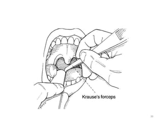
  • 38. INTERNAL APPROACH: PYRIFORM FOSSA • Patient is asked to open the mouth widely, and the tongue is grasped using a guaze pad or tongue blade. • A right angle forcep (e.g., Jackson-Krause) is covered with anesthetic-soaked guaze and is slid over the lateral posterior curvature of tongue and down into the pyriform sinuses bilaterally. • Cotton swabs are held in place for 5 minutes. 38
  • 39. 39
  • 40. D. TRACHEA AND VOCAL CORDS Translaryngeal block Patient selection – A translaryngeal block results in anesthesia of the trachea below the vocal cords. – However, injection of local anesthetic usually stimulates the cough reflex, and this block should be avoided in patients in whom coughing is undesirable. Drug – 3-4 ml of 4% lidocaine 40
  • 41. – Patient lies supine with neck slightly extended – Cricothyroid memb. is identified.
  • 42. Position – A 22 gauge or smaller needle is inserted into midline until air can be freely aspirated . – 3ml of LA is injected rapidly – Needle should be removed rapidly since the pt usually coughs vigorously. – This helps in spreading the LA under the vocal cord thus causing rapid anaesthesia of trachea &carina.
  • 44. 44
  • 45. Potential problem – Coughing – Bleeding from thyroid gland & its blood vessel. – Tracheal injury. – Subcutaneous emphysema.
  • 46. SPRAY AS YOU GO TECHNIQUE • Noninvasive and involves injecting local anesthetics through the suction port of the FOB. • It requires attaching a triple stopcock to the proximal portion of the suction port in order to connect oxygen tubing to flow at 2 to 4 L/min. • Under direct vision through the bronchoscope, targeted areas are sprayed with aliquots of 0.2 to 1.0 mL of 2% to 4% lidocaine. The physician then waits 30 to 60 seconds before advancing to deeper structures and repeating the maneuver. 46
  • 47. LABAT TECHNIQUE • Method of anesthetizing the vocal cords and trachea. • This technique requires a laryngeal mirror, a head lamp, and a Labat needle. • Sitting position, mouth wide open, the tongue is pulled outward. 47
  • 48. • In a right hand–dominant physician the left hand holds the laryngeal mirror over the oropharynx in order to identify the vocal cords. The right hand then holds the Labat needle and syringe and drips local anesthetic (4 mL of 2% lidocaine) over the vocal cords.
  • 49. The most difficult part of Awake intubation is – the decision to do it. THANKS