SlideShare a Scribd company logo
Biomedical analysis
BIOENGINEERING
Group members
Alfredo Ruggeri Associate professor
Enrico Grisan Post doc
Alfredo Giani (2003-) Post doc
Massimo De Luca (2004-) PhD student
Fabio Scarpa (2006-) PhD student
Lorenzo Marafatto (2005-) Fellowship
Marco Foracchia (-2003) PhD student
Biomedical images processing and analysis
BIOENGINEERING
Collaborations
J. Jaroszewski - Cornea Bank Berlin, Clinic of Ophthalmology, University
School of Medicine, Berlin, Germany
A. Neubauer - Dept. of Ophthalmology, Ludwig Maximilians University,
Munich, Germany
P. Gain - Ophthalmology Department, Bellevue Hospital, Saint-Etienne,
France
S. Klyce – Eye Center, Louisiana State University, New Orleans (LO), USA
S. Piermarocchi – Dept. of Ophthalmology, University of Padova
D. Ponzin - Veneto Eye Bank Foundation, Venice, Italy
A. Pocobelli - Eye Bank, S. Giovanni-Addolorata Hospital, Rome, Italy
A. Bezerianos - Dept. of Medical Physics, University of Patras, Greece
G. Barbaro - Nidek Technologies, Padova, Italy
P. Favaro - Siemens Corporate Research, Princeton (NJ), USA
Biomedical images processing and analysis
BIOENGINEERING
Grants
University of Padova: € 60.000 (shared)
University of Padova: € 15.000 (shared)
Ministry of University: € 20.000
CARIPARO Bank Foundation: € 40.000
Nidek Technologies: € 25.000 (+ 4 PhD fellowships)
TESI Imaging: under negotiation
Biomedical images processing and analysis
BIOENGINEERING
Publications
Biomedical images processing and analysis
0
1
2
3
4
5
6
7
8
2001 2002 2003 2004 2005
Intl Journals
Intl Conf Papers
Intl Conf Abstracts
In
BIOENGINEERING
Biomedical images processing and analysis
1.Cell contour recognition for in-vivo
microscopy of corneal endothelium
BIOENGINEERING
1. Contour extraction
Cell contour recognition
Artificial Neural Network with
weight-filters arrays
Mathematical morphology
2. Contour completion
Connection of floating facing boundaries
BIOENGINEERING
• The ENDO software is a module of the
system for ophthalmology.
Cell contour recognition
BIOENGINEERING
Cell contour recognition
Human visual processing is very powerful and
complex …
Kanisza triangles Kanisza square
BIOENGINEERING
Cell contours appear nice and clear on
a broad view….
Human visual processing is very powerful and
complex …
Cell contour recognition
… but local gray-scale
values do not give all the
information necessary to
identify all cell contours:
false contours
missed contours
… but local gray-scale
values do not give all the
information necessary to
identify all cell contours:
… but local gray-scale
values do not give all the
information necessary to
identify all cell contours:
BIOENGINEERING
A glimpse of tomorrow …
BIOENGINEERING
Biomedical images processing and analysis
2.Fourier analysis for the estimation of
endothelium cell density on eye bank images
Fully automatic technique in eye banks without cell contour detection.
 A repetitive pattern of cell
borders is clearly visible.
 Spatial frequency of this
pattern is proportional to cell
density.
 Frequency information is
available through Fourier
analysis.
BIOENGINEERING
A circular band
indicates that the
endothelium
image contains a
repetitive pattern
at a specific
frequency.
Spatial frequency is the radius of the band
Gray-scale image of 2D-DFT log-magnitude.
Frequency-based density estimation
Radius of circular band
can be used to estimate
cell density.
(Foracchia et al., Med Biol Eng Comput, 2004)
BIOENGINEERING
1500
2000
2500
3000
3500
1500 2000 2500 3000 3500
Manual density (cell/mm
2
)
Automatic
density
(cell/mm2)
Frequency-based density estimation
(Ruggeri et al., Br J Ophthalmol, 2005)
BIOENGINEERING
• The EyeBank software is a module of the
system for ophthalmology.
Nidek Technologies NAVIS-EyeBank system
BIOENGINEERING
3. Tracking techniques for vessel-like structure
Biomedical images processing and analysis
Applications to:
• vessels in retina
• nerves in cornea
Clinical parameters:
• length
• tortuosity
• bifurcations
• caliber course
• optic disc detection
BIOENGINEERING
Tracking techniques in retina
(Foracchia et al., Med Image Anal, 2005)
BIOENGINEERING
Tracking techniques in retina
(Foracchia et al., IEEE TMI, 2004)
BIOENGINEERING
Tracking techniques in cornea
BIOENGINEERING
Biomedical images processing and analysis
4.Methodologies in eye fundus analysis for the
diagnosis of retinopathy
Hypertensive and diabetic retinopathies are characterized by
presence of fundus lesions.
Automatic and objective tools
for image analysis:
• patient screening
• disease assessment &
monitoring in time
• (new) drugs efficacy
BIOENGINEERING
Steps:
• Detection
• Classification
• Measurement
• Clinical
assessment
Eye fundus analysis
(Grisan et al., EMBEC’05 Conf., 2005)
BIOENGINEERING
Biomedical images processing and analysis
5. Design and realization of an adaptive optics
fundus camera
Flash
path
Wavefront
sensor
Retinal
Imaging
Eye
Image Processing
BIOENGINEERING
Adaptive optics fundus camera
Acquired
image Coma Defocus Astigmatism
Corrected
image
Image
Analysis
Mirror
update
Simulation system with creation of aberrated
image and correction system
(Grisan et al., IEEE EMBS Conf., 2005)
BIOENGINEERING
Biomedical images processing and analysis
6. Automatic cariotyping
BIOENGINEERING
Automatic cariotyping
1. Segmentation of single chromosomes
2. Classification and pairing
1. - image enhancement
- cluster segmentation
- touching and overlapping
elimination by cuts
2. - feature extraction (banding)
- classification

More Related Content

PDF
Puplication-Assesssment of Vitritis On OCT-Journel TVST
PDF
人工智慧於眼科白內障手術的應用 (AI in Cataract Surgery)
PPT
12539 Image Processing BV Extraction.ppt
PPT
Retinal Pattern Recognition
PDF
Computer aided detection for vertebral deformities diagnosis based on deep le...
PDF
Various cataract detection methods-A survey
PDF
Analysis on Glaucoma Detection
PPTX
The bionic eye
Puplication-Assesssment of Vitritis On OCT-Journel TVST
人工智慧於眼科白內障手術的應用 (AI in Cataract Surgery)
12539 Image Processing BV Extraction.ppt
Retinal Pattern Recognition
Computer aided detection for vertebral deformities diagnosis based on deep le...
Various cataract detection methods-A survey
Analysis on Glaucoma Detection
The bionic eye

Similar to Biomedical Images.ppt (20)

PPT
Retinal Recognition
PDF
Segmentation of the Blood Vessel and Optic Disc in Retinal Images Using EM Al...
PDF
Q01765102112
PDF
PDF
Biomedical Optical Imaging 1st Edition James G Fujimoto Daniel Farkas
PDF
BiswasSK_CV
PDF
Detection of Macular Edema by using Various Techniques of Feature Extraction ...
PDF
Optical Coherence Tomography: Technology and applications for neuroimaging
PPTX
MEDICAL IMAGE PROCESSING
PDF
Glaucoma Detection from Retinal Images
PDF
AI in Ophthalmology | Startup Landscape
PPTX
Biomedical Optical Imaging
PDF
Optical biopsy
PDF
The Importance of Medical Multimedia
PDF
Eye Blink and Hand Gesture detection For Aid of Paralyzed Patients
PDF
Detection of Glaucoma using Optic Disk and Incremental Cup Segmentation from ...
PDF
Measuring Thicknesses of Films by Optical Coherence Tomography (OCT)
PDF
To Improve the Recognition Rate with High Security with Ear/Iris Biometric Re...
PDF
Inferior posterior staphyloma: choroidal maps and macular complications
Retinal Recognition
Segmentation of the Blood Vessel and Optic Disc in Retinal Images Using EM Al...
Q01765102112
Biomedical Optical Imaging 1st Edition James G Fujimoto Daniel Farkas
BiswasSK_CV
Detection of Macular Edema by using Various Techniques of Feature Extraction ...
Optical Coherence Tomography: Technology and applications for neuroimaging
MEDICAL IMAGE PROCESSING
Glaucoma Detection from Retinal Images
AI in Ophthalmology | Startup Landscape
Biomedical Optical Imaging
Optical biopsy
The Importance of Medical Multimedia
Eye Blink and Hand Gesture detection For Aid of Paralyzed Patients
Detection of Glaucoma using Optic Disk and Incremental Cup Segmentation from ...
Measuring Thicknesses of Films by Optical Coherence Tomography (OCT)
To Improve the Recognition Rate with High Security with Ear/Iris Biometric Re...
Inferior posterior staphyloma: choroidal maps and macular complications
Ad

More from breenaawan (20)

PPT
physicochemical_properties_of_drug_molecules.ppt
PPT
THE physicochemical properties AND ACTIVITY.ppt
PPTX
the significance of microbial growth.pptx
PPTX
the Hypersensitivity types with detail .pptx
PPTX
The sterilization and difference between narrown/broad spectrum antibiotics.pptx
PPTX
The antibodies with details in microbiology.pptx
PPTX
introduction to microbiology and basic terminology.pptx
PPTX
Role Of Skin And Mucous Membrane In Non Specific Resistance.pptx
PPTX
INTRODUCTION TO MICROBIOLOGY AND MICROORGANISMS (1).pptx
PPTX
Pharmacology of Major anticancerdrugs.pptx
PPTX
microbiology the health`chp 3 topic 1.pptx
PPTX
Aseptic and infectionmicrobiology the full unit 4.pptx
PPT
dnareplicationrepairundergrads-140407025100-phpapp02.ppt
PPTX
pharmacologyofdiabetesmellitus-191005121724 (1).pptx
PPT
The pharmacology of Gastrointestinal tract.ppt
PPTX
oxidativephos-140111054455-phpapp01.pptx
PPTX
unit 3 Defence Mechanism of the Body.pptx
PPT
unit 2 The control of microbial growth .ppt
PPT
Microbial growth control170713022721.ppt
PPTX
Asthma pharmacology drugs in detail2.pptx
physicochemical_properties_of_drug_molecules.ppt
THE physicochemical properties AND ACTIVITY.ppt
the significance of microbial growth.pptx
the Hypersensitivity types with detail .pptx
The sterilization and difference between narrown/broad spectrum antibiotics.pptx
The antibodies with details in microbiology.pptx
introduction to microbiology and basic terminology.pptx
Role Of Skin And Mucous Membrane In Non Specific Resistance.pptx
INTRODUCTION TO MICROBIOLOGY AND MICROORGANISMS (1).pptx
Pharmacology of Major anticancerdrugs.pptx
microbiology the health`chp 3 topic 1.pptx
Aseptic and infectionmicrobiology the full unit 4.pptx
dnareplicationrepairundergrads-140407025100-phpapp02.ppt
pharmacologyofdiabetesmellitus-191005121724 (1).pptx
The pharmacology of Gastrointestinal tract.ppt
oxidativephos-140111054455-phpapp01.pptx
unit 3 Defence Mechanism of the Body.pptx
unit 2 The control of microbial growth .ppt
Microbial growth control170713022721.ppt
Asthma pharmacology drugs in detail2.pptx
Ad

Recently uploaded (20)

PPTX
Acid Base Disorders educational power point.pptx
PPT
CHAPTER FIVE. '' Association in epidemiological studies and potential errors
PPTX
CME 2 Acute Chest Pain preentation for education
DOC
Adobe Premiere Pro CC Crack With Serial Key Full Free Download 2025
PPTX
15.MENINGITIS AND ENCEPHALITIS-elias.pptx
PPTX
SKIN Anatomy and physiology and associated diseases
PPTX
ca esophagus molecula biology detailaed molecular biology of tumors of esophagus
PPTX
DENTAL CARIES FOR DENTISTRY STUDENT.pptx
DOCX
NEET PG 2025 | Pharmacology Recall: 20 High-Yield Questions Simplified
PPT
Management of Acute Kidney Injury at LAUTECH
PDF
Therapeutic Potential of Citrus Flavonoids in Metabolic Inflammation and Ins...
PPTX
surgery guide for USMLE step 2-part 1.pptx
PPTX
post stroke aphasia rehabilitation physician
PPTX
Imaging of parasitic D. Case Discussions.pptx
DOCX
RUHS II MBBS Microbiology Paper-II with Answer Key | 6th August 2025 (New Sch...
PPT
ASRH Presentation for students and teachers 2770633.ppt
PPTX
CEREBROVASCULAR DISORDER.POWERPOINT PRESENTATIONx
PPTX
NEET PG 2025 Pharmacology Recall | Real Exam Questions from 3rd August with D...
PPTX
Chapter-1-The-Human-Body-Orientation-Edited-55-slides.pptx
PDF
Human Health And Disease hggyutgghg .pdf
Acid Base Disorders educational power point.pptx
CHAPTER FIVE. '' Association in epidemiological studies and potential errors
CME 2 Acute Chest Pain preentation for education
Adobe Premiere Pro CC Crack With Serial Key Full Free Download 2025
15.MENINGITIS AND ENCEPHALITIS-elias.pptx
SKIN Anatomy and physiology and associated diseases
ca esophagus molecula biology detailaed molecular biology of tumors of esophagus
DENTAL CARIES FOR DENTISTRY STUDENT.pptx
NEET PG 2025 | Pharmacology Recall: 20 High-Yield Questions Simplified
Management of Acute Kidney Injury at LAUTECH
Therapeutic Potential of Citrus Flavonoids in Metabolic Inflammation and Ins...
surgery guide for USMLE step 2-part 1.pptx
post stroke aphasia rehabilitation physician
Imaging of parasitic D. Case Discussions.pptx
RUHS II MBBS Microbiology Paper-II with Answer Key | 6th August 2025 (New Sch...
ASRH Presentation for students and teachers 2770633.ppt
CEREBROVASCULAR DISORDER.POWERPOINT PRESENTATIONx
NEET PG 2025 Pharmacology Recall | Real Exam Questions from 3rd August with D...
Chapter-1-The-Human-Body-Orientation-Edited-55-slides.pptx
Human Health And Disease hggyutgghg .pdf

Biomedical Images.ppt

  • 2. BIOENGINEERING Group members Alfredo Ruggeri Associate professor Enrico Grisan Post doc Alfredo Giani (2003-) Post doc Massimo De Luca (2004-) PhD student Fabio Scarpa (2006-) PhD student Lorenzo Marafatto (2005-) Fellowship Marco Foracchia (-2003) PhD student Biomedical images processing and analysis
  • 3. BIOENGINEERING Collaborations J. Jaroszewski - Cornea Bank Berlin, Clinic of Ophthalmology, University School of Medicine, Berlin, Germany A. Neubauer - Dept. of Ophthalmology, Ludwig Maximilians University, Munich, Germany P. Gain - Ophthalmology Department, Bellevue Hospital, Saint-Etienne, France S. Klyce – Eye Center, Louisiana State University, New Orleans (LO), USA S. Piermarocchi – Dept. of Ophthalmology, University of Padova D. Ponzin - Veneto Eye Bank Foundation, Venice, Italy A. Pocobelli - Eye Bank, S. Giovanni-Addolorata Hospital, Rome, Italy A. Bezerianos - Dept. of Medical Physics, University of Patras, Greece G. Barbaro - Nidek Technologies, Padova, Italy P. Favaro - Siemens Corporate Research, Princeton (NJ), USA Biomedical images processing and analysis
  • 4. BIOENGINEERING Grants University of Padova: € 60.000 (shared) University of Padova: € 15.000 (shared) Ministry of University: € 20.000 CARIPARO Bank Foundation: € 40.000 Nidek Technologies: € 25.000 (+ 4 PhD fellowships) TESI Imaging: under negotiation Biomedical images processing and analysis
  • 5. BIOENGINEERING Publications Biomedical images processing and analysis 0 1 2 3 4 5 6 7 8 2001 2002 2003 2004 2005 Intl Journals Intl Conf Papers Intl Conf Abstracts In
  • 6. BIOENGINEERING Biomedical images processing and analysis 1.Cell contour recognition for in-vivo microscopy of corneal endothelium
  • 7. BIOENGINEERING 1. Contour extraction Cell contour recognition Artificial Neural Network with weight-filters arrays Mathematical morphology 2. Contour completion Connection of floating facing boundaries
  • 8. BIOENGINEERING • The ENDO software is a module of the system for ophthalmology. Cell contour recognition
  • 9. BIOENGINEERING Cell contour recognition Human visual processing is very powerful and complex … Kanisza triangles Kanisza square
  • 10. BIOENGINEERING Cell contours appear nice and clear on a broad view…. Human visual processing is very powerful and complex … Cell contour recognition … but local gray-scale values do not give all the information necessary to identify all cell contours: false contours missed contours … but local gray-scale values do not give all the information necessary to identify all cell contours: … but local gray-scale values do not give all the information necessary to identify all cell contours:
  • 12. BIOENGINEERING Biomedical images processing and analysis 2.Fourier analysis for the estimation of endothelium cell density on eye bank images Fully automatic technique in eye banks without cell contour detection.  A repetitive pattern of cell borders is clearly visible.  Spatial frequency of this pattern is proportional to cell density.  Frequency information is available through Fourier analysis.
  • 13. BIOENGINEERING A circular band indicates that the endothelium image contains a repetitive pattern at a specific frequency. Spatial frequency is the radius of the band Gray-scale image of 2D-DFT log-magnitude. Frequency-based density estimation Radius of circular band can be used to estimate cell density. (Foracchia et al., Med Biol Eng Comput, 2004)
  • 14. BIOENGINEERING 1500 2000 2500 3000 3500 1500 2000 2500 3000 3500 Manual density (cell/mm 2 ) Automatic density (cell/mm2) Frequency-based density estimation (Ruggeri et al., Br J Ophthalmol, 2005)
  • 15. BIOENGINEERING • The EyeBank software is a module of the system for ophthalmology. Nidek Technologies NAVIS-EyeBank system
  • 16. BIOENGINEERING 3. Tracking techniques for vessel-like structure Biomedical images processing and analysis Applications to: • vessels in retina • nerves in cornea Clinical parameters: • length • tortuosity • bifurcations • caliber course • optic disc detection
  • 17. BIOENGINEERING Tracking techniques in retina (Foracchia et al., Med Image Anal, 2005)
  • 18. BIOENGINEERING Tracking techniques in retina (Foracchia et al., IEEE TMI, 2004)
  • 20. BIOENGINEERING Biomedical images processing and analysis 4.Methodologies in eye fundus analysis for the diagnosis of retinopathy Hypertensive and diabetic retinopathies are characterized by presence of fundus lesions. Automatic and objective tools for image analysis: • patient screening • disease assessment & monitoring in time • (new) drugs efficacy
  • 21. BIOENGINEERING Steps: • Detection • Classification • Measurement • Clinical assessment Eye fundus analysis (Grisan et al., EMBEC’05 Conf., 2005)
  • 22. BIOENGINEERING Biomedical images processing and analysis 5. Design and realization of an adaptive optics fundus camera Flash path Wavefront sensor Retinal Imaging Eye Image Processing
  • 23. BIOENGINEERING Adaptive optics fundus camera Acquired image Coma Defocus Astigmatism Corrected image Image Analysis Mirror update Simulation system with creation of aberrated image and correction system (Grisan et al., IEEE EMBS Conf., 2005)
  • 24. BIOENGINEERING Biomedical images processing and analysis 6. Automatic cariotyping
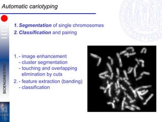
  • 25. BIOENGINEERING Automatic cariotyping 1. Segmentation of single chromosomes 2. Classification and pairing 1. - image enhancement - cluster segmentation - touching and overlapping elimination by cuts 2. - feature extraction (banding) - classification