Chest trauma
0 likes324 views

This document provides an overview of chest trauma, including the anatomy of the chest, common causes of chest injuries, signs and symptoms of specific chest injuries like pneumothorax, hemothorax, flail chest, and treatment approaches for these injuries at the basic life support level. It emphasizes the importance of rapid identification and treatment of chest trauma patients given the potential life-threatening nature of these injuries. Airway management and stabilization are key, along with monitoring for complications and rapid transport to advanced care.


Chest trauma
- 1. Chest Trauma Prepared By: Justin V Sebastian, MSc N, RN, PhD Scholar
- 2. Objectives ■ Anatomy of Thorax ■ Main Causes of Chest Injuries ■ S/S of Chest Injuries ■ Different Types of Chest Injuries ■ Treatments of Chest Injuries
- 3. Anatomy of the chest Two Lungs (right and left) Heart Diaphragm
- 4. Anatomy of the chest Pleural Space
- 5. Anatomy of the chest
- 6. Main Causes of Chest Trauma ■ Blunt Trauma- Blunt force to chest. ■ Penetrating Trauma- Projectile that enters chest causing small or large hole. ■ Compression Injury- Chest is caught between two objects and chest is compressed.
- 7. Injuries of chest ■ Simple/Closed Pneumothorax ■ Open Pneumothorax ■ Tension Pneumothorax ■ Flail Chest ■ Cardiac Tamponade ■ Traumatic Aortic Rupture ■ Traumatic Asphyxia ■ Diaphragmatic Rupture
- 8. Simple/Closed Pneumothorax ■ Opening in lung tissue that leaks air into chest cavity ■ Blunt trauma is main cause ■ May be spontaneous ■ Usually self correcting
- 9. S/S of Simple/Closed Pneumothorax ■ Chest Pain ■ Dyspnea ■ Tachypnea ■ Decreased Breath Sounds on Affected Side
- 10. ■ Diagnosis ▪ History of injury ▪ Physical examination ▪ Chest x ray ▪ Ct scan ▪ CBC ▪ Clotting studies ▪ Type and cross match ▪ Electrolytes ▪ O2 saturation ▪ Abg and ECG
- 11. Treatment for Simple/Closed Pneumothorax ■ ABC’s with C-spine control ■ Airway Assistance as needed ■ If not contraindicated transport in semi- sitting position ■ Provide supportive care ■ Contact Hospital and/or ALS unit as soon as possible
- 12. BLS Plus Care ■ Cardiac Monitor ■ IV access and Draw Blood Samples ■ Provide Airway Management which includes possible Intubation ■ Monitor for Development of Tension Pneumothorax
- 13. Open Pneumothorax ■ Opening in chest cavity that allows air to enter pleural cavity ■ Causes the lung to collapse due to increased pressure in pleural cavity ■ Can be life threatening and can deteriorate rapidly
- 21. S/S of Open Pneumothorax ■ Dyspnea ■ Sudden sharp pain ■ Subcutaneous Emphysema ■ Decreased lung sounds on affected side ■ Red Bubbles on Exhalation from wound
- 22. Subcutaneous Emphysema ■ Air collects in subcutaneous fat from pressure of air in pleural cavity ■ Can be seen from neck to groin area
- 23. Treatment for Open Pneumothorax ■ ABC’s with c-spine control as indicated ■ High Flow oxygen ■ Listen for decreased breath sounds on affected side ■ Apply occlusive dressing to wound ■ Notify Hospital and ALS unit as soon as possible
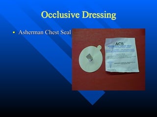
- 25. Occlusive Dressing ■ Asherman Chest Seal
- 26. BLS Plus Care ■ Monitor Heart Rhythm ■ Establish IV Access and Draw Blood Samples ■ Airway Control that may include Intubation ■ Monitor for Tension Pneumothorax
- 27. Tension Pneumothorax ■ Air builds in pleural space with no where for the air to escape ■ Results in collapse of lung on affected side that results in pressure on mediastium,the other lung, and great vessels
- 28. Tension Pneumothorax Each time we inhale, the lung collapses further. There is no place for the air to escape..
- 29. Tension Pneumothorax Each time we inhale, the lung collapses further. There is no place for the air to escape..
- 30. Tension Pneumothorax Heart is being compressed The trachea is pushed to the good side
- 31. S/S of Tension Pneumothorax ■ Anxiety/Restlessness ■ Severe Dyspnea ■ Absent Breath sounds on affected side ■ Tachypnea ■ Tachycardia ■ Poor Color ■ Accessory Muscle Use ■ JVD ■ Narrowing Pulse Pressures ■ Hypotension ■ Tracheal Deviation (late if seen at all)
- 32. Treatment of Tension Pneumothorax ■ ABC’s with c-spine as indicated ■ High Flow oxygen including BVM ■ Treat for S/S of Shock ■ Notify Hospital and ALS unit as soon as possible
- 33. BLS Plus Care ■ Monitor Cardiac Rhythm ■ Establish IV access and Draw Blood Samples ■ Airway control including Intubation ■ Needle Decompression of Affected Side
- 34. Needle Decompression ■ Locate 2-3 Intercostal space midclavicular line ■ Cleanse area using aseptic technique ■ Insert catheter ( 14g or larger) at least 3” in length over the top of the 3rd rib( nerve, artery, vein lie along bottom of rib) ■ Remove Stylette and listen for rush of air ■ Place Flutter valve over catheter ■ Reassess for Improvement
- 36. Flutter Valve ■ Asherman Chest Seal makes good Flutter Valve . ■ Also can use a Finger from a Latex Glove ■ Or A Condom works also
- 37. Hemothorax ■ Occurs when pleural space fills with blood ■ Usually occurs due to lacerated blood vessel in thorax ■ As blood increases, it puts pressure on heart and other vessels in chest cavity ■ Each Lung can hold 1.5 liters of blood
- 38. Hemothorax
- 39. Hemothorax
- 40. Hemothorax
- 41. Hemothorax
- 42. Hemothorax
- 43. Hemothorax May put pressure on the heart
- 44. Hemothorax Lots of blood vessels Where does the blood come from.
- 45. S/S of Hemothorax ■ Anxiety/Restlessness ■ Tachypnea ■ Signs of Shock ■ Frothy, Bloody Sputum ■ Diminished Breath Sounds on Affected Side ■ Tachycardia ■ Flat Neck Veins
- 46. Treatment for Hemothorax ■ ABC’s with c-spine control as indicated ■ Secure Airway assist ventilation if necessary ■ General Shock Care due to Blood loss ■ RAPID TRANSPORT ■ Contact Hospital and ALS Unit as soon as possible
- 47. BLS Plus Care ■ Monitor Cardiac Rhythm ■ Establish Large Bore IV preferably 2 and draw blood samples ■ Airway management to include Intubation ■ Rapid Transport ■ If Development of Hemo/Pneumothorax needle decompression may be indicated
- 48. Flail Chest The breaking of 2 or more ribs in 2 or more places
- 49. Flail Chest
- 50. S/S of Flail Chest ■ Anterior chest pain ■ Tenderness ■ Shortness of Breath ■ Paradoxical Movement ■ Bruising/Swelling ■ Crepitus( Grinding of bone ends on palpation)
- 51. Flail Chest is a True Emergency
- 52. Treatment of Flail Chest ■ ABC’s with c-spine control as indicated ■ High Flow oxygen that may include BVM ■ Monitor Patient for signs of Pneumothorax or Tension Pneumothorax ■ Use Gloved hand as splint till bulky dressing can be put on patient ■ Contact hospital and ALS Unit as soon as possible
- 53. Bulky Dressing for splint of Flail Chest ■ Use Trauma bandage and Triangular Bandages to splint ribs.
- 54. BLS Plus Care ■ Monitor Cardiac Rhythm ■ Establish IV access ■ Airway management to include Intubation ■ Observe for patient to develop Pneumothorax and even worse Tension Pneumothorax ■ If Tension Develops Needle Decompress affected side ■ Rapid Transport! Remember a True Emergency
- 55. Pericardial Tamponade Blood and fluids leak into the pericardial sac which surrounds the heart. As the pericardial sac fills, it causes the sac to expand until it cannot expand anymore pericardial sac
- 56. Pericardial Tamponade Once the pericardial sac can’t expand anymore, the fluid starts putting pressure on the heart Now the heart can’t fully expand and can’t pump effectively.
- 57. Pericardial Tamponade With poor pumping the blood pressure starts to drop. The heart rate starts to increase to compensate but is unable The patient’s level of conscious drops, and eventually the patient goes in cardiac arrest
- 58. S/S of Pericardial Tamponade ■ Distended Neck Veins ■ Increased Heart Rate ■ Respiratory Rate increases ■ Poor skin color ■ Narrowing Pulse Pressures ■ Hypotension ■ Death
- 59. Treatment of Pericardial Tamponade ■ ABC’s with c-spine control as indicated ■ High Flow oxygen which may include BVM ■ Treat S/S of shock ■ Rapid Transport ■ Notify Hospital and ALS Unit as soon as possible
- 60. BLS Plus Care ■ Cardiac Monitor ■ Large Bore IV access ■ Rapid Transport ■ What patient needs is pericardiocentesis
- 61. Pericardiocentesis ■ Using aseptic technique, Insert at least 3” needle at the angle of the Xiphoid Cartilage at the 7th rib ■ Advance needle at 45 degree towards the clavicle while aspirating syringe till blood return is seen ■ Continue to Aspirate till syringe is full then discard blood and attempt again till signs of no more blood ■ Closely monitor patient due to small amout of blood aspirated can cause a rapid change in blood pressure
- 62. Traumatic Aortic Rupture The heart, more or less, just hangs from the aortic arch Much like a big pendulum. If enough motion is placed on the heart (i.e.. Deceleration From a motor vehicle accident) the heart may tear away from the aorta.
- 63. Traumatic Aortic Rupture The chances of survival are very slim and are based on the degree of the tear. If there is just a small tear then the patient may survive. If the aorta is completely transected then the patient will die instantaneously
- 64. S/S Of Traumatic Aortic Rupture ■ Burning or Tearing Sensation in chest or shoulder blades ■ Rapidly dropping Blood Pressure ■ Pulse Rapidly Increasing ■ Rapid Loss of Consciousness
- 65. Treatment of Traumatic Aortic Rupture ■ ABC’s with c-spine control as indicated ■ High Flow oxygen that may include BVM ■ Treatment for Shock ■ RAPID TRANSPORT ■ Contact Hospital and ALS Unit As soon as possible
- 66. BLS Plus Care ■ Monitor Cardiac Rhythm ■ Large Bore IV therapy probably 2 and draw blood samples ■ Airway management that may include Intubation
- 67. Traumatic Asphyxia ■ Results from sudden compression injury to chest cavity ■ Can cause massive rupture of Vessels and organs of chest cavity ■ Ultimately Death
- 68. S/S of Traumatic Asphyxia ■ Severe Dyspnea ■ Distended Neck Veins ■ Bulging, Blood shot eyes ■ Swollen Tounge with cyanotic lips ■ Reddish-purple discoloration of face and neck ■ Petechiae
- 69. Treatment for Traumatic Asphyxia ■ ABC’s with c-spine control as indicated ■ High Flow oxygen including use of BVM ■ Treat for shock ■ Care for associated injuries ■ Rapid Transport ■ Contact Hospital and ALS Unit as soon as possible
- 70. BLS Plus Care ■ Cardiac Monitor ■ Establish IV Access and draw blood samples ■ Airway control including Intubation ■ Rapid transport
- 71. Diaphragmatic Rupture ■ A tear in the Diaphragm that allows the abdominal organs enter the chest cavity ■ More common on Left side due to liver helps protect the right side of diaphragm ■ Associated with multipile injury patients
- 73. S/S of Diaphragmatic Rupture ■ Abdominal Pain ■ Shortness of Air ■ Decreased Breath Sounds on side of rupture ■ Bowel Sounds heard in chest cavity
- 74. Treatment of Diaphragmatic Rupture ■ ABC’s with c-spine control as indicated ■ High Flow oxygen which may include BVM ■ Treat Associated Injuries ■ Rapid Transport ■ Contact Hospital and ALS Unit as soon as possible
- 75. BLS Plus Care ■ Cardiac Monitor ■ Establish IV access and draw blood samples ■ Airway management including Intubation ■ Observe for Pneumothorax due to compression on lung by abdominal contents ■ Possible insertion of NG tube to help decompress the stomach to relieve pressure ■ Rapid transport
- 76. Summary Chest Injuries are common and often life threatening in trauma patients. So, Rapid identification and treatment of these patients is paramount to patient survival. Airway management is very important and aggressive management is sometimes needed for proper management of most chest injuries.
- 77. The END ■ Questions? ■ Comments ■ Criticisms ■ Slide Remarks ■ If not thank You