Welcome to
Seminar
Presenters
Chronic Liver Disease
Outline
• Anatomy and Physiology
• Pathology
• Epidemiology
• Classification
• Etiology
• Approach to CLD
• Complications
• Investigations
• Treatment
• Prognosis
Anatomy & Physiology of Liver
Chronic Liver Disease- Pediatrics Gastroenterology.pptx
Functions of Liver
Synthesis:
- Albumin
- Coagulation factors
Glucose homeostasis;
Glycogenolysis & Gluconeogenesis
Storage:
- Glycogen
- Iron
- Cu, vitamins
Catabolism of hormones
and other serum proteins
Bile excretion
Pathology
Definition:
Liver Disease present over many months without progressive
improvement back to normal liver.
Chronic Liver Disease
Epidemiology
Epidemiology
Cirrhosis and chronic liver disease were the 10th leading cause of death for men
and 12th for women in the United States in 2010, killing about 27,000 people
each year.
 CLD is called as “National Disease of Japan” in 21st century. Fifty
thousands of people died from CLD this days.
 Approximately 1 in 679 or 0.15% or 400,000 people got Chronic Liver
Disease in USA from 1976-1980.
 90 out of 600 (15%) patients admitted in pediatric Gastroenterology
ward, BSMMU, were diagnosed as CLD in 2012.
Epidemiology (Cont.)
Source: (Digestive diseases in the United States: Epidemiology and Impact – NIH Publication No. 94-1447,
NIDDK, 1994)
Classification of CLD
1. Chronic hepatitis:
Symptomatic, biochemical or serologic evidence of continuing or relapsing hepatic
disease for more than 6 months, with histologically documented inflammation.
2. Cirrhosis:
Is a diffuse parenchymal disease of liver characterized by hepatocellular infiltration and
necrosis resulting in loss of hepatic architecture , formation of regenerative nodules
with fibrosis and abnormal vascular shunt formation.
Chronic Liver Disease- Pediatrics Gastroenterology.pptx
Pathogenesis of CLD
Chronic inflammation of liver
Inflammatory cytokines(TNF,lymphotoxin,IL-1,TGF,PDGF) are produced by stimulated Kupffer cell,
hepatocyte, endothelial cell.
Activation of stellate cell
Transformation of stellate cell into myofibroblast like cell which produce collagen(type I & III).
Collagen deposition in lobule, creating- 1. Broad septal band
2.Increased vascular resistance.
3. Fibrosis & regeneration of remaining hepatocytes
Fibrosis + loss of architecture+ formation of regenerative nodule.
Cirrhosis of liver
Classification
Chronic Hepatitis
Classification (Older)
1. Chr. Persistent
2. Chr. Lobular
3. Chr. Active
Etiology
Clinical grade
Histological severity
Stage
Classification (new) based on
Classification
Classification (cont.)
 Clinical grade-depends on S. ALT level
Mild : <100 IU/L
Moderate: 100-400 IU/L
Severe: > 400 IU/L
 Histological scoring
Knodell/HAI
Ishak score
Knodell score( HAI)
• Composed of summation of 4 individual scores:
• 1. Periportal necrosis with or without bridging necrosis.
• 2. Intra lobular degeneration and focal necrosis.
• 3. Portal inflammation.
• 4.Fibrosis.
Classification (cont.)
1.Periportal necrosis with or without bridging necrosis ( Score 0-10)
None 0
Mild piecemeal necrosis 1
Moderate piecemeal necrosis 3
Marked piecemeal necrosis 4
Moderate piecemeal necrosis+ bridging necrosis 5
Marked piecemeal necrosis + bridging necrosis 6
Interlobular necrosis 10
2.Inter lobular degeneration and focal necrosis
None 0
Mild(involvement of <1/3 of lobules or nodules) 1
Moderate(involvement of 1/3 to 2/3 of lobules or nodules) 3
Marked(involvement of >2/3 of lobules or nodules) 4
Classification (cont.)
Portal inflammation
No portal inflammation 0
Mild (sprinkling of inflammatory cells in <1/3 of portal tracts) 1
Moderate (increased inflammatory cells in1/3 to 2/3 of portal tracts) 3
Marked(dense packing of inflammatory cells in >2/3 of portal tracts) 4
Fibrosis
No fibrosis 0
Fibrous portal expansion 1
Bridging fibrosis(portal-portal to portal-central linkage) 3
Classification (cont.)
Interpretation
HAI (Total Score 18) Diagnosis
1-3 Minimal
4-8 Mild
9-12 Moderate
13-18 Severe
Scoring By Fibrosis Diagnosis
0 None
1 Mild
2 Moderate
3 Severe
4 Cirrhosis
Classification (cont.)
Classification of Cirrhosis
• Pathological:
Micro nodular
Macro nodular
Mixed
Biliary
Classification (cont.)
Normal Liver Microscopy
Fibrosis
Regenerating Nodule
Cirrhosis
Classification (cont.)
Micronodular
Thick regular septa.
Regenerating small nodules < 3mm
Involvement of every nodule
Seen in –
Haemachromatosis
Cholestasis
Hepatic venous outflow obstruction
Classification (cont.)
Macronodular
Septa and nodules (>3 mm) of variable sizes
Normal lobules in larger nodules
Secondary to chronic viral hepatitis
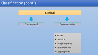
Clinical
Compensated Decompensated
Ascites
Jaundice
Encephalopathy
Fetor hepaticus
Coagulopathy
Classification (cont.)
Biliary cirrhosis
Bile stasis
General reduction of bile duct
Increased connective tissue within and extending from portal tracts
Lobular architecture preserved.
Seen in- Biliary atresia, CF, PFIC.
Classification (cont.)
Etiology
1. Infectious
2. Metabolic
3. Inflammatory
4. Biliary malformation
5. Vascular
6. Toxic
7. Nutritional
8. Miscellaneous
Etiology
Chronic hepatitis B with or without
D virus
Chr. hepatitis C
CMV
Herpes Simplex
virus
Rubella
Neonatal sepsis
Etiology- Infectious
• Alfa -1 antitrypsin deficiency
• Fructosemisa
• Wilson’s disease
• Cystic fibrosis
• Hemochromatosis
• Niemann- pick disease type D
• Tyrosinemia
• Galactosemia
• Gauccher’s disease
• Glycogen storage disease
Etiology- Metabolic
Autoimmune
chronic hepatitis
Primary
sclerosing
cholangitis
Autoimmune hepatitis is a chronic hepatitis of
unknown etiology.
 First described in the 1950s,
 Characterized generally by 3 features:
Hepatitis
Auto antibodies
High serum globulin concentration
Etiology- Inflammatory
Hepatotoxic drugs:
Isoniazid,
Methyldopa,
Nitrofurantoin,
Dantroline,
MTX,
Amidarone,
Propuylthiouracil, etc.
Toxin found in nature- e.g. mushroom
Organic solvents
Etiology- Toxin Mediated
• Intrahepatic biliary hypoplasia
• Biliary atresia
• Alagille syndrome
• Choledochal cyst
Etiology- Biliary Malformation
• Budd- Chiari syndrome
• CCF
• Congestive pericarditis
• Veno-occlusive liver disease
Etiology- Vascular
Etiology- Vascular
TPN
Hypervitaminosis A
Malnutrition
Etiology- Nutritional
ICC
Histocytosis X
Neonatal hepatitis
Cerebrohepatorenal syndrome
Etiology- Miscellaneous
Approach to a child with CLD
History
1. Duration of illness.
2. History of jaundice.
3. History of blood transfusion or parenteral injection.
4. H/O ingestion of any hepatotoxic drugs.
5. H/O bleeding manifestation (Hematemesis, melaena ).
6. Sign of encephalopathy (Altered sleep rhythm).
History
7. H/O umbilical sepsis or catheterization in neonatal period.
8. Color of urine, stool.
9. H/O fatigue, weight loss, anorexia, abdominal pain.
10. Recurrent chest infection or chronic diarrhoea.
11. H/O consanguinity, family history, sibling affection.
12. Religion (middle class hindu), copper made utensils.
13. Immunization history.
Physical sign- General
Hand Signs
Leukonychia Dupytren’s
contracture
Palmar
Erythema
Clubbing Asterixis
Skin Changes
Jaundice Spider angioma Gynecomastia
Scratch mark Bruising &
Bleeding
Xanthoma
Physical sign – Abdomen
1. Abdominal Distension.
2. Caput medusa.
3. Ascites.
4. Liver- size, consistency.
5. Splenomegaly.
6. Testicular atrophy.
7. Rectal examination.
Special presentations
HCV Small vessel vasculitis, peripheral neuropathy, GN, NS.
Autoimmune hepatitis Polyarthritis, allergic capillaritis, generalized
lymphadenopathy, GN, Hashimoto's thyroiditis, DM
Wilsons disease KF ring, sunflower cataract, neuro-psychiatric sign
Metabolic disease Cataract, facial dysmorphism, developmental delay,
convulsion
Complications
Complication
•Coagulopathy
•Hypoalbumenia
•Ascites, Electrolyte imbalance
•Hypoglycemia
Due to
parenchymal
dysfunction
•Variceal bleeding
•Hypersplenism
Due to portal
hypertension
•Sepsis, pneumonia, UTI, SBP
Infections
Complication (Cont)
Complication (Cont)
•Porto-systemic encephalopathy
•Hepato-renal syndrome
•Hepato-pulmonary syndrome
Systemic complication:
Hepatocellular carcinoma
Encephalopathy
Complication (Cont)
• Reversible decrease in neurologic function caused by liver disease
• Established mechanism
a. Accumulation of un-metabolized NH3 due to
• Poor hepatic function
• Poor systemic shunting.
b. Possible mechanism: False neurotransmitter, GABA,
Benzodiazepine receptor activation etc.
Encephalopathy- Grading
Complication (Cont)
• Thrombocytopenia
• Thrombocytopathy
• Decrease synthesis of vit K – dependent factors
• DIC
Coagulopathy
Complication (Cont)
Hepato-renal syndrome:
Complication (Cont)
Hepato-renal syndrome is defined as functional renal failure in patient with end stage
liver disease. The hallmark is intense renal vaso-constriction with co-existing systemic
vaso-dilatation, may be mediated by hemodynamic, humoral or neurogenic mechanism.
Diagnosis:
1. Oliguria (< 1 ml/kg/day).
2. Urine sodium (< 10 mEq/L).
3. Fractional Na+ excretion(<1%).
4. Urine: Plasma creatinine ratio-<10.
5. Exclusion of other kidney pathology.
Treatment:
Liver transplantation.
Hepato-renal syndrome:
Complication (Cont)
Hepato-pulmonary syndrome:
Complication (Cont)
Is characterized by typical triad of hypoxemia, intrapulmonary vascular dilations and liver
disease. There is intrapulmonary right to left shunting of blood which results in systemic
desaturation.
Clinical feature:
1. Shortness of breath.
2. Exercise in tolerance.
3. Cyanosis- lips & fingers. In a child with CLD.
4. Clubbing.
5. O2 saturation < 96%.
Treatment:
Liver transplantation.
Investigations
To Establish Diagnosis
Investigations
A. Evaluation of liver function:
i. S. bilirubin level
ii. S. ALT
iii. S. AST level
iv. Alkaline phosphatase level
v. S. protein
vi. Prothrombin time
vii. Blood sugar
B. Abdominal ultrasonography
C. Endoscopy of upper GIT
D. Liver biopsy
Investigations (Cont.)
To Determine the Etiology
Chronic Hepatitis
HBsAg positive
1. Look for markers of active
Viral replication (HBeAg, HBV,
DNA)
2. Look for co–infection
(anti – HDV lgM)
HBsAg negative
Autoantibodies (+ve)
AIH
Autoantibodies (-ve)
1. Anti – HCV and HCV
2. Rule out Wilson
disease
3. Antitrypsin deficiency
4. Drug induced
Investigations (Cont.)
Infections Screening:
1. HBV : HBsAg, HBeAg/antibody, HBV-DNA
2. HCV : HCV antibody, HCV- RNA
3. CMV : CMV antibody, Urine CMV antigen
4. EBV : Serology
5. Bacterial Cholangitis : Blood culture.
Wilsons disease:
24-hr urinary cu excretion after penicillamine challenge liver copper concentration.
Investigations (Cont)
Alfa 1- antitrypsin deficiency:
S. alpha 1- antitrypsin level
Glycogen storage disease:
lactic acid level, FBS, uric acid level, liver and muscle tissue enzyme level.
Autoimmune chr. hepatitis:
ANA, Anti SMA, ALKM1,Immunoglobulin level.
Investigations (Cont)
Tyrosinemia:
Serum amino acid level, Urine organic acid (succinyl acetone)
Hemochromatosis:
Serum iron, TIBC, Ferritin
Galactosemia:
Red blood cell galactose -1 phosphate uridyl transferase level.
Urinary non-glucose reducing sugar.
Investigations (Cont)
Cystic fibrosis:
Sweat chloride test, Genotype study
Ascitic fluid: Cytology, Albumin, Culture
Others:
• USG
• Endoscopy
• Proctoscopy
• Biopsy
Investigations (Cont)
Endoscopy of upper GIT:
White, pink, red, cherry red varices
Conn’s grading system:
Investigations (Cont)
Gr.1: Visible only during inspiration
Gr.2: Visible during both inspiration & expiration
Gr.3 Projectile into lumen <50%
Gr.4: Projectile into lumen >50%
varices
F. After initial investigations , the following may be needed:
i. HBV DNA
ii. HCV RNA
iii. MRCP
iv. ERCP sigmoidoscopy or colonoscopy
v. Bone marrow aspiration for abnormal storage materials
vi. Enzyme activity in leukocytes or fibroblasts for specific liver disease
Investigations (Cont.)
Treatment
A. No specific treatment that can reverse cirrhosis
B. Supportive: Nutritional:
a) High energy protein rich diet if there is no hepatic encephalopathy.
b) CHO should be taken adequately
c) Fat should be restricted if there is cholestasis
d) Vitamin and mineral supplementation
C. Rest
D. Clear bowel regularly.
E. Cholestasis: Pruritus: Ursodeoxycolic acid.
Treatment
Treatment (Cont.)
Etiology Treatment
Viral hepatitis (B, C, D) Adefovir, lamivudine, INF
Iron overload Venesection, deferoxamine
Wilsons Disease Copper chelator
Alfa 1-antitrypsin deficiency Transplant
Type IV glycogen disease Transplant
Galactosemia and tyrosinemia Transplant
Budd-Chiari syndrome Relieve main block/Transplant
Autoimmune hepatitis Immunosuppressive drug
Treatment of Encephalopathy
Treatment (Cont.)
• Avoid Precipitating Factors
• Fluid 60 ml/kg/day
• Protein restriction
• Gut sterilization and laxatives
• Systemic antibiotics
Treatment of Ascites
Treatment (Cont.)
• Bed rest
• Diet: Salt restriction (Na+ 1-2 mEq/Kg/d)
• Diuretics: Spironolactone, frusemide
• Fluid restriction: if S. Na+ <120 mEq/l
• Paracentesis: Refractory cases.
• Shunts: TIPSS
Treatment of SBP
Treatment (Cont.)
• Third generation cephalosporin + Flucloxacillin
• Amoxycillin-clavulanic acid
• Ciprofloxacin
• Ofloxacin
• Duration: 5-8 days
• Prophylactic- Norfloxacin, Cotrimoxazole
Treatment of Portal Hypertension
Immediate Management
• ABC Management: Fluid resuscitation, Blood Product
• Sclerotherapy
• Band Ligation
• Balloon Tamponade
• TIPS
• Pharmacotherapy: Vasopressin, Octreotide.
Treatment (Cont.)
Band Ligation
Sclerotherapy
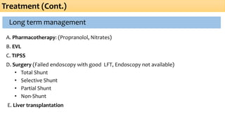
Long term management
A. Pharmacotherapy: (Propranolol, Nitrates)
B. EVL
C. TIPSS
D. Surgery (Failed endoscopy with good LFT, Endoscopy not available)
• Total Shunt
• Selective Shunt
• Partial Shunt
• Non-Shunt
E. Liver transplantation
Treatment (Cont.)
Treatment of complication (Cont.)
Treatment (Cont.)
Coagulopathy
• H2 –blocker
• Inj- Vit-K
• Recombinant active human factor VII
• FFP
• Platelet transfusion
• Desmopressin ( VII, VIII, IX, XII)
Treatment of complication (Cont.)
Treatment (Cont.)
• Give oxygen inhalation
• Somatostatin
• Liver transplantation
• TIPS
Hepatopulmonary Syndrome
Hepato-renal Syndrome
• Vasoconstrictors (Terlipressin) & Albumin
• TIPS
• Extracorporeal albumin dialysis
• Liver transplantation
• Prevention
o IV albumin (1.5gm/kg at infection dx &
1 gm/kg iv 48 hrs. later)
o Long term oral norfloxacin-SBP
prophylaxis
Treatment (Cont.)
Antifibrogenic therapy
• Inhibitors of HSC (Hepatic Stellate Cell) activation:
Antioxidants.
• Antifibrogenic agents:
TGF-β ( transforming growth factor), Endothelin receptor inhibitor.
• Scar degradation agents:
Metalloproteinases
Definitive Treatment
Treatment (Cont.)
It is the definitive treatment for patients with decompensated cirrhosis
• Obstructive biliary tract disease
• Metabolic disorders
• Acute hepatitis: Fulminant Hepatic Failure
• Chronic hepatitis with cirrhosis: Hepatitis B or C, Autoimmune
Liver Transplantation
Definitive Treatment
Treatment (Cont.)
• Intrahepatic cholestasis: idiopathic neonatal hepatitis, Alagille syndrome, familial
intrahepatic cholestasis (Byler disease)
• Miscellaneous: cryptogenic cirrhosis, congenital hepatic fibrosis, Caroli disease, cystic
fibrosis, polycystic liver disease, cirrhosis induced by total parenteral nutrition (TPN)
• Primary liver tumors.
Liver Transplantation
Definitive Treatment
Treatment (Cont.)
Timing for liver transplantation
• Persistent rise of bilirubin ( >9mg/dl)
• Prolongation of PT
• Persistent fall in S. Albumin (<35g/l)
Liver Transplantation
Out- come: 5 years survival 70%
10 years survival 65%
Cirrhosis- grading (Child- Pugh) For Prognosis
Parameter 1 Point 2 Point 3 Point
Ascites None Moderate Severe
Encephalopathy None 1-2 3-4
Bilirubin <2 2-3 >3
Albumin >3.5 2.8- 3.5 <2.8
PT <16 17-21 >22
Interpretation & Survival in Cirrhosis
• A and B Good risk
• C Bad risk
Total score Child class 1-year 5-year 10-year Hepatic
Death
<7 A 82 45 25 43
7-9 B 62 20 7 72
10-15 C 42 20 0 85
Cirrhosis- grading (Child- Pugh) For Prognosis
Prognosis
• Patient with hepatic failure, persistent jaundice, huge ascites not respond with drug,
persistent hypotension, liver size small, prognosis bad.
• If S. Albumin is less than 25gm/L, S. Sodium < 120mEq/L, unrelated to diuretic therapy,
it is a grave condition.
• Extensive necrosis and inflammatory infiltration of liver are poor prognosis.
• Compensated cirrhosis become decompensated 10% per year.
• 1 year survival rate of cirrhotic patient following the first episode of spontaneous
bacterial peritonitis 30-45% and first episode of encephalopathy 40%.
Chronic Liver Disease- Pediatrics Gastroenterology.pptx

More Related Content

PPTX
Approac h to cholestatic jaundice
PPTX
Chronic liver disease in the children .pptx
PDF
Obstructive jaundice , PBC .pdf
PPTX
Approach to cholestatic jaundice
PPT
Lecture 1-8th-nov-07
PPTX
Primary Biliary Cirrhosis/ Primary Biliary Cholangitis
PPTX
MCS 300 Abdomen and GasTrointestine .powerpoint
PPTX
chronic liver diseasein children2025.pptx
Approac h to cholestatic jaundice
Chronic liver disease in the children .pptx
Obstructive jaundice , PBC .pdf
Approach to cholestatic jaundice
Lecture 1-8th-nov-07
Primary Biliary Cirrhosis/ Primary Biliary Cholangitis
MCS 300 Abdomen and GasTrointestine .powerpoint
chronic liver diseasein children2025.pptx

Similar to Chronic Liver Disease- Pediatrics Gastroenterology.pptx (20)

PPTX
chronic liver disease in children2025.pptx
PPTX
Chronic liver disease in children22.pptx
PPTX
Cirrhosis
PPT
Liver disease
PPTX
Cirrhosis of liver .pptx
PPTX
Primary Biliary cholangitis, lecture class for MBBS
PPT
Cirrhosis and Its Complications
PPTX
Gastro intestinal Nursing Chronic Liver Disease.pptx
PPTX
FINAL CLD SEMINAR on liver cancer .pptx
PPTX
DISORDERS OF LIVER, PANCREAS AND BILIARY TRACT
PPTX
LIVER CIRRHOSIS MEDICINE
PPTX
Cirrhosis liver disease unraveling the mystery.pptx
PDF
ALCOHOLIC LIVER DISEASE
PPTX
Liver cirrhosis (Causes, clinical manifestation, complications, diagnosis, tr...
PPTX
Fulminant hepatic failure
PPTX
Liver transplant
PPTX
PPT on Neonatal cholestasis by Dr.ajay k chourasia
PDF
CKD MANAGEMENT.pdf
PPTX
Presentation (1).pptx medicine cirrhosis
PPTX
Neonatal cholestasis syndrome
chronic liver disease in children2025.pptx
Chronic liver disease in children22.pptx
Cirrhosis
Liver disease
Cirrhosis of liver .pptx
Primary Biliary cholangitis, lecture class for MBBS
Cirrhosis and Its Complications
Gastro intestinal Nursing Chronic Liver Disease.pptx
FINAL CLD SEMINAR on liver cancer .pptx
DISORDERS OF LIVER, PANCREAS AND BILIARY TRACT
LIVER CIRRHOSIS MEDICINE
Cirrhosis liver disease unraveling the mystery.pptx
ALCOHOLIC LIVER DISEASE
Liver cirrhosis (Causes, clinical manifestation, complications, diagnosis, tr...
Fulminant hepatic failure
Liver transplant
PPT on Neonatal cholestasis by Dr.ajay k chourasia
CKD MANAGEMENT.pdf
Presentation (1).pptx medicine cirrhosis
Neonatal cholestasis syndrome

Recently uploaded (20)

PDF
495958952-Techno-Obstetric-sminiOSCE.pdf
PPTX
DIARRHOEA IN CHILDREN presented to COG.ppt
PPTX
Hyperthyroidism, Thyrotoxicosis, Grave's Disease with MCQs.pptx
PPTX
Critical Issues in Periodontal Research- An overview
PDF
Glaucoma Definition, Introduction, Etiology, Epidemiology, Clinical Presentat...
PDF
Tackling Intensified Climatic Civil and Meteorological Aviation Weather Chall...
PPTX
etomidate and ketamine action mechanism.pptx
PPTX
Tuberculosis : NTEP and recent updates (2024)
PPTX
Method of organizing health promotion and education and counselling activitie...
PDF
NCM-107-LEC-REVIEWER.pdf 555555555555555
PPTX
المحاضرة الثالثة Urosurgery (Inflammation).pptx
PPTX
presentation on dengue and its management
PPT
intrduction to nephrologDDDDDDDDDy lec1.ppt
PPTX
01. cell injury-2018_11_19 -student copy.pptx
PPTX
IND is a submission to the food and drug administration (FDA), requesting per...
PPTX
Peripheral Arterial Diseases PAD-WPS Office.pptx
PDF
Integrating Traditional Medicine with Modern Engineering Solutions (www.kiu....
PDF
Gonadotropin-releasing hormone agonist versus HCG for oocyte triggering in an...
PPTX
SUMMARY OF EAR, NOSE AND THROAT DISORDERS INCLUDING DEFINITION, CAUSES, CLINI...
PPTX
Local Anesthesia Local Anesthesia Local Anesthesia
495958952-Techno-Obstetric-sminiOSCE.pdf
DIARRHOEA IN CHILDREN presented to COG.ppt
Hyperthyroidism, Thyrotoxicosis, Grave's Disease with MCQs.pptx
Critical Issues in Periodontal Research- An overview
Glaucoma Definition, Introduction, Etiology, Epidemiology, Clinical Presentat...
Tackling Intensified Climatic Civil and Meteorological Aviation Weather Chall...
etomidate and ketamine action mechanism.pptx
Tuberculosis : NTEP and recent updates (2024)
Method of organizing health promotion and education and counselling activitie...
NCM-107-LEC-REVIEWER.pdf 555555555555555
المحاضرة الثالثة Urosurgery (Inflammation).pptx
presentation on dengue and its management
intrduction to nephrologDDDDDDDDDy lec1.ppt
01. cell injury-2018_11_19 -student copy.pptx
IND is a submission to the food and drug administration (FDA), requesting per...
Peripheral Arterial Diseases PAD-WPS Office.pptx
Integrating Traditional Medicine with Modern Engineering Solutions (www.kiu....
Gonadotropin-releasing hormone agonist versus HCG for oocyte triggering in an...
SUMMARY OF EAR, NOSE AND THROAT DISORDERS INCLUDING DEFINITION, CAUSES, CLINI...
Local Anesthesia Local Anesthesia Local Anesthesia

Chronic Liver Disease- Pediatrics Gastroenterology.pptx

  • 3. Outline • Anatomy and Physiology • Pathology • Epidemiology • Classification • Etiology • Approach to CLD • Complications • Investigations • Treatment • Prognosis
  • 7. Functions of Liver Synthesis: - Albumin - Coagulation factors Glucose homeostasis; Glycogenolysis & Gluconeogenesis Storage: - Glycogen - Iron - Cu, vitamins Catabolism of hormones and other serum proteins Bile excretion
  • 9. Definition: Liver Disease present over many months without progressive improvement back to normal liver. Chronic Liver Disease
  • 11. Epidemiology Cirrhosis and chronic liver disease were the 10th leading cause of death for men and 12th for women in the United States in 2010, killing about 27,000 people each year.
  • 12.  CLD is called as “National Disease of Japan” in 21st century. Fifty thousands of people died from CLD this days.  Approximately 1 in 679 or 0.15% or 400,000 people got Chronic Liver Disease in USA from 1976-1980.  90 out of 600 (15%) patients admitted in pediatric Gastroenterology ward, BSMMU, were diagnosed as CLD in 2012. Epidemiology (Cont.) Source: (Digestive diseases in the United States: Epidemiology and Impact – NIH Publication No. 94-1447, NIDDK, 1994)
  • 13. Classification of CLD 1. Chronic hepatitis: Symptomatic, biochemical or serologic evidence of continuing or relapsing hepatic disease for more than 6 months, with histologically documented inflammation. 2. Cirrhosis: Is a diffuse parenchymal disease of liver characterized by hepatocellular infiltration and necrosis resulting in loss of hepatic architecture , formation of regenerative nodules with fibrosis and abnormal vascular shunt formation.
  • 15. Pathogenesis of CLD Chronic inflammation of liver Inflammatory cytokines(TNF,lymphotoxin,IL-1,TGF,PDGF) are produced by stimulated Kupffer cell, hepatocyte, endothelial cell. Activation of stellate cell Transformation of stellate cell into myofibroblast like cell which produce collagen(type I & III). Collagen deposition in lobule, creating- 1. Broad septal band 2.Increased vascular resistance. 3. Fibrosis & regeneration of remaining hepatocytes Fibrosis + loss of architecture+ formation of regenerative nodule. Cirrhosis of liver
  • 17. Chronic Hepatitis Classification (Older) 1. Chr. Persistent 2. Chr. Lobular 3. Chr. Active Etiology Clinical grade Histological severity Stage Classification (new) based on Classification
  • 18. Classification (cont.)  Clinical grade-depends on S. ALT level Mild : <100 IU/L Moderate: 100-400 IU/L Severe: > 400 IU/L  Histological scoring Knodell/HAI Ishak score
  • 19. Knodell score( HAI) • Composed of summation of 4 individual scores: • 1. Periportal necrosis with or without bridging necrosis. • 2. Intra lobular degeneration and focal necrosis. • 3. Portal inflammation. • 4.Fibrosis. Classification (cont.)
  • 20. 1.Periportal necrosis with or without bridging necrosis ( Score 0-10) None 0 Mild piecemeal necrosis 1 Moderate piecemeal necrosis 3 Marked piecemeal necrosis 4 Moderate piecemeal necrosis+ bridging necrosis 5 Marked piecemeal necrosis + bridging necrosis 6 Interlobular necrosis 10 2.Inter lobular degeneration and focal necrosis None 0 Mild(involvement of <1/3 of lobules or nodules) 1 Moderate(involvement of 1/3 to 2/3 of lobules or nodules) 3 Marked(involvement of >2/3 of lobules or nodules) 4 Classification (cont.)
  • 21. Portal inflammation No portal inflammation 0 Mild (sprinkling of inflammatory cells in <1/3 of portal tracts) 1 Moderate (increased inflammatory cells in1/3 to 2/3 of portal tracts) 3 Marked(dense packing of inflammatory cells in >2/3 of portal tracts) 4 Fibrosis No fibrosis 0 Fibrous portal expansion 1 Bridging fibrosis(portal-portal to portal-central linkage) 3 Classification (cont.)
  • 22. Interpretation HAI (Total Score 18) Diagnosis 1-3 Minimal 4-8 Mild 9-12 Moderate 13-18 Severe Scoring By Fibrosis Diagnosis 0 None 1 Mild 2 Moderate 3 Severe 4 Cirrhosis Classification (cont.)
  • 23. Classification of Cirrhosis • Pathological: Micro nodular Macro nodular Mixed Biliary Classification (cont.)
  • 24. Normal Liver Microscopy Fibrosis Regenerating Nodule Cirrhosis Classification (cont.)
  • 25. Micronodular Thick regular septa. Regenerating small nodules < 3mm Involvement of every nodule Seen in – Haemachromatosis Cholestasis Hepatic venous outflow obstruction Classification (cont.) Macronodular Septa and nodules (>3 mm) of variable sizes Normal lobules in larger nodules Secondary to chronic viral hepatitis
  • 27. Biliary cirrhosis Bile stasis General reduction of bile duct Increased connective tissue within and extending from portal tracts Lobular architecture preserved. Seen in- Biliary atresia, CF, PFIC. Classification (cont.)
  • 29. 1. Infectious 2. Metabolic 3. Inflammatory 4. Biliary malformation 5. Vascular 6. Toxic 7. Nutritional 8. Miscellaneous Etiology
  • 30. Chronic hepatitis B with or without D virus Chr. hepatitis C CMV Herpes Simplex virus Rubella Neonatal sepsis Etiology- Infectious
  • 31. • Alfa -1 antitrypsin deficiency • Fructosemisa • Wilson’s disease • Cystic fibrosis • Hemochromatosis • Niemann- pick disease type D • Tyrosinemia • Galactosemia • Gauccher’s disease • Glycogen storage disease Etiology- Metabolic
  • 32. Autoimmune chronic hepatitis Primary sclerosing cholangitis Autoimmune hepatitis is a chronic hepatitis of unknown etiology.  First described in the 1950s,  Characterized generally by 3 features: Hepatitis Auto antibodies High serum globulin concentration Etiology- Inflammatory
  • 34. • Intrahepatic biliary hypoplasia • Biliary atresia • Alagille syndrome • Choledochal cyst Etiology- Biliary Malformation
  • 35. • Budd- Chiari syndrome • CCF • Congestive pericarditis • Veno-occlusive liver disease Etiology- Vascular
  • 39. Approach to a child with CLD
  • 40. History 1. Duration of illness. 2. History of jaundice. 3. History of blood transfusion or parenteral injection. 4. H/O ingestion of any hepatotoxic drugs. 5. H/O bleeding manifestation (Hematemesis, melaena ). 6. Sign of encephalopathy (Altered sleep rhythm).
  • 41. History 7. H/O umbilical sepsis or catheterization in neonatal period. 8. Color of urine, stool. 9. H/O fatigue, weight loss, anorexia, abdominal pain. 10. Recurrent chest infection or chronic diarrhoea. 11. H/O consanguinity, family history, sibling affection. 12. Religion (middle class hindu), copper made utensils. 13. Immunization history.
  • 44. Skin Changes Jaundice Spider angioma Gynecomastia Scratch mark Bruising & Bleeding Xanthoma
  • 45. Physical sign – Abdomen 1. Abdominal Distension. 2. Caput medusa. 3. Ascites. 4. Liver- size, consistency. 5. Splenomegaly. 6. Testicular atrophy. 7. Rectal examination.
  • 46. Special presentations HCV Small vessel vasculitis, peripheral neuropathy, GN, NS. Autoimmune hepatitis Polyarthritis, allergic capillaritis, generalized lymphadenopathy, GN, Hashimoto's thyroiditis, DM Wilsons disease KF ring, sunflower cataract, neuro-psychiatric sign Metabolic disease Cataract, facial dysmorphism, developmental delay, convulsion
  • 48. Complication •Coagulopathy •Hypoalbumenia •Ascites, Electrolyte imbalance •Hypoglycemia Due to parenchymal dysfunction •Variceal bleeding •Hypersplenism Due to portal hypertension •Sepsis, pneumonia, UTI, SBP Infections
  • 50. Complication (Cont) •Porto-systemic encephalopathy •Hepato-renal syndrome •Hepato-pulmonary syndrome Systemic complication: Hepatocellular carcinoma
  • 51. Encephalopathy Complication (Cont) • Reversible decrease in neurologic function caused by liver disease • Established mechanism a. Accumulation of un-metabolized NH3 due to • Poor hepatic function • Poor systemic shunting. b. Possible mechanism: False neurotransmitter, GABA, Benzodiazepine receptor activation etc.
  • 53. • Thrombocytopenia • Thrombocytopathy • Decrease synthesis of vit K – dependent factors • DIC Coagulopathy Complication (Cont)
  • 54. Hepato-renal syndrome: Complication (Cont) Hepato-renal syndrome is defined as functional renal failure in patient with end stage liver disease. The hallmark is intense renal vaso-constriction with co-existing systemic vaso-dilatation, may be mediated by hemodynamic, humoral or neurogenic mechanism. Diagnosis: 1. Oliguria (< 1 ml/kg/day). 2. Urine sodium (< 10 mEq/L). 3. Fractional Na+ excretion(<1%). 4. Urine: Plasma creatinine ratio-<10. 5. Exclusion of other kidney pathology. Treatment: Liver transplantation.
  • 56. Hepato-pulmonary syndrome: Complication (Cont) Is characterized by typical triad of hypoxemia, intrapulmonary vascular dilations and liver disease. There is intrapulmonary right to left shunting of blood which results in systemic desaturation. Clinical feature: 1. Shortness of breath. 2. Exercise in tolerance. 3. Cyanosis- lips & fingers. In a child with CLD. 4. Clubbing. 5. O2 saturation < 96%. Treatment: Liver transplantation.
  • 58. To Establish Diagnosis Investigations A. Evaluation of liver function: i. S. bilirubin level ii. S. ALT iii. S. AST level iv. Alkaline phosphatase level v. S. protein vi. Prothrombin time vii. Blood sugar B. Abdominal ultrasonography C. Endoscopy of upper GIT D. Liver biopsy
  • 60. Chronic Hepatitis HBsAg positive 1. Look for markers of active Viral replication (HBeAg, HBV, DNA) 2. Look for co–infection (anti – HDV lgM) HBsAg negative Autoantibodies (+ve) AIH Autoantibodies (-ve) 1. Anti – HCV and HCV 2. Rule out Wilson disease 3. Antitrypsin deficiency 4. Drug induced Investigations (Cont.)
  • 61. Infections Screening: 1. HBV : HBsAg, HBeAg/antibody, HBV-DNA 2. HCV : HCV antibody, HCV- RNA 3. CMV : CMV antibody, Urine CMV antigen 4. EBV : Serology 5. Bacterial Cholangitis : Blood culture. Wilsons disease: 24-hr urinary cu excretion after penicillamine challenge liver copper concentration. Investigations (Cont)
  • 62. Alfa 1- antitrypsin deficiency: S. alpha 1- antitrypsin level Glycogen storage disease: lactic acid level, FBS, uric acid level, liver and muscle tissue enzyme level. Autoimmune chr. hepatitis: ANA, Anti SMA, ALKM1,Immunoglobulin level. Investigations (Cont)
  • 63. Tyrosinemia: Serum amino acid level, Urine organic acid (succinyl acetone) Hemochromatosis: Serum iron, TIBC, Ferritin Galactosemia: Red blood cell galactose -1 phosphate uridyl transferase level. Urinary non-glucose reducing sugar. Investigations (Cont) Cystic fibrosis: Sweat chloride test, Genotype study
  • 64. Ascitic fluid: Cytology, Albumin, Culture Others: • USG • Endoscopy • Proctoscopy • Biopsy Investigations (Cont)
  • 65. Endoscopy of upper GIT: White, pink, red, cherry red varices Conn’s grading system: Investigations (Cont) Gr.1: Visible only during inspiration Gr.2: Visible during both inspiration & expiration Gr.3 Projectile into lumen <50% Gr.4: Projectile into lumen >50%
  • 67. F. After initial investigations , the following may be needed: i. HBV DNA ii. HCV RNA iii. MRCP iv. ERCP sigmoidoscopy or colonoscopy v. Bone marrow aspiration for abnormal storage materials vi. Enzyme activity in leukocytes or fibroblasts for specific liver disease Investigations (Cont.)
  • 69. A. No specific treatment that can reverse cirrhosis B. Supportive: Nutritional: a) High energy protein rich diet if there is no hepatic encephalopathy. b) CHO should be taken adequately c) Fat should be restricted if there is cholestasis d) Vitamin and mineral supplementation C. Rest D. Clear bowel regularly. E. Cholestasis: Pruritus: Ursodeoxycolic acid. Treatment
  • 70. Treatment (Cont.) Etiology Treatment Viral hepatitis (B, C, D) Adefovir, lamivudine, INF Iron overload Venesection, deferoxamine Wilsons Disease Copper chelator Alfa 1-antitrypsin deficiency Transplant Type IV glycogen disease Transplant Galactosemia and tyrosinemia Transplant Budd-Chiari syndrome Relieve main block/Transplant Autoimmune hepatitis Immunosuppressive drug
  • 71. Treatment of Encephalopathy Treatment (Cont.) • Avoid Precipitating Factors • Fluid 60 ml/kg/day • Protein restriction • Gut sterilization and laxatives • Systemic antibiotics
  • 72. Treatment of Ascites Treatment (Cont.) • Bed rest • Diet: Salt restriction (Na+ 1-2 mEq/Kg/d) • Diuretics: Spironolactone, frusemide • Fluid restriction: if S. Na+ <120 mEq/l • Paracentesis: Refractory cases. • Shunts: TIPSS
  • 73. Treatment of SBP Treatment (Cont.) • Third generation cephalosporin + Flucloxacillin • Amoxycillin-clavulanic acid • Ciprofloxacin • Ofloxacin • Duration: 5-8 days • Prophylactic- Norfloxacin, Cotrimoxazole
  • 74. Treatment of Portal Hypertension Immediate Management • ABC Management: Fluid resuscitation, Blood Product • Sclerotherapy • Band Ligation • Balloon Tamponade • TIPS • Pharmacotherapy: Vasopressin, Octreotide. Treatment (Cont.) Band Ligation Sclerotherapy
  • 75. Long term management A. Pharmacotherapy: (Propranolol, Nitrates) B. EVL C. TIPSS D. Surgery (Failed endoscopy with good LFT, Endoscopy not available) • Total Shunt • Selective Shunt • Partial Shunt • Non-Shunt E. Liver transplantation Treatment (Cont.)
  • 76. Treatment of complication (Cont.) Treatment (Cont.) Coagulopathy • H2 –blocker • Inj- Vit-K • Recombinant active human factor VII • FFP • Platelet transfusion • Desmopressin ( VII, VIII, IX, XII)
  • 77. Treatment of complication (Cont.) Treatment (Cont.) • Give oxygen inhalation • Somatostatin • Liver transplantation • TIPS Hepatopulmonary Syndrome Hepato-renal Syndrome • Vasoconstrictors (Terlipressin) & Albumin • TIPS • Extracorporeal albumin dialysis • Liver transplantation • Prevention o IV albumin (1.5gm/kg at infection dx & 1 gm/kg iv 48 hrs. later) o Long term oral norfloxacin-SBP prophylaxis
  • 78. Treatment (Cont.) Antifibrogenic therapy • Inhibitors of HSC (Hepatic Stellate Cell) activation: Antioxidants. • Antifibrogenic agents: TGF-β ( transforming growth factor), Endothelin receptor inhibitor. • Scar degradation agents: Metalloproteinases
  • 79. Definitive Treatment Treatment (Cont.) It is the definitive treatment for patients with decompensated cirrhosis • Obstructive biliary tract disease • Metabolic disorders • Acute hepatitis: Fulminant Hepatic Failure • Chronic hepatitis with cirrhosis: Hepatitis B or C, Autoimmune Liver Transplantation
  • 80. Definitive Treatment Treatment (Cont.) • Intrahepatic cholestasis: idiopathic neonatal hepatitis, Alagille syndrome, familial intrahepatic cholestasis (Byler disease) • Miscellaneous: cryptogenic cirrhosis, congenital hepatic fibrosis, Caroli disease, cystic fibrosis, polycystic liver disease, cirrhosis induced by total parenteral nutrition (TPN) • Primary liver tumors. Liver Transplantation
  • 81. Definitive Treatment Treatment (Cont.) Timing for liver transplantation • Persistent rise of bilirubin ( >9mg/dl) • Prolongation of PT • Persistent fall in S. Albumin (<35g/l) Liver Transplantation Out- come: 5 years survival 70% 10 years survival 65%
  • 82. Cirrhosis- grading (Child- Pugh) For Prognosis Parameter 1 Point 2 Point 3 Point Ascites None Moderate Severe Encephalopathy None 1-2 3-4 Bilirubin <2 2-3 >3 Albumin >3.5 2.8- 3.5 <2.8 PT <16 17-21 >22
  • 83. Interpretation & Survival in Cirrhosis • A and B Good risk • C Bad risk Total score Child class 1-year 5-year 10-year Hepatic Death <7 A 82 45 25 43 7-9 B 62 20 7 72 10-15 C 42 20 0 85 Cirrhosis- grading (Child- Pugh) For Prognosis
  • 84. Prognosis • Patient with hepatic failure, persistent jaundice, huge ascites not respond with drug, persistent hypotension, liver size small, prognosis bad. • If S. Albumin is less than 25gm/L, S. Sodium < 120mEq/L, unrelated to diuretic therapy, it is a grave condition. • Extensive necrosis and inflammatory infiltration of liver are poor prognosis. • Compensated cirrhosis become decompensated 10% per year. • 1 year survival rate of cirrhotic patient following the first episode of spontaneous bacterial peritonitis 30-45% and first episode of encephalopathy 40%.