Comparison of ct and cbct
83 likes26,315 views

The document compares Computed Tomography (CT) and Cone Beam Computed Tomography (CBCT) in orthodontics, highlighting how each technology works, their indications, advantages, and disadvantages. CT offers high-contrast resolution and multiplanar imaging but has higher radiation exposure and cost, while CBCT provides rapid scanning and lower radiation doses, though with poorer soft tissue contrast. The conclusion indicates that while CBCT is a promising tool for orthodontics, it should be used judiciously when 2D imaging suffices.


Comparison of ct and cbct
- 2. COMPARISON OF CT AND CBCT DONE BY: AMRITHA JAMES,CRI GUIDED BY: DR. KARUNANATHI MDS, DR.SARANYA DEVI, MDS Department of orthodontics
- 3. contents Computed tomography Introduction How does a CT work? Image reconstruction in CT Artifacts Indications of CT Advantages and disadvantages of CT Cone beam computed tomography Introduction How does a CBCT work? Principle of CBCT Image formation Artifacts Indication of CBCT in orthodontics Advantages and disadvantages of CBCT CT vs. CBCT
- 4. CT CT scanner was invented by Godfrey Newbold Hounsfield in Hayes, England A CT scan makes use of computer- processed combinations of many X- ray images taken from different angles to produce cross-sectional (tomographic) images.
- 5. How does a CT work? A CT scanner consists of an x-ray tube that emits a finely collimated fan shaped x-ray beam directed through a patient to a series of scintillation detectors. These detectors measure the number of photons that exit the patient. The detectors form a continuous ring around the patient and the x- ray tube moves in a circle within the fixed detector ring. This information is used to construct a cross- sectional image of the patient.
- 7. Image reconstruction Many scans are progressively taken as the object is gradually passed through the gantry. They are combined together by the mathematical procedure known as tomographic reconstruction.
- 8. The x ray beam attenuation data is collected in a grid pattern called a matrix. Each square in the matrix is made up of pixel which represents the x ray attenuation of small finite volume of tissue(voxel) or volume element. Typical matrix in CT are 256*256 or 512*512 pixels.
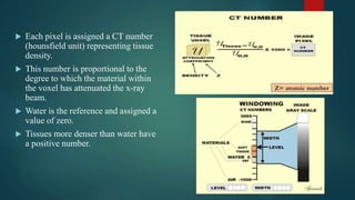
- 9. Each pixel is assigned a CT number (hounsfield unit) representing tissue density. This number is proportional to the degree to which the material within the voxel has attenuated the x-ray beam. Water is the reference and assigned a value of zero. Tissues more denser than water have a positive number.
- 10. Image display Areas of high density with high CT number appear white on the greyscale. Areas of low density with low CT number appear black on the greyscale.
- 11. 3 step process of CT imaging
- 12. Artifacts Aliasing Artifact or Streaks These appear as dark lines which radiate away from sharp corners. It occurs because it is impossible for the scanner to take enough projections of the object, which is usually metallic. Ring Artifact Probably the most common mechanical artifact, the image of one or many 'rings' appears within an image. This is due to a detector fault.
- 13. Noise Artifact This appears as gaining on the image and is caused by a low signal to noise ratio. This occurs more commonly when a thin slice thickness is used. It can also occur when the kV or mA is too low. Motion Artifact This is seen as blurring which is caused by patient movement. Beam Hardening This can give a 'cupped appearance'. It occurs when there is more attenuation in the center of the object than around the edge. This is easily corrected by filtration .
- 14. Indications of CT in dentistry 1. Evaluation of extent of any suspected pathology in the head and neck, including tumors, cysts and infection. 2. Determination of location and extent of facial fractures. 3. Radiographic pre surgical evaluation for implant placement.
- 15. ADVANTAGES OF CT 1. CT completely eliminates the superimposition of images of structures outside the area of interest. 2. Because of the inherent high-contrast resolution of CT, differences between tissues that differ in physical density by less than 1% can be distinguished. 3. Data from a single CT imaging procedure consisting of either multiple contiguous or one helical scan can be viewed as images in the axial, coronal, or sagittal planes, depending on the diagnostic task. This is referred to as multiplanar reformatted imaging.
- 16. DISADVANTAGES OF CT Time consuming Expensive for routine clinical use High radiation exposure Expensive equipment and hence is not always accessible
- 17. CBCT CBCT is a variation on traditional computed tomography (CT) Unlike traditional CT scanners, in CBCT an X-ray tube and detector panel rotate around the patient capturing data with a cone-shaped X-ray beam instead of the “slices” CTs are typically known for. Images are then reconstructed using algorithms to produce 3-dimensional images at high resolution. CBCT machine
- 18. NewTom 9000 / NewTom 3G i-CAT
- 19. HOW DOES A CBCT WORK? All CBCT scanners consists of an x ray source and detector mounted on a rotating gantry. During rotation of the gantry, the x ray source produces a divergent cone shaped radiation, while the receptor records the residual x rays after attenuation by patients tissues. The x ray source and detector moves through an arc of 180 to 360 degree to produce multiple planar projection images. Theses images constitute the raw primary data which is then reconstructed by a computer algorithm to generate cross sectional images.
- 21. Components of image production 1. X-ray generation 2. X- ray detection: Image sensor- PSP (photo stimulable phosphorus plates),CCD sensors, FPD (flat panel detector) 3. Image reconstruction
- 23. PRINCIPLE OF CBCT FIELD OF VIEW: Collimation of x ray beam by adjustment of FOV limits the radiation to one Radius Of Interest. These depend on the detector size and shape, beam projection geometry and the ability to collimate or not It is desirable to limit the field size to the smallest volume that can accommodate the region of interest.
- 24. REGION OF INTEREST BEYOND FOV: Data from two or more separate scans are superimposed and overlapped using reference points A software is then used to stich or blend the images together
- 25. VOXEL: The spatial resolution is determined by individual volume elements called voxels. These are cubic in nature equal in all dimensions The principle determinant of voxel size is the pixel size of the detector. Detectors with smaller pixel size capture fewer x-ray photons per voxel and result in more noise. To balance it out a good scanner has higher dosage of radiation All current CBCT machines have 12 bit detectors and are capable of identifying 4096 shades of gray .
- 26. IMAGE FORMATION Image formed can be visualized as 2D trans-axial, multi-planar reformatted 3D techniques such as surface reconstruction and volume rendering A combination of 2D and 3D techniques saggital(A), coronal (B), and axial (C) planes.
- 27. THE MECHANISM OF CBCT ACQUISITION
- 28. artifacts Any distortion or error in the image that is unrelated to the subject being studied is called an artifact. Occurs at the interface of the material with a completely different radiological property from the subject being imaged. They can be classified into 1. Inherent artifacts 2. Procedure related artifacts 3. Introduced artifacts
- 29. STREAKING ARTIFACT MOVEMENT ARTIFACT BEAM HARDENING METAL ARTIFACT
- 30. CBCT IN ORTHODONTICS Impacted Tooth Root Resorption associated with impacted teeth Developmental anomalies Root Fracture Evaluate the residual alveolar bone thickness Evaluating the bony defect caused by cleft palate
- 31. Temporomandibular Evaluation Nerve Mapping Ceph Tracing Airway Analysis
- 32. Advantages of CBCT Rapid scan time Beam limitation Image accuracy Reduction in patient radiation dose when compared to medical CT Interactive display modes Multiplanar reformatting 3 dimensional volume rendering Better images with good spatial resolution Economical, comfortable and safe
- 33. Disadvantages of cbct Poor contrast resolution, thus soft tissue cannot be viewed Artifacts Image noise
- 34. Ct vs cbct
- 35. CT CBCT 1. Traditional CT uses a high- output, rotating anode X-ray tube. 1. Cone beam tomography utilizes a low-power, medical fluoroscopy tube that provides continuous imaging throughout the scan.
- 36. CT CBCT 2. Traditional computerized tomography records data with a fan-shaped X-ray beam onto image detectors arranged in an arc around the patient. 2. The advanced cone beam technology uses a cone-shaped X-ray beam that transmits onto a solid-state area sensor for image capture.
- 37. CT CBCT 3. Produces a single slice image per scan. Each slice must overlap slightly in order to properly reconstruct the images. 3. Produces the complete volume image in a single rotation.
- 38. CT CBCT 4. Slower due to spiral motion. Scan time is longer. 4. The single-turn motion image capture used in CBCT is quicker than traditional spiral motion of CT. Average time for one cbct scan may vary from 7-30 seconds.
- 39. CT CBCT 5. Has high radiation dose. The average medical CT scan of the oral and maxillofacial area can reach levels of 1,200– 3,300 microsieverts. To collect adequate 5. Has lower radiation dose as a result of no overlap of slices. Radiation exposure using the standard full field of view from an i-CAT® CBCT machine is 36 microsieverts.
- 40. CT CBCT 6. To collect adequate formation, there is overlapping of radiation. 6. No overlap of slices
- 41. CT CBCT 7. Only one jaw can be visualized at one time. 7. Both jaws can be imaged at the same time.
- 42. medical CT 120 kV, 100 mAs CBCT 110 kV, 10 mAs CT CBCT 8. High-contrast resolution 8. Poor contrast resolution, thus soft tissue cannot be viewed.
- 43. CT CBCT 9. Cost is high 9. Cost of equipment is approximately 3-5 times less than traditional Medical CT CT CBCT 10. Can cause claustrophobia 10. The open design of the cone beam CTs virtually eliminates claustrophobia and greatly enhances patient comfort and acceptance
- 44. CT vs CBCT
- 45. CONCLUSION CBCT is the future of orthodontics and its applications in orthodontics seem almost limitless. CBCT imaging provides insight into treatment planning that is unachievable with other imaging methods, and allows clinicians to provide more predictable patient care, however CBCT should be used with careful consideration ,it should not be used where 2D imaging suffices.
- 47. references CBCT in Orthodontics: The Wave of Future Jiwanasha Manish Agrawal, Manish Suresh Agrawal, Lalita Girish Nanjannawar, Anita D Parushetti Facing the facts: dental CBCT vs. medical CT scans By Bruce Howerton, DDS, MS The clinical application of cone beam CT in orthodontics Moiz Ahmad Khana, Syed Sheeraz Hussainb Using cone beam technology in orthodontics Edward Lin Applications of CBCT in dental practice: A review of the literature Hadi Mohammed Alamri, BDS n Mitra Sadrameli, DMD and Mazen Abdullah Alshalhoob, BDS Mahtab Sadrameli, DMD, MAGD Mohammed Abdullah Alshehri, BDS, AEGD 2D / 3D Cone-Beam CT images or conventional radiography: Which is more reliable? Carolina Perez Couceiro**, Oswaldo de Vasconcellos Vilella***