SlideShare a Scribd company logo
Presentation on
COPD & Acute Exacerbation of COPD
INTERN DOCTOR
SUNIL KUMAR YADAV
13TH BATCH
MEDICINE DEPARTMENT
NMCTH
INTRODUCTION
COPD refers to chronic, slowly progressive disorder of airway obstruction that is not fully
reversible, with little or no variability. It includes chronic bronchitis and emphysema.
Chronic Bronchitis
A condition associated with excessive mucus production sufficient to cause cough with
expectoration on most days of a week for at least 3 months of the year for more than 2
consecutive years.
Emphysema
Emphysema is defined as the permanent, abnormal distention of the air spaces distal to the
terminal bronchiole with destruction of alveolar septa and without obvious fibrosis.
COPD and AE of COPD
Etiology of Chronic Bronchitis and Emphysema
1. Cigarette smoking
• Cigarette smoking is the most commonly identified factor associated with both chronic
bronchitis during life and extent of emphysema at post-mortem.
• Prolonged cigarette smoking impairs ciliary movement, inhibits function of alveolar
macrophages, and leads to hypertrophy and hyperplasia of mucus-secreting glands. It is
probable that smoke also inhibits antiproteases and causes polymorphonuclear leukocytes to
release proteolytic enzymes acutely. Inhaled cigarette smoke can produce an acute increase in
airways resistance due to vagally mediated, smooth-muscle constriction.
2. Air pollution
Air pollution with sulphur dioxide and particulate matter is associated with exacerbations of
chronic bronchitis, and the incidence and mortality rates for chronic bronchitis and
emphysema is probably higher in heavily industrialized urban areas.
3. Occupation: Occupations exposing workers to inorganic or organic dusts, or to noxious
gases.
4. Acute respiratory infections
5. Familial and genetic factors
6. α1-Antitrypsin (a1AT) deficiency
7. Cystic fibrosis
Pathophysiology
• The hallmark of chronic bronchitis is hypertrophy of the mucus-producing glands found in the
submucosaof large cartilaginous airways.
• Quantitation of this anatomic change is based on the ratio of the thickness of the submucosal
glands to that of the bronchial wall (Reid's index).
• Histologically there is goblet cell hyperplasia, mucosal and submucosal inflammatory cells,
edema, peribronchial fibrosis, intraluminal mucus plugs, and increased smooth muscle in small
airways.
• Inflammation in chronic bronchitis occurs at the alveolar epithelium and differs predominantly
eosinophilic inflammation of asthma by the predominance of neutrophils and the
peribronchiolar location of fibrotic changes.
COPD and AE of COPD
COPD and AE of COPD
Classification of Emphysema
Based on the site of damage:
1. Centri-acinar emphysema: Distension and damage of lung tissue is concentrated around the
respiratory bronchioles, whilst the more distal alveolar ducts and alveoli tend to be well
preserved. This form of emphysema is extremely common. Cigarette smoking.
2. Pan-acinar emphysema: This is less common. Here, distension and destruction appear to
involve the whole of the acinus, and in the extreme form the lung becomes a mass of bullae.
This type of emphysema occurs in alpha1-antitrypsin deficiency.
3. Irregular emphysema: There is scarring and damage affecting the lung parenchyma patchily
without particular regard for acinar structure .
COPD and AE of COPD
COPD and AE of COPD
Clinical Features of COPD
A. Symptoms
• Cough
• Sputum
• Breathlessness worsened with exertion
• Edema
• Morning headaches (due to hypercapnia)
B. Signs
1. Pulmonary signs
• Features of CO2 retention
- Central cyanosis
-Flapping tremors
-Bounding pulse
-Warm peripheries, Early monring headache and Papilloedema.
• Features of Cor pulmonale (RVF+ Pulmonary HTN)
-Peripheral edema
-Raised JVP
-Right ventricular heave
-Loud P2
-Enlarged liver (hepatomegaly)
2. Extra pulmonary signs
• Altered fat metabolism contributing to weight loss , and Muscular weakness reflecting
deconditioning and cellular changes in skeletal muscles.
• Increased prevalence of osteoporosis, and Increased circulating inflammatory markers.
• Impaired salt and water excretion leading to peripheral oedema.
COPD and AE of COPD
Diagnostic Tests
1. Chest X-ray: Best initial test.
• Increased anterior-posterior (AP diameter)
• Hyperinflation: >6 anterior ribs seen above diaphragm in mid clavicular line, or ≥ 10 Posterior
Ribs.
• Large central pulmonary arteries, Decreased peripheral vascular markings and Bulla.
• Tubular heart shadow
• Air trapping and flattened diaphragms
2. Pulmonary function test:
Most accurate test Decreased FEV1, decreased FVC, decreased FEV1/FVC ratio (<70%)
• Increased TLC because of increased residual volume.
• Incomplete improvement with bronchodilators (<12%)
• Little or no worsening with methacholine.
COPD and AE of COPD
3. ABG:
Acute exacerbations associated with increased PCO2 and hypoxia. Respiratory acidosis may be
present.
4. CBC (increased Hb, increased PCV)
5. ECG
• Right atrial and ventricular hypertrophy (cor
pulmonale)
•Poor progression of R wave
•Atrial fibrillation or multifocal atrial tachycardia.
6. High resolution (CT scan (HRCT))
COPD and AE of COPD
Differential Diagnosis of COPD
1. Pneumonia
2. Pneumothorax
3. Left ventricular failure
4. Pulmonary embolism
5. Lung cancer
6. Upper airway obstruction
7. Chronic asthma
8. Tuberculosis
9. Bronchiectasis
10. Congestive cardiac failure
11. Hypersensitivity pneumonitis
12. Interstitial lung disease.
Management
1. Non pharmacological
Smoking cessation and oxygen therapy are the only treatment modalities that delay the
progression of disease and improve mortality.
• Smoking cessation
• Use of domiciliary oxygen (at least 15 hours/day) Criteria for domiciliary oxygen use in COPD -
--PaO2 <55mmHg or SaO2 <88% -or-
-If there are signs of right sided heart failure/disease or hematocrit >55%, use when - PaO2
<60mmHg or SaO2 <90%
•Influenza and pneumococcal vaccination
-Influenza vaccine annually
-Pneumococcal vaccine once (Give only one lifetime booster if the patient was <65 years age at
first vaccination)
• Encourage exercise
• Treatment of poor nutrition and obesity
• Pulmonary rehabilitation
2. Pharmacological
• Anticholinergic agents (Ipratropium, triotropium): first line agents
• Shortacting beta agonists (Albuterol)
• Inhaled steroids
• Long acting beta agonists (Salmeterol)
• Treatment of co-existing infection: Azithromycin, IV ceftriaxone
3. Surgery
Lung volume reduction surgeries are done in selected
patients such pulmonectomy. lobectomy.
Indications for surgery are:
• Recurrent pneumothoraces
• Isolated bullous disease
Prognosis
COPD has a variable natural history but is usually pro- gressive. The prognosis is inversely
related to age and directly related to the post-bronchodilator FEV1. Other poor prognostic
indicators include weight loss and pul-monary hypertension. A composite comprising the body
mass index (B), the degree of airflow obstruction(O), a measurement of dyspnoea (D) and
exercise capacity (E) may assist in predicting death from respiratory and other causes (Box
19.33). Respiratory failure, cardiac disease and lung cancer represent common modes of
death.
COPD and AE of COPD
COPD and AE of COPD
Acute Exacerbation of copd
Characterised by
1. increased Symptoms.
2. deterioration in lungs function
3. Health Status
if ds. Progress then become More frequent; Are usually triggerded by
1. Bacteria
2. virus
3. change in Air quality
↓
Finally may lead to
1. Development of Respiratory failure
2. fluid Retention
3. Important case of Death.
Refferal to Hospital
1. presence of cynosis
2. peripheral Edema
3. Alternation in consciousness.
Difference between Blue bloaters and Pink puffers
Management of Acute Exacerbation of COPD
Note:
Management of AE-COPD is similar to that of acute asthma, as both are type Il respiratory
failure, except the oxygen flow is low in AE COPD.
Investigation
1. Pulmonary function test → FEV., peak flow
2. ABG
3. ECG
4. Chest x-ray
5. Sputum for culture.
6. CBC
7. Urine R/E
8. Electrolytes, urea/creatinine.
Treatment
1. Oxygen
• 24-28% via face mask, 2L/min by nasal prongs.
• Check ABG within 60 mins and adjust according to PaCO2 or pH.
•Keep Pa02 ≥ 7.5 KPa
• May require ventilation in coma, respiratory arrest.
Note:
O2 supply should be low flow because in COPD patients, hypoxia is the stimulus for respiration
instead of hypercarbia in normal people, so removal of hypoxia by high flow results in
cessation of respiration.
2. Nebulization
• Short acting B2 agonists (salbutomol) 5mg in 2-4 ml NS.
• 1st dose in 30 min. Repeat of necessary in 2-4 hours.
• Add ipratropium bromide if response is inadequate 0.5mg 4 hourly.
3. Corticosteroids
• IV hydrocortisone 200mg 6 hourly
• If oral intake possible, oral predisolone (30-60mg) 1mg/kg tapered over 1 week.
4. Monitoring
• Oxygen
• Serum potassium
5. Hydration
• IV fluids
• K+ supplementation may be needed as repeated doses of salbutamol can lower plasma K+.
6. Appropriates antibiotics
• If patient fails to improve
7. Add IV MgSO4 1.2-2g over 20 mins or,
• IV aminophylline 5mg/kg loading dose and 1mg/kg/hr maintainance dose over 20 minutes or
• Inj. adrenaline SC 0.3mg x ⅟ 2 hourly * 3 doses.
8. Monitoring of vitals and electrolytes.
9. Others
• Diuretics, if JVP elevated and oedema present
• If patient continues to deteriorate, respiratory stimulant like doxapram can be considered.
• Prophylactic subcutaneous low molecular weight heparin, for DIC.
Complications of COPD
1. Cor-pulmonale
2. Acute exacerbation
3. Respiratory failure
4. Pneumothorax
5. Mental status deterioration
6. Osteoporosis
7. Renal failure
THANK YOU

More Related Content

PPTX
Topic: Neurogenic shock and advance nursing care .
PPTX
IONTOPHORESIS
PPTX
Reusable resuscitator
PPTX
Pathophysiology of shock
PPTX
HUMIDIFICATION AND NEBULIZATION.pptx
PPT
Physiotherapy for Critically ill patients
Topic: Neurogenic shock and advance nursing care .
IONTOPHORESIS
Reusable resuscitator
Pathophysiology of shock
HUMIDIFICATION AND NEBULIZATION.pptx
Physiotherapy for Critically ill patients

What's hot (20)

PPTX
Hemorrhage & Shock
PDF
Pathophysiology of respiratory failure
PPTX
BRONCHIECTASIS
PPTX
Emergency management of anaphylactic shock
PPT
Pain managment with modalities 1
PPT
Chest Infections
PDF
Cardiopulmonary assessment
PPTX
Weaning from mechanical ventilation
PPTX
ultrsound.pptx
PPT
Understanding Anesthesia for physiotherapist
PPTX
Collateral ventilation
PPTX
Musculoskeletal disorders
PPTX
Pulmonary rehabilitation exercise prescription in copd
PPTX
Oxygen therapy devices [autosaved]
PPTX
localized breathing exs.pptx
PPTX
COPD COMPLETE POWER POINT AS PER GOLD....
PPTX
Humidification
PPTX
PPT
Muscle stiffness and spasm
PPTX
Broncho hygienic techniques.
Hemorrhage & Shock
Pathophysiology of respiratory failure
BRONCHIECTASIS
Emergency management of anaphylactic shock
Pain managment with modalities 1
Chest Infections
Cardiopulmonary assessment
Weaning from mechanical ventilation
ultrsound.pptx
Understanding Anesthesia for physiotherapist
Collateral ventilation
Musculoskeletal disorders
Pulmonary rehabilitation exercise prescription in copd
Oxygen therapy devices [autosaved]
localized breathing exs.pptx
COPD COMPLETE POWER POINT AS PER GOLD....
Humidification
Muscle stiffness and spasm
Broncho hygienic techniques.
Ad

Similar to COPD and AE of COPD (20)

PDF
Chronic obstractive pulmunary disease (COPD)
PDF
LEC 2 COPD internal medicine , davidson and macleod
PDF
L3 4 .copd
PPTX
Chronic obstructive pulmonary disease2
PPTX
Chronic obstructive pulmonary disease(copd)
PPTX
Chronic obstructive pulmonary disease 1
PPTX
CHRONIC OBSTRUCTIVE PULMONARY DISEASE and ATHSMA.pptx
PPT
Copd(留学生2009)
PPT
COPD & asthma.ppt
PPTX
1. COPD (Chronic Obstructive Pulmonary Disease)
PPTX
COPD.pptx
PPTX
copd-180104083331.pptx
PPTX
COPD.pptx
PPTX
COPD by Dr.Raafat AL-Awadhi nursing .pptx
PPTX
Chronic obstructive pulmonary disease (COPD).pptx
PPTX
COPD ppt
PPT
PPT
Copd(chronic obstructive pulmonary disease)
PPTX
Chronic obstructive pulmonary disease case
Chronic obstractive pulmunary disease (COPD)
LEC 2 COPD internal medicine , davidson and macleod
L3 4 .copd
Chronic obstructive pulmonary disease2
Chronic obstructive pulmonary disease(copd)
Chronic obstructive pulmonary disease 1
CHRONIC OBSTRUCTIVE PULMONARY DISEASE and ATHSMA.pptx
Copd(留学生2009)
COPD & asthma.ppt
1. COPD (Chronic Obstructive Pulmonary Disease)
COPD.pptx
copd-180104083331.pptx
COPD.pptx
COPD by Dr.Raafat AL-Awadhi nursing .pptx
Chronic obstructive pulmonary disease (COPD).pptx
COPD ppt
Copd(chronic obstructive pulmonary disease)
Chronic obstructive pulmonary disease case
Ad

More from Asraf Hussain (13)

PPTX
Pulmonary Embolism for Nurses.pptx
PPTX
Myocardial Infarction Nursing.pptx
PPT
Cardiac physiology for nurses.ppt
PPTX
Pulmonary Embolism
PPTX
Antiplatelet drugs
PPTX
Chronic diarrhea
PPTX
Electrolytes imbalance
PPTX
PPTX
Lower gi bleed
PPTX
Op poisning
PPTX
Upper GI Bleeding
PPTX
Acute respiratory distress syndrome
PPTX
Lung cancer for undergraduates
Pulmonary Embolism for Nurses.pptx
Myocardial Infarction Nursing.pptx
Cardiac physiology for nurses.ppt
Pulmonary Embolism
Antiplatelet drugs
Chronic diarrhea
Electrolytes imbalance
Lower gi bleed
Op poisning
Upper GI Bleeding
Acute respiratory distress syndrome
Lung cancer for undergraduates

Recently uploaded (20)

PDF
Copy of OB - Exam #2 Study Guide. pdf
PPTX
ANATOMY OF MEDULLA OBLANGATA AND SYNDROMES.pptx
PPTX
surgery guide for USMLE step 2-part 1.pptx
PPTX
the psycho-oncology for psychiatrists pptx
PPTX
Post Op complications in general surgery
PDF
TISSUE LECTURE (anatomy and physiology )
PPT
Copy-Histopathology Practical by CMDA ESUTH CHAPTER(0) - Copy.ppt
PPTX
Electrolyte Disturbance in Paediatric - Nitthi.pptx
PDF
SEMEN PREPARATION TECHNIGUES FOR INTRAUTERINE INSEMINATION.pdf
PPTX
Human Reproduction: Anatomy, Physiology & Clinical Insights.pptx
PPT
Rheumatology Member of Royal College of Physicians.ppt
PPTX
antibiotics rational use of antibiotics.pptx
PPTX
regulatory aspects for Bulk manufacturing
PPTX
2 neonat neotnatology dr hussein neonatologist
PDF
شيت_عطا_0000000000000000000000000000.pdf
PPTX
MANAGEMENT SNAKE BITE IN THE TROPICALS.pptx
DOCX
PEADIATRICS NOTES.docx lecture notes for medical students
PDF
Extended-Expanded-role-of-Nurses.pdf is a key for student Nurses
PPT
STD NOTES INTRODUCTION TO COMMUNITY HEALT STRATEGY.ppt
PPTX
CHEM421 - Biochemistry (Chapter 1 - Introduction)
Copy of OB - Exam #2 Study Guide. pdf
ANATOMY OF MEDULLA OBLANGATA AND SYNDROMES.pptx
surgery guide for USMLE step 2-part 1.pptx
the psycho-oncology for psychiatrists pptx
Post Op complications in general surgery
TISSUE LECTURE (anatomy and physiology )
Copy-Histopathology Practical by CMDA ESUTH CHAPTER(0) - Copy.ppt
Electrolyte Disturbance in Paediatric - Nitthi.pptx
SEMEN PREPARATION TECHNIGUES FOR INTRAUTERINE INSEMINATION.pdf
Human Reproduction: Anatomy, Physiology & Clinical Insights.pptx
Rheumatology Member of Royal College of Physicians.ppt
antibiotics rational use of antibiotics.pptx
regulatory aspects for Bulk manufacturing
2 neonat neotnatology dr hussein neonatologist
شيت_عطا_0000000000000000000000000000.pdf
MANAGEMENT SNAKE BITE IN THE TROPICALS.pptx
PEADIATRICS NOTES.docx lecture notes for medical students
Extended-Expanded-role-of-Nurses.pdf is a key for student Nurses
STD NOTES INTRODUCTION TO COMMUNITY HEALT STRATEGY.ppt
CHEM421 - Biochemistry (Chapter 1 - Introduction)

COPD and AE of COPD

  • 1. Presentation on COPD & Acute Exacerbation of COPD INTERN DOCTOR SUNIL KUMAR YADAV 13TH BATCH MEDICINE DEPARTMENT NMCTH
  • 2. INTRODUCTION COPD refers to chronic, slowly progressive disorder of airway obstruction that is not fully reversible, with little or no variability. It includes chronic bronchitis and emphysema. Chronic Bronchitis A condition associated with excessive mucus production sufficient to cause cough with expectoration on most days of a week for at least 3 months of the year for more than 2 consecutive years. Emphysema Emphysema is defined as the permanent, abnormal distention of the air spaces distal to the terminal bronchiole with destruction of alveolar septa and without obvious fibrosis.
  • 4. Etiology of Chronic Bronchitis and Emphysema 1. Cigarette smoking • Cigarette smoking is the most commonly identified factor associated with both chronic bronchitis during life and extent of emphysema at post-mortem. • Prolonged cigarette smoking impairs ciliary movement, inhibits function of alveolar macrophages, and leads to hypertrophy and hyperplasia of mucus-secreting glands. It is probable that smoke also inhibits antiproteases and causes polymorphonuclear leukocytes to release proteolytic enzymes acutely. Inhaled cigarette smoke can produce an acute increase in airways resistance due to vagally mediated, smooth-muscle constriction. 2. Air pollution Air pollution with sulphur dioxide and particulate matter is associated with exacerbations of chronic bronchitis, and the incidence and mortality rates for chronic bronchitis and emphysema is probably higher in heavily industrialized urban areas.
  • 5. 3. Occupation: Occupations exposing workers to inorganic or organic dusts, or to noxious gases. 4. Acute respiratory infections 5. Familial and genetic factors 6. α1-Antitrypsin (a1AT) deficiency 7. Cystic fibrosis
  • 6. Pathophysiology • The hallmark of chronic bronchitis is hypertrophy of the mucus-producing glands found in the submucosaof large cartilaginous airways. • Quantitation of this anatomic change is based on the ratio of the thickness of the submucosal glands to that of the bronchial wall (Reid's index). • Histologically there is goblet cell hyperplasia, mucosal and submucosal inflammatory cells, edema, peribronchial fibrosis, intraluminal mucus plugs, and increased smooth muscle in small airways. • Inflammation in chronic bronchitis occurs at the alveolar epithelium and differs predominantly eosinophilic inflammation of asthma by the predominance of neutrophils and the peribronchiolar location of fibrotic changes.
  • 9. Classification of Emphysema Based on the site of damage: 1. Centri-acinar emphysema: Distension and damage of lung tissue is concentrated around the respiratory bronchioles, whilst the more distal alveolar ducts and alveoli tend to be well preserved. This form of emphysema is extremely common. Cigarette smoking. 2. Pan-acinar emphysema: This is less common. Here, distension and destruction appear to involve the whole of the acinus, and in the extreme form the lung becomes a mass of bullae. This type of emphysema occurs in alpha1-antitrypsin deficiency. 3. Irregular emphysema: There is scarring and damage affecting the lung parenchyma patchily without particular regard for acinar structure .
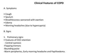
  • 12. Clinical Features of COPD A. Symptoms • Cough • Sputum • Breathlessness worsened with exertion • Edema • Morning headaches (due to hypercapnia) B. Signs 1. Pulmonary signs • Features of CO2 retention - Central cyanosis -Flapping tremors -Bounding pulse -Warm peripheries, Early monring headache and Papilloedema.
  • 13. • Features of Cor pulmonale (RVF+ Pulmonary HTN) -Peripheral edema -Raised JVP -Right ventricular heave -Loud P2 -Enlarged liver (hepatomegaly) 2. Extra pulmonary signs • Altered fat metabolism contributing to weight loss , and Muscular weakness reflecting deconditioning and cellular changes in skeletal muscles. • Increased prevalence of osteoporosis, and Increased circulating inflammatory markers. • Impaired salt and water excretion leading to peripheral oedema.
  • 15. Diagnostic Tests 1. Chest X-ray: Best initial test. • Increased anterior-posterior (AP diameter) • Hyperinflation: >6 anterior ribs seen above diaphragm in mid clavicular line, or ≥ 10 Posterior Ribs. • Large central pulmonary arteries, Decreased peripheral vascular markings and Bulla. • Tubular heart shadow • Air trapping and flattened diaphragms 2. Pulmonary function test: Most accurate test Decreased FEV1, decreased FVC, decreased FEV1/FVC ratio (<70%) • Increased TLC because of increased residual volume. • Incomplete improvement with bronchodilators (<12%) • Little or no worsening with methacholine.
  • 17. 3. ABG: Acute exacerbations associated with increased PCO2 and hypoxia. Respiratory acidosis may be present. 4. CBC (increased Hb, increased PCV) 5. ECG • Right atrial and ventricular hypertrophy (cor pulmonale) •Poor progression of R wave •Atrial fibrillation or multifocal atrial tachycardia. 6. High resolution (CT scan (HRCT))
  • 19. Differential Diagnosis of COPD 1. Pneumonia 2. Pneumothorax 3. Left ventricular failure 4. Pulmonary embolism 5. Lung cancer 6. Upper airway obstruction 7. Chronic asthma 8. Tuberculosis 9. Bronchiectasis 10. Congestive cardiac failure 11. Hypersensitivity pneumonitis 12. Interstitial lung disease.
  • 20. Management 1. Non pharmacological Smoking cessation and oxygen therapy are the only treatment modalities that delay the progression of disease and improve mortality. • Smoking cessation • Use of domiciliary oxygen (at least 15 hours/day) Criteria for domiciliary oxygen use in COPD - --PaO2 <55mmHg or SaO2 <88% -or- -If there are signs of right sided heart failure/disease or hematocrit >55%, use when - PaO2 <60mmHg or SaO2 <90% •Influenza and pneumococcal vaccination -Influenza vaccine annually -Pneumococcal vaccine once (Give only one lifetime booster if the patient was <65 years age at first vaccination) • Encourage exercise • Treatment of poor nutrition and obesity • Pulmonary rehabilitation
  • 21. 2. Pharmacological • Anticholinergic agents (Ipratropium, triotropium): first line agents • Shortacting beta agonists (Albuterol) • Inhaled steroids • Long acting beta agonists (Salmeterol) • Treatment of co-existing infection: Azithromycin, IV ceftriaxone
  • 22. 3. Surgery Lung volume reduction surgeries are done in selected patients such pulmonectomy. lobectomy. Indications for surgery are: • Recurrent pneumothoraces • Isolated bullous disease Prognosis COPD has a variable natural history but is usually pro- gressive. The prognosis is inversely related to age and directly related to the post-bronchodilator FEV1. Other poor prognostic indicators include weight loss and pul-monary hypertension. A composite comprising the body mass index (B), the degree of airflow obstruction(O), a measurement of dyspnoea (D) and exercise capacity (E) may assist in predicting death from respiratory and other causes (Box 19.33). Respiratory failure, cardiac disease and lung cancer represent common modes of death.
  • 25. Acute Exacerbation of copd Characterised by 1. increased Symptoms. 2. deterioration in lungs function 3. Health Status if ds. Progress then become More frequent; Are usually triggerded by 1. Bacteria 2. virus 3. change in Air quality ↓ Finally may lead to 1. Development of Respiratory failure 2. fluid Retention 3. Important case of Death.
  • 26. Refferal to Hospital 1. presence of cynosis 2. peripheral Edema 3. Alternation in consciousness.
  • 27. Difference between Blue bloaters and Pink puffers
  • 28. Management of Acute Exacerbation of COPD Note: Management of AE-COPD is similar to that of acute asthma, as both are type Il respiratory failure, except the oxygen flow is low in AE COPD. Investigation 1. Pulmonary function test → FEV., peak flow 2. ABG 3. ECG 4. Chest x-ray 5. Sputum for culture. 6. CBC 7. Urine R/E 8. Electrolytes, urea/creatinine.
  • 29. Treatment 1. Oxygen • 24-28% via face mask, 2L/min by nasal prongs. • Check ABG within 60 mins and adjust according to PaCO2 or pH. •Keep Pa02 ≥ 7.5 KPa • May require ventilation in coma, respiratory arrest. Note: O2 supply should be low flow because in COPD patients, hypoxia is the stimulus for respiration instead of hypercarbia in normal people, so removal of hypoxia by high flow results in cessation of respiration. 2. Nebulization • Short acting B2 agonists (salbutomol) 5mg in 2-4 ml NS. • 1st dose in 30 min. Repeat of necessary in 2-4 hours. • Add ipratropium bromide if response is inadequate 0.5mg 4 hourly.
  • 30. 3. Corticosteroids • IV hydrocortisone 200mg 6 hourly • If oral intake possible, oral predisolone (30-60mg) 1mg/kg tapered over 1 week. 4. Monitoring • Oxygen • Serum potassium 5. Hydration • IV fluids • K+ supplementation may be needed as repeated doses of salbutamol can lower plasma K+. 6. Appropriates antibiotics • If patient fails to improve
  • 31. 7. Add IV MgSO4 1.2-2g over 20 mins or, • IV aminophylline 5mg/kg loading dose and 1mg/kg/hr maintainance dose over 20 minutes or • Inj. adrenaline SC 0.3mg x ⅟ 2 hourly * 3 doses. 8. Monitoring of vitals and electrolytes. 9. Others • Diuretics, if JVP elevated and oedema present • If patient continues to deteriorate, respiratory stimulant like doxapram can be considered. • Prophylactic subcutaneous low molecular weight heparin, for DIC.
  • 32. Complications of COPD 1. Cor-pulmonale 2. Acute exacerbation 3. Respiratory failure 4. Pneumothorax 5. Mental status deterioration 6. Osteoporosis 7. Renal failure