SlideShare a Scribd company logo
University of Calicut
Sixth Semester BSc Zoology Degree Programme
Zoology Core Course XIII Theory
Reproductive Biology, Developmental
Biology and Teratology
Course Code: ZO6B13T
Rahana Moideen Koya V.K
MSc., MPhil, MEd, MA
Assistant Professor
Department of Zoology
Farook College , Kozhikode
rahanamoideenkoya@gmail.com
Embryonic development of amphioxus
Amphioxus-Branchiostoma lanceolatum is a primitive marine
chodate Suphylum –Cephalochodata
It is oviparous,indirect devt.free living ciliated larva.early devt.
Some similarity –Echinodermata same time vertebrate also.
Devpt of Amphioxus
Gonads-Sexes separate-no well marked sexual
dimorphism.
Testes and ovaries are located on both sides of the body
bet body wall ,bordering the atrium and the muscle
groups(myotomes).About28 pairs on each side
Upon sexual maturity the sperms and ova are discharged
into atrium by repture of body wall .then atriopore.to sea
water.
Ovum-0.01mm-0.12mm in diameter.nucleus towards one
side of egg ,animal pole.outside plasma membrane thin
mucopolysaccharides vitalline mem.
Sperm-very small head middle piece,tail.head is spherical
middle piece very short.
V
Devpt of Amphioxus
Fertilization-External sea water. Female spawn through out
summer and always towards evening ,while the animals are
swimming.Males shed sperms into water near the female.One
of these sperms penetrate vitelline membrane enters egg
near the vegetal pole of the egg.As soon as sperm reaches
into the egg the vitelline membrane is lifted off from the
surface of of the egg to form fertilization membrane. The
space bet.plasma membrane and fertilization membrane
–Perivitelline space. Viscous fluid.
The entry of sperm into the egg stimulates the second
maturation division release of second polar body. Which
remains attached to to the animal pole in the perivitelline
space.Sperm and egg nuclei enlarge to form male and
female pronuclei.They migrate towards equator of the egg
and fuse to form zygote nucleus.fusion of pronuclei slightly
above the equatorial plane of egg
Embryonic development of amphioxus
1. Fertilization : External.
2. Types of Egg: According to amount of yolk
oligolecithal. According to distribution ;
homolecithal.
3. Types of cleavage: Equal holoblastic cleavage.
Cleavage
Devpt of Amphioxus
Meridional-Bisects both poles,through animal vegetal axisof
egg .
Devpt of Amphioxus
Cleavage-Holoblastic almost equal.
First.Meridional 60-90 min,after
fertilization.animal to vegetal pole.
Second-45 min.after first.meridional right
angles to first.
Third-30 min.after second.divide 4
blastomeres in the latitudinal (horizontal)plane
at right angles to the first two cleavage
furrows. 8 blastomeres..But it is slightly above
the equator .So macro-micromeres .
• Fourth cleavage-Meridional and is double
cleavage. Ie.two in number ,and the
furrows pass from the animal pole to the
vegetal pole at right angles to each other
to form 16 blastomeres.
•
Gradually cleavage becomes irregular micromeres divide more
rapidly ,the synchronous character of the division declines and
increase in no of blastomeres become arithmetical than
geometrical.
Devpt of Amphioxus
• Blastulation-As the cleavage proceeds
more and more blastomeres are produced
and semifluid material accumulate in the
centre of mass of cells.this serves to push
all the blastomeres outward,so that they
become arranged in single layer called
blastoderm ,central cavity blastocoel.fluid
or jelly inside. 200 cells when fully
formed.which are arranged in the form of
columnar epithelium around the the
blastocoel.the cells at vegetal pole are
larger.coeloblastula.
• Blastulation:
Blastula stage is a ball of cells that
excceds more than 256 blastomeres. The
blstoderm formed of one cell layer thick.
The blastocoele enclosed almost the
central cavity .
• Gastrulation:
This occurred as a result of different cell
movements simply by epiboly ,
invagination & involution.
• Gastrulation-Morphogenetic movt.or
formative movt..Gastrulation begins about
800 cells in the embryo bet 9th
and 10 th
cleavage. The main processes involved in
the gastrulation are.1.Invagination
• 2.Involution(embolic movts-movt.of cells
from the surface in to the interior of the
embryo.
• 3.epibolic movts.
• 4.Antero-posterior elongation of the
embryo.
• Invgination-Flattening of the blastoderm at the vegetal
pole
• The endodermal plate gradually invaginate or bend
inwards into the blastocoel –so the embryo bome cup
shaped with a large cavity. The new cavity –archenteron
opens to ext.by blastopore.
• The cup shaped embryo is double walled –ext.lining
consists of presumptive epidermis and presumptive Ns
named as ectoderm
• Internal layer –mesoderm and endoderm ,both of these
layers remain continuous with other over the rim of the
gastrula.
• The circular rim of the blastopore is called the lip.
• The prospective notochord lies in the dorsal lip and the
prospective mesoderm lies in the ventral lip.
• Involution-Along with the invagination of the
endodermal plate the prospective notochord and
mesodermal material from the rim of the cup
shaped embryo involute (rolling over of cells
from the surface in to the interior of the
embryo.)into the interior of the
gastrula.Blastopore is very broad in its initial
stage.soon become contract reduced.As more
material is shifted from the surface to the interior
of the gastrula ,endoderm comes into contact
with the opposite epidermal wall by replacing the
blastocoel by the new cavity ,the archenteron.
• Epiboly-Deals with the movt. Of ectodermal cells
over the surface of the embryo.Initially
blastopore is a wide and roughly triangular
opening and the ventrolateral lips of the
blastopore tend to become continuous.They
begin to grow dorsally and the dorsal lip become
arched. The blastopore becomes an oval
aperture.
• Its dorsolateral and ventrolateral lips grow
towards one another and blastopore becomes
circular.Oval blastopore becomes small circular
opening.
• Antero-posterior elongation of the embryo-
• After constriction of the rim of blastopore ,the gastrula
exhibit a general elongation in the ant-post direction . All
the presumptive areas participate in elongation.
Externally the neural ectoderm lies in the mid dorsal line
above the notochord and epidermal ectoderm occupies
the remainder. The notochordal cells form a long
median band just below the neurectoderm .The lateral
horns of the mesoderm crescent converge towards the
dorsal side of the embryo and come to lie on both sides
of the presumptive notochord .The gastrula now consists
of Outer epiblast consisting of neural and epidermal
ectoderm and an inner hypoblast comprising prospective
notochord,mesoderm &endoderm.
Devpt of Amphioxus
Devpt of Amphioxus
Devpt of Amphioxus
Devpt of Amphioxus
Blastula
Gastrulation
Gastrula of Amphioxus
Devpt of Amphioxus
Devpt of Amphioxus
• Neurulation—Formation of neural
tube.The prospective neural ectoderm
lying on the middorsal line of
gastrula.These neural ectodermal cells
become separated from the cells of
epidermal ectoderm in the form of an
elongated plate called neural plate or
medullary plate.This plate gradually sinks
and gets separated from the epidermal
ectoderm.
Devpt of Amphioxus
Devpt of Amphioxus
Devpt of Amphioxus
Devpt of Amphioxus
Devpt of Amphioxus
Devpt of Amphioxus
Devpt of Amphioxus
Devpt of Amphioxus
Devpt of Amphioxus
• Diagram of amphioxus embryology. The gastrula and neurula stages
are shown in side and cross-sectional views (left and right of each
pair, respectively). The broken line on the side view of the
late-neurula stage indicates the level of the frontal section (marked
by the arrow). Arrowheads 1–3 show migration of the non-neural
ectoderm across the neuroectoderm (first phase of neurulation),
and arrowheads 3 and 4 indicate the curling up of the neural plate
into the neural tube (second phase of neurulation). During the
mid-neurula stage, somites (SO) and notochord (stippled) arise from
the gut by evagination and upfolding, respectively. For the
late-neurula stage, the approximate positions of the endostyle (EN),
pronephric kidney (PN), and rudiment of the possible
adenohypophysis (AP) are indicated near the anterior end; the
frontal section of the tail-bud region shows the notochord (NC), the
neurenteric canal (NEC), and a nascent somite (SO*); the cross
sections show the ventral mesoderm (VM) evaginating down from
the somites and the formation of the heart tube (HT) midventrally
between the gut and the visceral mesothelium.
•
• Somites are blocks of mesodermSomites are blocks
of mesoderm that are located on either side of the neural
tubeSomites are blocks of mesoderm that are located on either
side of the neural tube in the developing vertebrate embryo.
Somites are precursor populations of cells that give rise to
important structures associated with the vertebrate body plan
and will eventually differentiate into dermis, skeletal muscle,
cartilage, tendons, and vertebrae. Somites also determine the
migratory paths of neural crest cells and of the axons of
spinal nerves.
• Neural crest cells (NCC) are
multipotent cells induced at the border of
the neural plate that subsequently migrate
throughout the embryo and
later differentiate into multiple cell types
contributing to most of the peripheral
nervous system and the cranio-facial
cartilage and bones, as well as pigment
and endocrine cells.

More Related Content

PPTX
O.b. c 7 motivation concepts
PPTX
Types of egg, Classification of eggs, Structure of frog egg, Structure of chi...
PPT
Single nucleotide polymorphism, (SNP)
PPTX
Cancer Immunotherapy and Gene Therapy
PPTX
Final chick embryonic-development-ppt
PPTX
Single nucleotide polymorphisms (sn ps), haplotypes,
PPTX
Cleavage: Definition, types, and mechanism
O.b. c 7 motivation concepts
Types of egg, Classification of eggs, Structure of frog egg, Structure of chi...
Single nucleotide polymorphism, (SNP)
Cancer Immunotherapy and Gene Therapy
Final chick embryonic-development-ppt
Single nucleotide polymorphisms (sn ps), haplotypes,
Cleavage: Definition, types, and mechanism

What's hot (20)

PPTX
morphogenetic movements-developmental biology
PPTX
Development of frog/ embryogenesis
PPTX
Development of chick
PPTX
vittelogenesis.pptx
PPTX
Development of chick embryo- structure of egg,cleavages,fate map and primitiv...
PDF
Cleavage
PPTX
Cleavage, Types of cleavage
PPTX
Comparative Anatomy of Digestive System of Vertebrates
PPTX
Axis formation in birds and mammals
PPT
Gastrulation in frog
PPTX
Activation of egg metabolism
PPTX
Torsion in gastropoda
PPTX
Comparative Anatomy of Respiratory System of Vertebrates
PPTX
Structure and types of eggs
PDF
embryonic-induction.pdf
PPTX
Skull typpes
PPTX
Biochemistry of fertilization.
PPTX
block to polyspermy.pptx
PPTX
Larval form of mollusca
morphogenetic movements-developmental biology
Development of frog/ embryogenesis
Development of chick
vittelogenesis.pptx
Development of chick embryo- structure of egg,cleavages,fate map and primitiv...
Cleavage
Cleavage, Types of cleavage
Comparative Anatomy of Digestive System of Vertebrates
Axis formation in birds and mammals
Gastrulation in frog
Activation of egg metabolism
Torsion in gastropoda
Comparative Anatomy of Respiratory System of Vertebrates
Structure and types of eggs
embryonic-induction.pdf
Skull typpes
Biochemistry of fertilization.
block to polyspermy.pptx
Larval form of mollusca
Ad

Similar to Devpt of Amphioxus (20)

PPTX
PPTX
Development of chick.pptx
PPTX
Gastrulation in Frog.pptx
PPTX
Descriptive embryology and developmental biology lecture .pptx
PPT
06 - Ectodermal Derivtives.ppt for medical
PPTX
Development of oral cavity and face .ppt by dr. samidha arora
PPT
ANATOMICAL DESCRIPTION OF DERIVATIVES OF GERM LAYERS.ppt
PDF
Third week of development.pdf
PPTX
embryonic development in chick.pptx
PPT
Gastrulation
PPTX
Embryonic development
PPTX
Chick Dev.pptx
PPTX
Growth and development in animals
PPT
Journey of an embryo...development biology
PPTX
DEVELOPMENT OF DIGESTIVE SYSTEM.pptx
PPT
Development of chick
PPTX
embryo f.pptx best ppt for all hard workers here you go
PPTX
Development of face by revath
PPTX
Axis and pattern formation in amphibia
PPTX
General Embryology
Development of chick.pptx
Gastrulation in Frog.pptx
Descriptive embryology and developmental biology lecture .pptx
06 - Ectodermal Derivtives.ppt for medical
Development of oral cavity and face .ppt by dr. samidha arora
ANATOMICAL DESCRIPTION OF DERIVATIVES OF GERM LAYERS.ppt
Third week of development.pdf
embryonic development in chick.pptx
Gastrulation
Embryonic development
Chick Dev.pptx
Growth and development in animals
Journey of an embryo...development biology
DEVELOPMENT OF DIGESTIVE SYSTEM.pptx
Development of chick
embryo f.pptx best ppt for all hard workers here you go
Development of face by revath
Axis and pattern formation in amphibia
General Embryology
Ad

More from RAHANAMOIDEENKOYAVK (19)

PPTX
biosphere reseves-converted (1).pptx
PPTX
vulva formation in c.elegans.pptx
PPTX
vulva formation in c.elegans.pptx
PPTX
vulva formation in c.elegans.pptx
PPTX
vulva formation in c.elegans.pptx
PPTX
regenerationsat-.pptx
PDF
declaration of np.pdf
PPTX
cloning_dolly.pptx
PPTX
PPTX
Enzymes msc
PPTX
Suicide inhibitors
PPTX
Michaelis Menten Reaction
PPTX
Sex Differentiation TDF
PPTX
Physiology of smell
PPTX
Mutation bsc
PPTX
Water absorption.minerals,vitamines
PPTX
Polygenicinheritance.,atavism ,multiple allelism
PPTX
Control of digestion,Different mechanisms for absorption.
PPT
Parasitism in relation to mankind
biosphere reseves-converted (1).pptx
vulva formation in c.elegans.pptx
regenerationsat-.pptx
declaration of np.pdf
cloning_dolly.pptx
Enzymes msc
Suicide inhibitors
Michaelis Menten Reaction
Sex Differentiation TDF
Physiology of smell
Mutation bsc
Water absorption.minerals,vitamines
Polygenicinheritance.,atavism ,multiple allelism
Control of digestion,Different mechanisms for absorption.
Parasitism in relation to mankind

Recently uploaded (20)

PPT
6.1 High Risk New Born. Padetric health ppt
PDF
CHAPTER 3 Cell Structures and Their Functions Lecture Outline.pdf
PDF
ELS_Q1_Module-11_Formation-of-Rock-Layers_v2.pdf
PDF
Lymphatic System MCQs & Practice Quiz – Functions, Organs, Nodes, Ducts
PPTX
Vitamins & Minerals: Complete Guide to Functions, Food Sources, Deficiency Si...
PPTX
Introduction to Cardiovascular system_structure and functions-1
PDF
The scientific heritage No 166 (166) (2025)
PDF
Formation of Supersonic Turbulence in the Primordial Star-forming Cloud
PPTX
Microbiology with diagram medical studies .pptx
PPTX
2. Earth - The Living Planet earth and life
PPTX
C1 cut-Methane and it's Derivatives.pptx
PPTX
Fluid dynamics vivavoce presentation of prakash
PPTX
famous lake in india and its disturibution and importance
PDF
Unveiling a 36 billion solar mass black hole at the centre of the Cosmic Hors...
PPT
POSITIONING IN OPERATION THEATRE ROOM.ppt
PPTX
Taita Taveta Laboratory Technician Workshop Presentation.pptx
PPT
protein biochemistry.ppt for university classes
PPTX
Introduction to Fisheries Biotechnology_Lesson 1.pptx
PDF
Cosmic Outliers: Low-spin Halos Explain the Abundance, Compactness, and Redsh...
PPTX
neck nodes and dissection types and lymph nodes levels
6.1 High Risk New Born. Padetric health ppt
CHAPTER 3 Cell Structures and Their Functions Lecture Outline.pdf
ELS_Q1_Module-11_Formation-of-Rock-Layers_v2.pdf
Lymphatic System MCQs & Practice Quiz – Functions, Organs, Nodes, Ducts
Vitamins & Minerals: Complete Guide to Functions, Food Sources, Deficiency Si...
Introduction to Cardiovascular system_structure and functions-1
The scientific heritage No 166 (166) (2025)
Formation of Supersonic Turbulence in the Primordial Star-forming Cloud
Microbiology with diagram medical studies .pptx
2. Earth - The Living Planet earth and life
C1 cut-Methane and it's Derivatives.pptx
Fluid dynamics vivavoce presentation of prakash
famous lake in india and its disturibution and importance
Unveiling a 36 billion solar mass black hole at the centre of the Cosmic Hors...
POSITIONING IN OPERATION THEATRE ROOM.ppt
Taita Taveta Laboratory Technician Workshop Presentation.pptx
protein biochemistry.ppt for university classes
Introduction to Fisheries Biotechnology_Lesson 1.pptx
Cosmic Outliers: Low-spin Halos Explain the Abundance, Compactness, and Redsh...
neck nodes and dissection types and lymph nodes levels

Devpt of Amphioxus

  • 1. University of Calicut Sixth Semester BSc Zoology Degree Programme Zoology Core Course XIII Theory Reproductive Biology, Developmental Biology and Teratology Course Code: ZO6B13T
  • 2. Rahana Moideen Koya V.K MSc., MPhil, MEd, MA Assistant Professor Department of Zoology Farook College , Kozhikode rahanamoideenkoya@gmail.com Embryonic development of amphioxus
  • 3. Amphioxus-Branchiostoma lanceolatum is a primitive marine chodate Suphylum –Cephalochodata It is oviparous,indirect devt.free living ciliated larva.early devt. Some similarity –Echinodermata same time vertebrate also.
  • 5. Gonads-Sexes separate-no well marked sexual dimorphism. Testes and ovaries are located on both sides of the body bet body wall ,bordering the atrium and the muscle groups(myotomes).About28 pairs on each side Upon sexual maturity the sperms and ova are discharged into atrium by repture of body wall .then atriopore.to sea water. Ovum-0.01mm-0.12mm in diameter.nucleus towards one side of egg ,animal pole.outside plasma membrane thin mucopolysaccharides vitalline mem. Sperm-very small head middle piece,tail.head is spherical middle piece very short. V
  • 7. Fertilization-External sea water. Female spawn through out summer and always towards evening ,while the animals are swimming.Males shed sperms into water near the female.One of these sperms penetrate vitelline membrane enters egg near the vegetal pole of the egg.As soon as sperm reaches into the egg the vitelline membrane is lifted off from the surface of of the egg to form fertilization membrane. The space bet.plasma membrane and fertilization membrane –Perivitelline space. Viscous fluid.
  • 8. The entry of sperm into the egg stimulates the second maturation division release of second polar body. Which remains attached to to the animal pole in the perivitelline space.Sperm and egg nuclei enlarge to form male and female pronuclei.They migrate towards equator of the egg and fuse to form zygote nucleus.fusion of pronuclei slightly above the equatorial plane of egg
  • 9. Embryonic development of amphioxus 1. Fertilization : External. 2. Types of Egg: According to amount of yolk oligolecithal. According to distribution ; homolecithal. 3. Types of cleavage: Equal holoblastic cleavage.
  • 12. Meridional-Bisects both poles,through animal vegetal axisof egg .
  • 14. Cleavage-Holoblastic almost equal. First.Meridional 60-90 min,after fertilization.animal to vegetal pole. Second-45 min.after first.meridional right angles to first. Third-30 min.after second.divide 4 blastomeres in the latitudinal (horizontal)plane at right angles to the first two cleavage furrows. 8 blastomeres..But it is slightly above the equator .So macro-micromeres .
  • 15. • Fourth cleavage-Meridional and is double cleavage. Ie.two in number ,and the furrows pass from the animal pole to the vegetal pole at right angles to each other to form 16 blastomeres. •
  • 16. Gradually cleavage becomes irregular micromeres divide more rapidly ,the synchronous character of the division declines and increase in no of blastomeres become arithmetical than geometrical.
  • 18. • Blastulation-As the cleavage proceeds more and more blastomeres are produced and semifluid material accumulate in the centre of mass of cells.this serves to push all the blastomeres outward,so that they become arranged in single layer called blastoderm ,central cavity blastocoel.fluid or jelly inside. 200 cells when fully formed.which are arranged in the form of columnar epithelium around the the blastocoel.the cells at vegetal pole are larger.coeloblastula.
  • 19. • Blastulation: Blastula stage is a ball of cells that excceds more than 256 blastomeres. The blstoderm formed of one cell layer thick. The blastocoele enclosed almost the central cavity . • Gastrulation: This occurred as a result of different cell movements simply by epiboly , invagination & involution.
  • 20. • Gastrulation-Morphogenetic movt.or formative movt..Gastrulation begins about 800 cells in the embryo bet 9th and 10 th cleavage. The main processes involved in the gastrulation are.1.Invagination • 2.Involution(embolic movts-movt.of cells from the surface in to the interior of the embryo. • 3.epibolic movts. • 4.Antero-posterior elongation of the embryo.
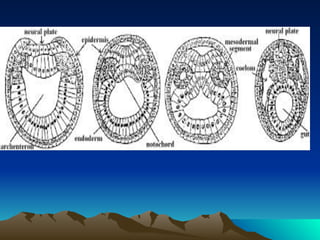
  • 21. • Invgination-Flattening of the blastoderm at the vegetal pole • The endodermal plate gradually invaginate or bend inwards into the blastocoel –so the embryo bome cup shaped with a large cavity. The new cavity –archenteron opens to ext.by blastopore. • The cup shaped embryo is double walled –ext.lining consists of presumptive epidermis and presumptive Ns named as ectoderm • Internal layer –mesoderm and endoderm ,both of these layers remain continuous with other over the rim of the gastrula. • The circular rim of the blastopore is called the lip. • The prospective notochord lies in the dorsal lip and the prospective mesoderm lies in the ventral lip.
  • 22. • Involution-Along with the invagination of the endodermal plate the prospective notochord and mesodermal material from the rim of the cup shaped embryo involute (rolling over of cells from the surface in to the interior of the embryo.)into the interior of the gastrula.Blastopore is very broad in its initial stage.soon become contract reduced.As more material is shifted from the surface to the interior of the gastrula ,endoderm comes into contact with the opposite epidermal wall by replacing the blastocoel by the new cavity ,the archenteron.
  • 23. • Epiboly-Deals with the movt. Of ectodermal cells over the surface of the embryo.Initially blastopore is a wide and roughly triangular opening and the ventrolateral lips of the blastopore tend to become continuous.They begin to grow dorsally and the dorsal lip become arched. The blastopore becomes an oval aperture. • Its dorsolateral and ventrolateral lips grow towards one another and blastopore becomes circular.Oval blastopore becomes small circular opening.
  • 24. • Antero-posterior elongation of the embryo- • After constriction of the rim of blastopore ,the gastrula exhibit a general elongation in the ant-post direction . All the presumptive areas participate in elongation. Externally the neural ectoderm lies in the mid dorsal line above the notochord and epidermal ectoderm occupies the remainder. The notochordal cells form a long median band just below the neurectoderm .The lateral horns of the mesoderm crescent converge towards the dorsal side of the embryo and come to lie on both sides of the presumptive notochord .The gastrula now consists of Outer epiblast consisting of neural and epidermal ectoderm and an inner hypoblast comprising prospective notochord,mesoderm &endoderm.
  • 34. • Neurulation—Formation of neural tube.The prospective neural ectoderm lying on the middorsal line of gastrula.These neural ectodermal cells become separated from the cells of epidermal ectoderm in the form of an elongated plate called neural plate or medullary plate.This plate gradually sinks and gets separated from the epidermal ectoderm.
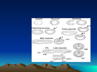
  • 44. • Diagram of amphioxus embryology. The gastrula and neurula stages are shown in side and cross-sectional views (left and right of each pair, respectively). The broken line on the side view of the late-neurula stage indicates the level of the frontal section (marked by the arrow). Arrowheads 1–3 show migration of the non-neural ectoderm across the neuroectoderm (first phase of neurulation), and arrowheads 3 and 4 indicate the curling up of the neural plate into the neural tube (second phase of neurulation). During the mid-neurula stage, somites (SO) and notochord (stippled) arise from the gut by evagination and upfolding, respectively. For the late-neurula stage, the approximate positions of the endostyle (EN), pronephric kidney (PN), and rudiment of the possible adenohypophysis (AP) are indicated near the anterior end; the frontal section of the tail-bud region shows the notochord (NC), the neurenteric canal (NEC), and a nascent somite (SO*); the cross sections show the ventral mesoderm (VM) evaginating down from the somites and the formation of the heart tube (HT) midventrally between the gut and the visceral mesothelium. •
  • 45. • Somites are blocks of mesodermSomites are blocks of mesoderm that are located on either side of the neural tubeSomites are blocks of mesoderm that are located on either side of the neural tube in the developing vertebrate embryo. Somites are precursor populations of cells that give rise to important structures associated with the vertebrate body plan and will eventually differentiate into dermis, skeletal muscle, cartilage, tendons, and vertebrae. Somites also determine the migratory paths of neural crest cells and of the axons of spinal nerves.
  • 46. • Neural crest cells (NCC) are multipotent cells induced at the border of the neural plate that subsequently migrate throughout the embryo and later differentiate into multiple cell types contributing to most of the peripheral nervous system and the cranio-facial cartilage and bones, as well as pigment and endocrine cells.