SlideShare a Scribd company logo
Different states of
unconsciousness
Dr. Sachit Koirala
Resident, 2nd year
Department of Surgery, KISTMCTH
• Determine the disease causing diminished state of
unconsciousness
• Determine the direction in which it is evolving
To protect the brain against irreversible damage
Most common causes of diminished consciousness
1. Metabolic and diffuse disorders (65%)
• Drug poisoning
• Ischemia or anoxia
• Hepatic encephalopathy
• Encephalomyelitis and encephalitis
• Subarachnoid hemorrhage
• Endocrine disorders (including diabetes)
• Acid-base disorders
• Uremic encephalopathy
Most common causes of diminished consciousness
2. Supratentorial mass lesions (20%)
• Intracerebral hematoma
• Subdural hematoma
• Cerebral infarct
• Brain tumor
• Brain abscess
• Epidural hematoma
• Thalamic infarct
• Pituitary apoplexy
Most common causes of diminished consciousness
3. Subtentorial lesions (13%)
• Brainstem infarct
• Pontine hemorrhage
• Cerebellar hemorrhage
• Cerebellar tumor
• Cerebellar infarct
4. Psychiatric disorders (3%)
Consciousness
State of awareness of self and environment
AND
normal responsiveness to external stimulation and inner need.
Normal consciousness may fluctuate during the day
BUT
the normal individual can be brought immediately to a state of full
alertness and function.
Confusion
Inability to think with customary speed, clarity, and coherence.
Some degree of inattentiveness and disorientation i.e. Distractibility
and imperceptiveness.
“Clouding of sensorium”
Results most often from process that influences the brain globally, such
as a toxic or metabolic disturbance or dementia or sleep deprivation.
Salient features of a confused individual:
• Severely confused and inattentive persons are unable to do more
than carry out the simplest commands.
• Speech may be limited to a few words or phrases but some confused
individuals are voluble (~Talkative).
• Disoriented in time and place
• Do not grasp their immediate situation
• Do not understand the predicament of their own confusion
• May misidentify people or objects.
• These illusions may lead to fear or agitation.
• Loss of capacity to recall events of the past hours or days
• Deficit in working memory
Delirium = Confusion (Adams and Victor)
Delirium: Acute confusional state (Harrison’s)
Drowsiness
Inability to sustain a wakeful state without application of external
stimuli.
Sustain alertness for at least some brief period, without the further
necessity of external stimuli
Some degree of inattentiveness and mild confusion are present,
improving with arousal.
The patient shifts positions somewhat naturally and without
prompting.
The lids droop; there may be snoring, the jaw and limb muscles are
slack, and the limbs are relaxed.
Stupor
A state in which the patient can be roused only by vigorous and repeated
stimuli
Arousal cannot be sustained without repeated stimulation.
Responses to spoken commands are either absent or slow and inadequate.
Restless or stereotyped motor activity is common.
Reduction or elimination of the natural shifting of body positions.
When left unstimulated, these patients quickly drift back into a deep sleep-
like state.
The eyes are usually found to be displaced outward and upward, a feature
that is shared with sleep.
Vegetative state
• Person is awake but is showing no signs of awareness.
• Preserved autonomic and respiratory function without cognition
• Open their eyes
• Wake up and fall asleep at regular intervals
• Have basic reflexes (such as blinking when they're startled by a loud
noise or withdrawing their hand when it's squeezed hard)
• Able to regulate their heartbeat and breathing without assistance.
BUT
the patient regains neither awareness nor purposeful behavior.
Persistent Vegetative State
• Vegetative state for 3 months after non traumatic brain injury
• Vegetative state for 12 months after traumatic brain injury
• Also known as Apallic syndrome or Neocortical death
Multi-Society Task Force on PVS concluded that the outcome from a
vegetative state is better in traumatic as compared to nontraumatic
cases.
Vegetative state vs Coma
• A coma is when a person shows no signs of being awake and no signs
of being aware
• A vegetative state is when a person is awake but is showing no signs
of awareness
Minimally conscious state
• Patient is capable of some rudimentary behavior
• Following a simple command, gesturing, or producing single words or
brief phrases
• Demonstrate a degree of awareness
• Inconsistent from one examination to other
• Transitional or permanent state
• Continuum of vegetative state. Less severe but profound dementia
Locked in syndrome (Deefferented state)
• Little or no disturbance of consciousness
• Inability to respond adequately with motor activity and speech
• Most often caused by a large lesion of the ventral pons (basis pontis),
usually as a result of occlusion of the basilar artery
• Spare both the somatosensory pathways, and the ascending neuronal
systems responsible for arousal and wakefulness
• Spare certain midbrain elements that allow the eyelids to be raised in
wakefulness
• Interrupts the descending corticobulbar and corticospinal tracts
• Can respond by voluntary vertical gaze and blinking
Akinetic mutism
• Inert (akinetic) and silent (mutism)
• Bilateral lesions in anterior part of frontal lobe or thalamus
• Motor and sensory pathways are intact
• Profoundly apathetic
• Marked delay in verbal and motor response (Abulia)
• Intense stimulation  May speak normally and relate the events
Catatonia
• Unresponsive
• Can simulate stupor, light coma or akinetic mutism
• No signs of structural brain damage (Normal pupillary reaction,
normal reflexes)
• Oculocephalic reflex is muted.
• Resistance to eye opening
• Waxy flexibility to passive limb movement
• Long periods of uncomfortable limb positions (Catalepsy)
• EEG shows normal alpha activity that is attenuated by stimulation
Different states of unconsciousness
Electroencephalogram (EEG) in states of
impaired consciousness
Confusion/Drowsiness
• Disappearance of normal alpha rhythm
• Replacement by random slow waves of low to moderate voltage
Stupor
• Regular slow waves (2-3 Hz) of high voltage
Deep coma
• Slow, low voltage waves
• Intermittent suppression of electrical activity
Brain death
• Complete absence of electrical activity
EEG in impaired consciousness due to
metabolic disorders
Hepatic or renal failure (Uremia)
• Slow waves
• High voltage
Coma due to sedatives intoxication
• Beta activity replaces normal rhythm
Delta pattern
Triphasic waves
Deeply comatose patients
• Diffuse transient alpha activity
• Also known as alpha coma
• Can be mistaken for normal activity
BUT
• No reactivity to sensory stimuli
Associated with pontine or diffuse cortical lesions
Poor prognosis
Alpha coma
Anatomy and neurophysiology of alertness
and coma
Reticular Activating System (RAS)
• Indistinct boundaries
• Neurons are interspersed
• Locus coeruleus: Upper dorsolateral pons of the brainstem. Activated
directly by orexin from the lateral hypothalamus. Nor-epinephrine
• Raphe nuclei: midline throughout the brainstem within the pons, midbrain,
and medulla. Serotonergic
• Tuberomamillary nucleus: located within the posterior aspect of the
hypothalamus. Histaminergic
• Lateral and dorsal pedunculopontine tegmentum: Primarily cholinergic
neurons in neighboring groups within the midbrain and pons. Cholinergic
neurons project to the thalamus and cortex.
Diffusion tensor tactography
RAS receives collaterals from the spinothalamic and trigeminal–
thalamic pathways
Project to the whole of the cerebral cortex, not just to the sensory
cortex of parietal lobe
Cerebral cortex also modulates afferent sensory stimulus towards the
RAS through corticofugal projections
Pathologic basis of vegetative state
• Diffuse cerebral injury as a result of closed head trauma
• Widespread necrosis of the cortex after cardiac arrest or other form
of anoxia
• Thalamic necrosis
Prominent pathologic changes are often in the thalamic and
subthalamic nuclei rather than solely in the cortex.
In traumatic cases, the pathologic findings are often of diffuse
subcortical white matter degeneration (described as diffuse axonal
injury), prominent thalamic degeneration, and ischemic damage in the
cortex.
Anatomic findings indicate that PVS is a state in which the cortex is
either diffusely injured or effectively disconnected and isolated from
the thalamus, or the thalamic nuclei are destroyed
Pathologic anatomy of coma
Two broad processes
1. Structural, or morphologic, consisting either of a discrete structural
lesion in the upper brainstem and lower diencephalon (which may
be primary or secondary to compression) or of more widespread
destructive changes throughout the hemispheres.
2. Metabolic or submicroscopic, resulting in suppression of neuronal
activity in the cerebrum and reticular activating system.
Structural or morphological
1. A large mass in one cerebral hemisphere— tumor; abscess; massive
infarct; or intracerebral, subdural, or epidural hemorrhage.
Cause coma by secondary compression of the midbrain and central
thalamic region of the RAS.
Displacement or direct compression of these structures by the
advancing medical temporal lobe which is forced into the tentorial
opening.
OR
cerebellar lesion may compress the adjacent upper brainstem reticular
region by displacing it forward and upward
Structural or morphological
2. Destructive lesion is located immediately within the thalamus or midbrain,
in which case the neurons of the RAS are damaged directly.
Brainstem stroke from basilar artery occlusion, thalamic and upper brainstem
hemorrhages, and some forms of traumatic damage.
3. Widespread bilateral damage to the cortex and cerebral white matter, the
result of traumatic damage (contusions, diffuse axonal injury), bilateral
ischemic strokes or hemorrhages, encephalitis, meningitis, hypoxia, or global
ischemia.
The coma in these cases results from interruption of thalamocortical impulses
or from generalized destruction of cortical neurons. It is only if the cerebral
lesions are bilateral and extensive that consciousness is impaired. Many of
the diseases in this category also cause severe thalamic damage.
Coma in concussion
• Transient but large increase in intracranial pressure
• 200 to 700 lb/in2 (10000-36000 mmHg)
• Lasting a few thousandths of a second
• Vibration set up in the skull and transmitted to the brain was for many
years thought to be the basis of the abrupt paralysis of nervous function
that characterizes concussive head injury (commotio cerebri).
• Sudden swirling motion of the brain induced by acceleration or
deceleration from a blow to the head produces a rotation (torque) of the
cerebral hemispheres around the axis of the upper brainstem.
If severe: multiple shearing lesions or hemorrhages in diencephalon and
brain stem.
‘In the largest group of cases of coma, no structural lesion is
revealed by any technique of conventional pathology.’
Metabolic and toxic encephalopathies
• Cerebral Blood Flow (CBF): 55mL/min/100g
• Reduction in cerebral blood flow
• Cerebral blood flow may stay near normal while metabolism is greatly
reduced
Clinical approach to comatose patient
• Often, comatose patient is brought to the hospital and little pertinent
medical information is available.
• BLS/ACLS
• If history or suspicion of trauma: ATLS (ABCDE) approach.
• Patient’s airway is cleared and blood pressure is restored.
• With hypotension, placement of a central venous line and administration
of fluids, pressor agents and blood.
• If respirations are shallow or labored, or if there is emesis with a threat of
aspiration, tracheal intubation and mechanical ventilation are instituted.
• An oropharyngeal airway is otherwise adequate in a comatose patient who
is breathing normally.
• Deeply comatose patients with shallow respirations require endotracheal
intubation.
• The patient with a head injury may also have suffered a fracture of the
cervical vertebrae, in which case caution must be exercised in moving the
head and neck.
• Circumstances in which the person was found.
• Previous health, whether there was a history of diabetes, a head
injury, convulsion, alcohol or drug use, or a prior episode of coma or
attempted suicide.
• Sedatives, antiepileptic drugs, opiates, certain antibiotics,
antidepressants, and antipsychotic drugs.
General examination : Vitals
Temperature
Fever
• Systemic infection (eg. Pneumonia, meningitis, viral encephalitis)
• Temperature >107.6F: Heat stroke, anticholinergic drug intoxication
• Central fever is rare
Hypothermia
Alcohol or barbiturate intoxication
Drowning
Cold exposure
Advanced TB meningitis
General examination : Vitals
Respiration
Slow breathing: opiate or barbiturate intoxication, hypothyroidism
Deep rapid (Kussmaul respiration) : pneumonia, diabetic or uremic
acidosis, pulmonary edema, or the less-common occurrence of an
intracranial disease that causes central neurogenic hyperventilation.
Slow, irregular, or cyclic (Cheyne-Stokes respiration) : Raised ICP
General examination : Vitals
Respiration
Apneustic breathing (Pause 2-3s after inspiration) : Low pontine lesion
Biot breathing/Ataxia of breathing (chaotic, being irregularly interrupted
and each breath varying in rate and depth) : Dorsal medulla lesion
Cheyne-Stokes respiration
• Isolation of the brainstem respiratory centers from the cerebrum,
rendering them more sensitive than usual to carbon dioxide
(hyperventilation drive)
• Signifies bilateral dysfunction of cerebral structures
• Intoxication or a metabolic derangement
• Bilateral structural lesions such as subdural hematomas
General examination : Vitals
Blood pressure
Hypertension: cerebral hemorrhage, hypertensive encephalopathy and in
children with markedly elevated intracranial pressure.
Hypotension: Alcohol or barbiturate intoxication, DKA, internal
hemorrhage, myocardial infarction, dissecting aortic aneurysm,
septicemia, Addison disease, or massive brain trauma.
General examination : Vitals
Heart rate
Bradycardia
• Heart block from medications such as tricyclic antidepressants or
anticonvulsants
• If combined with periodic breathing and hypertension, an increase in
intracranial pressure.
Neurological examination
Inspection
• Predominant postures of the limbs and body
• Presence or absence of spontaneous movements on one side
• Position of the head and eyes
• Rate, depth, and rhythm of respiration
Brainstem function
Pupillary size and reactivity
Ocular movements
Vestibulo-ocular reflexes
Pattern of breathing
Pupillary reaction
• Loss of light reaction usually precedes enlargement of pupil
• Unilaterally enlarged (“Hutchinson”) pupil : Early indicator of
stretching or compression of the third nerve. Presence of an overlying
ipsilateral hemispheral mass.
• The light-unreactive pupil continues to enlarge to a size of 6 to 9 mm
diameter and soon, there is slight outward deviation of the eye.
• As midbrain displacement continues, both pupils dilate and become
unreactive to light as a result of compression of the oculomotor
nuclei in the rostral midbrain.
Pupillary reaction
• Pontine tegmental lesions (eg. Pontine hemorrhage) : extremely
miotic pupils (<1 mm in diameter) with barely perceptible reaction to
strong light.
• Ipsilateral pupillary dilatation from pinching the side of the neck (the
ciliospinal reflex) is usually lost in brainstem lesions.
• The Horner syndrome (miosis, ptosis, and reduced facial sweating):
ipsilateral to a lesion of the brainstem or hypothalamus or as a sign of
dissection of the internal carotid artery.
Coma caused by drug intoxications and intrinsic metabolic disorder
• Pupillary reactions are usually spared
Exceptions
• Serum concentration of opiates that are high enough to cause coma
 Pinpoint pupils
• Systemic poisoning with atropine or with drugs that have atropinic
qualities, especially the tricyclic antidepressants or SSRI: dilatation
and relative unresponsiveness to light
Different states of unconsciousness
Movements of eye, eyelids and corneal
reaction
• Stupor or light coma of metabolic origin:
Eyes rove conjugately from side to side in seemingly random fashion.
Disappears as coma deepens  motionless and exotropic
• Lateral and slight downward deviation: third-nerve palsy on that side
• Medial deviation : Sixth-nerve palsy on that side
Movements of eye, eyelids and corneal
reaction
Conjugate deviation of the eyes away from the side of the paralysis: large
cerebral lesion (looking toward the lesion)
Toward the side of the paralysis: unilateral pontine lesion (looking away from
the lesion)
Retraction and convergence nystagmus: lesions in the tegmentum of the
mid-brain
Ocular bobbing: lesions of the tegmentum of the pons
Down and inward (looking at the nose): hematomas or ischemic lesions of
the thalamus and upper midbrain (a variant of Parinaud syndrome)
“Ocular dipping,” (eyes move down slowly and return rapidly to the
meridian) : Anoxia and drug intoxications
Vestibulo-ocular reflex
Elicitation of these ocular reflexes in a comatose patient:
1. evidence of unimpeded function of the midbrain and pontine
tegmental structures that integrate bi-ocular eye movements
2. intactness of the CN nerves (III, IV, and VI) on both sides
Caloric test / vestibulo-ocular test
More vigorous stimulus for ocular movements in coma is accomplished
by irrigation of one ear with 10 mL of cold water (or room-temperature
water if the patient is still arousable).
Causes slow conjugate deviation of the eyes toward the irrigated ear,
followed in a few seconds by compensatory nystagmus (fast
component away from the stimulated side).
In comatose patients, the fast “corrective” phase of nystagmus that is
mediated by the frontal lobes is lost and the eyes are tonically
deflected.
Reduction in the frequency and eventual loss of spontaneous blinking
 Loss of response to touching the eyelashes  Lack of response to
corneal touch (the corneal reflex afferent limb travels in the trigeminal
nerve and efferent limb travels in facial nerve) : signs of deepening
coma
Sequentially increasing stimuli
• calling his name
• simple commands
• noxious stimuli such as tickling the nares, supraorbital or sternal
pressure, pinching the side of the neck or inner parts of the arms or
thighs, or applying pressure to the knuckles
Vocalization may persist in stupor and will be the first response to be
lost as coma appears.
Grimacing and deft avoidance movements of stimulated parts
of the body are preserved in stupor.
Yawning and spontaneous shifting of body positions indicate a minimal
degree of unresponsiveness.
Hemiplegia
• Lack of restless movements of the limbs
• Inadequate protective movements in response to painful stimuli.
• The weakened limbs are usually slack and, if lifted from the bed, they
“fall flail.”
• The hemiplegic leg lies in a position of external rotation
• Affected thigh appears wider and flatter than the nonhemiplegic one.
In expiration, the cheek and lips puff out on the paralyzed side of the
face.
A lesion in one cerebral hemisphere causes the eyes to be turned away
from the paralyzed side (toward the lesion).
The opposite occurs with brainstem lesions.
In most cases, a hemiplegia and an accompanying Babinski sign are
indicative of a contralateral hemispheral lesion.
Posturing
Decerebrate rigidity
• Opisthotonos
• clenching of the jaws
• stiff extension of the limbs
• internal rotation of the arms and plantar flexion of the feet
Lesions that interrupt the fibers of the red nucleus lead to inhibition of
the rubrospinal tract (To flexor muscles) and constant excitation of the
vestibulospinal and pontine reticulospinal tracts (To extensor muscles)
Posturing
Decorticate rigidity
• Arms in flexion and adduction and legs extended.
• Lesions at a more rostral level of the nervous system—in the cerebral
white matter or internal capsule and thalamus.
If preceded by decorticate or decerebrate postures, the coma is
profound and usually progresses to brain death.
Clinical signs of raised ICP
• History of headache before the onset of coma
• Vomiting,
• Severe hypertension beyond the patient’s static level
• Unexplained bradycardia
• Retinal hemorrhages
• Papilledema
Develops within 12 to 24 h in cases of brain trauma and hemorrhage
If seen at presentation with coma, signifies brain tumor or abscess
(lesion of longer duration)
Absence of papilledema does not exclude the presence of increased
intracranial pressure, particularly in the elderly.
Acute Hydrocephalus
• Subarachnoid hemorrhage or from obstruction of the ventricular
system by a tumor in the posterior fossa
• Abulia  Stupor  Coma
• Pupils: small and the tone in the legs is usually increased or there may
be extensor posturing.
Classification of Coma
1.Diseases that cause no focal or lateralizing neurologic signs, usually
with normal brainstem functions. CT scan and cellular content of the
CSF are normal
A. Exogenous intoxications: alcohols, barbiturates and other sedative
drugs, opiates
B. Endogenous metabolic disturbances: anoxia, diabetic acidosis,
uremia, hepatic failure, nonketotic hyperosmolar hyperglycemia, hypo-
and hypernatremia, hypoglycemia, addisonian crisis, profound
nutritional deficiency, carbon monoxide poisoning, thyroid states,
hypercalcemia
C. Severe systemic infections: pneumonia, peritonitis, typhoid fever,
malaria, septicemia, Waterhouse- Friderichsen syndrome
Classification of Coma
D. Circulatory collapse (shock) from any cause
E. Postseizure states
F. Hypertensive encephalopathy and eclampsia
G. Hyperthermia and hypothermia
H. Concussion
I. Acute hydrocephalus
Classification of Coma
2. Diseases that cause meningeal irritation and an excess of white
blood cells (WBCs) or red blood cells (RBCs) in the CSF, usually without
focal or lateralizing cerebral or brainstem signs
A. Subarachnoid hemorrhage from ruptured aneurysm, arteriovenous
malformation, and cerebral trauma.
B. Acute bacterial meningitis
C. Viral meningoencephalitis
D. Neoplastic meningeal infiltration
E. Parasitic meningitis
F. Pituitary apoplexy
Classification of Coma
3. Diseases that cause focal brainstem or lateralizing cerebral signs, with or
without changes in the CSF.
A. Hemispheral hemorrhage or massive cerebral infarction
B. Brainstem infarction caused by basilar artery thrombosis or embolism
C. Brain abscess, subdural empyema, herpes encephalitis
D. Epidural and subdural hemorrhage and brain contusion
E. Brain tumor
F. Cerebellar and pontine hemorrhage
G. Multiple focal cerebral lesions that cumulate to cause generalized brain
dysfunction: multiple embolic infarction caused by bacterial endocarditis,
TTP, sagittal sinus thrombosis, diffuse fat embolism.
Different states of unconsciousness
Thank you

More Related Content

PPTX
Disorder of consciousness
PPT
PPTX
Epilepsy
PPTX
3. EPILEPSY AND SEIZURES.1234566666667pptx
PPTX
9 coma
PDF
9-coma-150428134911-conversion-gate01.pdf
PPTX
Epilepsy – A Modern Day Perspective
PPTX
seizureandepilepsy-drsanatan-200801144813.pptx
Disorder of consciousness
Epilepsy
3. EPILEPSY AND SEIZURES.1234566666667pptx
9 coma
9-coma-150428134911-conversion-gate01.pdf
Epilepsy – A Modern Day Perspective
seizureandepilepsy-drsanatan-200801144813.pptx

Similar to Different states of unconsciousness (20)

PPTX
Confusional States
PPTX
PPTX
Approach to a Child in Coma2006 (2).pptx
PPT
Approach to Coma.ppt
PPTX
Altered consciousness, gcs, coma
PPTX
ALTERED SENSORIUM AND COMA Consciousness
PPTX
epilepsy management.powerpoint presentation
PPT
Epilepsy
PPTX
Epilepsy Clinical Features, Pathophysiology & Management.pptx
PPTX
Approach to Seizure elderly adults.pptx
PPTX
DizzinessanddistconcsJune2013.pptx
PPTX
seizure disorder_015608.pptx
PPTX
APPROACH TO A COMATOSE CHILD-SP [Autosaved].pptx
PDF
AETIOLOGY, PATHOPHYSIOLOGY, PRESENTATION AND MANAGEMENT OF ANTIEPILEPTIC DRUG...
PPTX
Seizure.pptx
PPTX
PPT
Copy of COMA cNS lecture for under gra.ppt
PPT
Epilepsy.....
Confusional States
Approach to a Child in Coma2006 (2).pptx
Approach to Coma.ppt
Altered consciousness, gcs, coma
ALTERED SENSORIUM AND COMA Consciousness
epilepsy management.powerpoint presentation
Epilepsy
Epilepsy Clinical Features, Pathophysiology & Management.pptx
Approach to Seizure elderly adults.pptx
DizzinessanddistconcsJune2013.pptx
seizure disorder_015608.pptx
APPROACH TO A COMATOSE CHILD-SP [Autosaved].pptx
AETIOLOGY, PATHOPHYSIOLOGY, PRESENTATION AND MANAGEMENT OF ANTIEPILEPTIC DRUG...
Seizure.pptx
Copy of COMA cNS lecture for under gra.ppt
Epilepsy.....
Ad

More from KIST Surgery (20)

PPTX
surgical non specific infection
PPTX
Splenic Injury.pptx
PPT
Cleft lip & palate.ppt
PPTX
Fistula in ano
PPTX
Pancreatic Cystic Neoplasm
PPTX
Journal club : Gallstone pancreatitis
PPTX
Hydatid Cyst
PPTX
Mesenteric Ischemia
PPTX
Raised intracranial pressure
PPTX
Mesenteric Ischemia
PPTX
PRE OPERATION PREPARATION
PPTX
MENINGIOMA
PPTX
Optimization Of High Risk Surgical Patients
PPTX
Intestinal ishaemia
PPTX
Intestinal Obstruction
PPTX
Intravenous Fluids In Surgical Patients
PPTX
Intracranial hematomas
PPTX
Journal club - Hernioplasty
PPTX
Journal club LCBDE+LC vs ERCP+LC
PPTX
Breast disorders
surgical non specific infection
Splenic Injury.pptx
Cleft lip & palate.ppt
Fistula in ano
Pancreatic Cystic Neoplasm
Journal club : Gallstone pancreatitis
Hydatid Cyst
Mesenteric Ischemia
Raised intracranial pressure
Mesenteric Ischemia
PRE OPERATION PREPARATION
MENINGIOMA
Optimization Of High Risk Surgical Patients
Intestinal ishaemia
Intestinal Obstruction
Intravenous Fluids In Surgical Patients
Intracranial hematomas
Journal club - Hernioplasty
Journal club LCBDE+LC vs ERCP+LC
Breast disorders
Ad

Recently uploaded (20)

PDF
شيت_عطا_0000000000000000000000000000.pdf
PPTX
ONCOLOGY Principles of Radiotherapy.pptx
PPTX
2 neonat neotnatology dr hussein neonatologist
PPTX
Cardiovascular - antihypertensive medical backgrounds
PPTX
Human Reproduction: Anatomy, Physiology & Clinical Insights.pptx
PDF
Extended-Expanded-role-of-Nurses.pdf is a key for student Nurses
DOCX
PEADIATRICS NOTES.docx lecture notes for medical students
PDF
Oral Aspect of Metabolic Disease_20250717_192438_0000.pdf
PPTX
PRESENTACION DE TRAUMA CRANEAL, CAUSAS, CONSEC, ETC.
PDF
Transcultural that can help you someday.
PPT
nephrology MRCP - Member of Royal College of Physicians ppt
PDF
focused on the development and application of glycoHILIC, pepHILIC, and comm...
PPTX
NRPchitwan6ab2802f9.pptxnepalindiaindiaindiapakistan
PPTX
Neuropathic pain.ppt treatment managment
PPTX
surgery guide for USMLE step 2-part 1.pptx
PDF
TISSUE LECTURE (anatomy and physiology )
PPT
Copy-Histopathology Practical by CMDA ESUTH CHAPTER(0) - Copy.ppt
PPTX
Post Op complications in general surgery
PPT
STD NOTES INTRODUCTION TO COMMUNITY HEALT STRATEGY.ppt
PPTX
Clinical approach and Radiotherapy principles.pptx
شيت_عطا_0000000000000000000000000000.pdf
ONCOLOGY Principles of Radiotherapy.pptx
2 neonat neotnatology dr hussein neonatologist
Cardiovascular - antihypertensive medical backgrounds
Human Reproduction: Anatomy, Physiology & Clinical Insights.pptx
Extended-Expanded-role-of-Nurses.pdf is a key for student Nurses
PEADIATRICS NOTES.docx lecture notes for medical students
Oral Aspect of Metabolic Disease_20250717_192438_0000.pdf
PRESENTACION DE TRAUMA CRANEAL, CAUSAS, CONSEC, ETC.
Transcultural that can help you someday.
nephrology MRCP - Member of Royal College of Physicians ppt
focused on the development and application of glycoHILIC, pepHILIC, and comm...
NRPchitwan6ab2802f9.pptxnepalindiaindiaindiapakistan
Neuropathic pain.ppt treatment managment
surgery guide for USMLE step 2-part 1.pptx
TISSUE LECTURE (anatomy and physiology )
Copy-Histopathology Practical by CMDA ESUTH CHAPTER(0) - Copy.ppt
Post Op complications in general surgery
STD NOTES INTRODUCTION TO COMMUNITY HEALT STRATEGY.ppt
Clinical approach and Radiotherapy principles.pptx

Different states of unconsciousness

  • 1. Different states of unconsciousness Dr. Sachit Koirala Resident, 2nd year Department of Surgery, KISTMCTH
  • 2. • Determine the disease causing diminished state of unconsciousness • Determine the direction in which it is evolving To protect the brain against irreversible damage
  • 3. Most common causes of diminished consciousness 1. Metabolic and diffuse disorders (65%) • Drug poisoning • Ischemia or anoxia • Hepatic encephalopathy • Encephalomyelitis and encephalitis • Subarachnoid hemorrhage • Endocrine disorders (including diabetes) • Acid-base disorders • Uremic encephalopathy
  • 4. Most common causes of diminished consciousness 2. Supratentorial mass lesions (20%) • Intracerebral hematoma • Subdural hematoma • Cerebral infarct • Brain tumor • Brain abscess • Epidural hematoma • Thalamic infarct • Pituitary apoplexy
  • 5. Most common causes of diminished consciousness 3. Subtentorial lesions (13%) • Brainstem infarct • Pontine hemorrhage • Cerebellar hemorrhage • Cerebellar tumor • Cerebellar infarct 4. Psychiatric disorders (3%)
  • 6. Consciousness State of awareness of self and environment AND normal responsiveness to external stimulation and inner need. Normal consciousness may fluctuate during the day BUT the normal individual can be brought immediately to a state of full alertness and function.
  • 7. Confusion Inability to think with customary speed, clarity, and coherence. Some degree of inattentiveness and disorientation i.e. Distractibility and imperceptiveness. “Clouding of sensorium” Results most often from process that influences the brain globally, such as a toxic or metabolic disturbance or dementia or sleep deprivation.
  • 8. Salient features of a confused individual: • Severely confused and inattentive persons are unable to do more than carry out the simplest commands. • Speech may be limited to a few words or phrases but some confused individuals are voluble (~Talkative). • Disoriented in time and place • Do not grasp their immediate situation • Do not understand the predicament of their own confusion • May misidentify people or objects. • These illusions may lead to fear or agitation. • Loss of capacity to recall events of the past hours or days • Deficit in working memory
  • 9. Delirium = Confusion (Adams and Victor) Delirium: Acute confusional state (Harrison’s)
  • 10. Drowsiness Inability to sustain a wakeful state without application of external stimuli. Sustain alertness for at least some brief period, without the further necessity of external stimuli Some degree of inattentiveness and mild confusion are present, improving with arousal. The patient shifts positions somewhat naturally and without prompting. The lids droop; there may be snoring, the jaw and limb muscles are slack, and the limbs are relaxed.
  • 11. Stupor A state in which the patient can be roused only by vigorous and repeated stimuli Arousal cannot be sustained without repeated stimulation. Responses to spoken commands are either absent or slow and inadequate. Restless or stereotyped motor activity is common. Reduction or elimination of the natural shifting of body positions. When left unstimulated, these patients quickly drift back into a deep sleep- like state. The eyes are usually found to be displaced outward and upward, a feature that is shared with sleep.
  • 12. Vegetative state • Person is awake but is showing no signs of awareness. • Preserved autonomic and respiratory function without cognition • Open their eyes • Wake up and fall asleep at regular intervals • Have basic reflexes (such as blinking when they're startled by a loud noise or withdrawing their hand when it's squeezed hard) • Able to regulate their heartbeat and breathing without assistance. BUT the patient regains neither awareness nor purposeful behavior.
  • 13. Persistent Vegetative State • Vegetative state for 3 months after non traumatic brain injury • Vegetative state for 12 months after traumatic brain injury • Also known as Apallic syndrome or Neocortical death Multi-Society Task Force on PVS concluded that the outcome from a vegetative state is better in traumatic as compared to nontraumatic cases.
  • 14. Vegetative state vs Coma • A coma is when a person shows no signs of being awake and no signs of being aware • A vegetative state is when a person is awake but is showing no signs of awareness
  • 15. Minimally conscious state • Patient is capable of some rudimentary behavior • Following a simple command, gesturing, or producing single words or brief phrases • Demonstrate a degree of awareness • Inconsistent from one examination to other • Transitional or permanent state • Continuum of vegetative state. Less severe but profound dementia
  • 16. Locked in syndrome (Deefferented state) • Little or no disturbance of consciousness • Inability to respond adequately with motor activity and speech • Most often caused by a large lesion of the ventral pons (basis pontis), usually as a result of occlusion of the basilar artery • Spare both the somatosensory pathways, and the ascending neuronal systems responsible for arousal and wakefulness • Spare certain midbrain elements that allow the eyelids to be raised in wakefulness • Interrupts the descending corticobulbar and corticospinal tracts • Can respond by voluntary vertical gaze and blinking
  • 17. Akinetic mutism • Inert (akinetic) and silent (mutism) • Bilateral lesions in anterior part of frontal lobe or thalamus • Motor and sensory pathways are intact • Profoundly apathetic • Marked delay in verbal and motor response (Abulia) • Intense stimulation  May speak normally and relate the events
  • 18. Catatonia • Unresponsive • Can simulate stupor, light coma or akinetic mutism • No signs of structural brain damage (Normal pupillary reaction, normal reflexes) • Oculocephalic reflex is muted. • Resistance to eye opening • Waxy flexibility to passive limb movement • Long periods of uncomfortable limb positions (Catalepsy) • EEG shows normal alpha activity that is attenuated by stimulation
  • 20. Electroencephalogram (EEG) in states of impaired consciousness Confusion/Drowsiness • Disappearance of normal alpha rhythm • Replacement by random slow waves of low to moderate voltage Stupor • Regular slow waves (2-3 Hz) of high voltage Deep coma • Slow, low voltage waves • Intermittent suppression of electrical activity Brain death • Complete absence of electrical activity
  • 21. EEG in impaired consciousness due to metabolic disorders Hepatic or renal failure (Uremia) • Slow waves • High voltage Coma due to sedatives intoxication • Beta activity replaces normal rhythm Delta pattern Triphasic waves
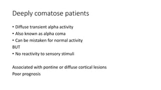
  • 22. Deeply comatose patients • Diffuse transient alpha activity • Also known as alpha coma • Can be mistaken for normal activity BUT • No reactivity to sensory stimuli Associated with pontine or diffuse cortical lesions Poor prognosis
  • 24. Anatomy and neurophysiology of alertness and coma Reticular Activating System (RAS) • Indistinct boundaries • Neurons are interspersed • Locus coeruleus: Upper dorsolateral pons of the brainstem. Activated directly by orexin from the lateral hypothalamus. Nor-epinephrine • Raphe nuclei: midline throughout the brainstem within the pons, midbrain, and medulla. Serotonergic • Tuberomamillary nucleus: located within the posterior aspect of the hypothalamus. Histaminergic • Lateral and dorsal pedunculopontine tegmentum: Primarily cholinergic neurons in neighboring groups within the midbrain and pons. Cholinergic neurons project to the thalamus and cortex.
  • 26. RAS receives collaterals from the spinothalamic and trigeminal– thalamic pathways Project to the whole of the cerebral cortex, not just to the sensory cortex of parietal lobe Cerebral cortex also modulates afferent sensory stimulus towards the RAS through corticofugal projections
  • 27. Pathologic basis of vegetative state • Diffuse cerebral injury as a result of closed head trauma • Widespread necrosis of the cortex after cardiac arrest or other form of anoxia • Thalamic necrosis Prominent pathologic changes are often in the thalamic and subthalamic nuclei rather than solely in the cortex.
  • 28. In traumatic cases, the pathologic findings are often of diffuse subcortical white matter degeneration (described as diffuse axonal injury), prominent thalamic degeneration, and ischemic damage in the cortex. Anatomic findings indicate that PVS is a state in which the cortex is either diffusely injured or effectively disconnected and isolated from the thalamus, or the thalamic nuclei are destroyed
  • 29. Pathologic anatomy of coma Two broad processes 1. Structural, or morphologic, consisting either of a discrete structural lesion in the upper brainstem and lower diencephalon (which may be primary or secondary to compression) or of more widespread destructive changes throughout the hemispheres. 2. Metabolic or submicroscopic, resulting in suppression of neuronal activity in the cerebrum and reticular activating system.
  • 30. Structural or morphological 1. A large mass in one cerebral hemisphere— tumor; abscess; massive infarct; or intracerebral, subdural, or epidural hemorrhage. Cause coma by secondary compression of the midbrain and central thalamic region of the RAS. Displacement or direct compression of these structures by the advancing medical temporal lobe which is forced into the tentorial opening. OR cerebellar lesion may compress the adjacent upper brainstem reticular region by displacing it forward and upward
  • 31. Structural or morphological 2. Destructive lesion is located immediately within the thalamus or midbrain, in which case the neurons of the RAS are damaged directly. Brainstem stroke from basilar artery occlusion, thalamic and upper brainstem hemorrhages, and some forms of traumatic damage. 3. Widespread bilateral damage to the cortex and cerebral white matter, the result of traumatic damage (contusions, diffuse axonal injury), bilateral ischemic strokes or hemorrhages, encephalitis, meningitis, hypoxia, or global ischemia. The coma in these cases results from interruption of thalamocortical impulses or from generalized destruction of cortical neurons. It is only if the cerebral lesions are bilateral and extensive that consciousness is impaired. Many of the diseases in this category also cause severe thalamic damage.
  • 32. Coma in concussion • Transient but large increase in intracranial pressure • 200 to 700 lb/in2 (10000-36000 mmHg) • Lasting a few thousandths of a second • Vibration set up in the skull and transmitted to the brain was for many years thought to be the basis of the abrupt paralysis of nervous function that characterizes concussive head injury (commotio cerebri). • Sudden swirling motion of the brain induced by acceleration or deceleration from a blow to the head produces a rotation (torque) of the cerebral hemispheres around the axis of the upper brainstem. If severe: multiple shearing lesions or hemorrhages in diencephalon and brain stem.
  • 33. ‘In the largest group of cases of coma, no structural lesion is revealed by any technique of conventional pathology.’
  • 34. Metabolic and toxic encephalopathies • Cerebral Blood Flow (CBF): 55mL/min/100g • Reduction in cerebral blood flow • Cerebral blood flow may stay near normal while metabolism is greatly reduced
  • 35. Clinical approach to comatose patient • Often, comatose patient is brought to the hospital and little pertinent medical information is available. • BLS/ACLS • If history or suspicion of trauma: ATLS (ABCDE) approach.
  • 36. • Patient’s airway is cleared and blood pressure is restored. • Patient’s airway is cleared and blood pressure is restored. • With hypotension, placement of a central venous line and administration of fluids, pressor agents and blood. • If respirations are shallow or labored, or if there is emesis with a threat of aspiration, tracheal intubation and mechanical ventilation are instituted. • An oropharyngeal airway is otherwise adequate in a comatose patient who is breathing normally. • Deeply comatose patients with shallow respirations require endotracheal intubation. • The patient with a head injury may also have suffered a fracture of the cervical vertebrae, in which case caution must be exercised in moving the head and neck.
  • 37. • Circumstances in which the person was found. • Previous health, whether there was a history of diabetes, a head injury, convulsion, alcohol or drug use, or a prior episode of coma or attempted suicide. • Sedatives, antiepileptic drugs, opiates, certain antibiotics, antidepressants, and antipsychotic drugs.
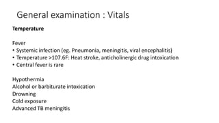
  • 38. General examination : Vitals Temperature Fever • Systemic infection (eg. Pneumonia, meningitis, viral encephalitis) • Temperature >107.6F: Heat stroke, anticholinergic drug intoxication • Central fever is rare Hypothermia Alcohol or barbiturate intoxication Drowning Cold exposure Advanced TB meningitis
  • 39. General examination : Vitals Respiration Slow breathing: opiate or barbiturate intoxication, hypothyroidism Deep rapid (Kussmaul respiration) : pneumonia, diabetic or uremic acidosis, pulmonary edema, or the less-common occurrence of an intracranial disease that causes central neurogenic hyperventilation. Slow, irregular, or cyclic (Cheyne-Stokes respiration) : Raised ICP
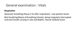
  • 40. General examination : Vitals Respiration Apneustic breathing (Pause 2-3s after inspiration) : Low pontine lesion Biot breathing/Ataxia of breathing (chaotic, being irregularly interrupted and each breath varying in rate and depth) : Dorsal medulla lesion
  • 41. Cheyne-Stokes respiration • Isolation of the brainstem respiratory centers from the cerebrum, rendering them more sensitive than usual to carbon dioxide (hyperventilation drive) • Signifies bilateral dysfunction of cerebral structures • Intoxication or a metabolic derangement • Bilateral structural lesions such as subdural hematomas
  • 42. General examination : Vitals Blood pressure Hypertension: cerebral hemorrhage, hypertensive encephalopathy and in children with markedly elevated intracranial pressure. Hypotension: Alcohol or barbiturate intoxication, DKA, internal hemorrhage, myocardial infarction, dissecting aortic aneurysm, septicemia, Addison disease, or massive brain trauma.
  • 43. General examination : Vitals Heart rate Bradycardia • Heart block from medications such as tricyclic antidepressants or anticonvulsants • If combined with periodic breathing and hypertension, an increase in intracranial pressure.
  • 44. Neurological examination Inspection • Predominant postures of the limbs and body • Presence or absence of spontaneous movements on one side • Position of the head and eyes • Rate, depth, and rhythm of respiration
  • 45. Brainstem function Pupillary size and reactivity Ocular movements Vestibulo-ocular reflexes Pattern of breathing
  • 46. Pupillary reaction • Loss of light reaction usually precedes enlargement of pupil • Unilaterally enlarged (“Hutchinson”) pupil : Early indicator of stretching or compression of the third nerve. Presence of an overlying ipsilateral hemispheral mass. • The light-unreactive pupil continues to enlarge to a size of 6 to 9 mm diameter and soon, there is slight outward deviation of the eye. • As midbrain displacement continues, both pupils dilate and become unreactive to light as a result of compression of the oculomotor nuclei in the rostral midbrain.
  • 47. Pupillary reaction • Pontine tegmental lesions (eg. Pontine hemorrhage) : extremely miotic pupils (<1 mm in diameter) with barely perceptible reaction to strong light. • Ipsilateral pupillary dilatation from pinching the side of the neck (the ciliospinal reflex) is usually lost in brainstem lesions. • The Horner syndrome (miosis, ptosis, and reduced facial sweating): ipsilateral to a lesion of the brainstem or hypothalamus or as a sign of dissection of the internal carotid artery.
  • 48. Coma caused by drug intoxications and intrinsic metabolic disorder • Pupillary reactions are usually spared Exceptions • Serum concentration of opiates that are high enough to cause coma  Pinpoint pupils • Systemic poisoning with atropine or with drugs that have atropinic qualities, especially the tricyclic antidepressants or SSRI: dilatation and relative unresponsiveness to light
  • 50. Movements of eye, eyelids and corneal reaction • Stupor or light coma of metabolic origin: Eyes rove conjugately from side to side in seemingly random fashion. Disappears as coma deepens  motionless and exotropic • Lateral and slight downward deviation: third-nerve palsy on that side • Medial deviation : Sixth-nerve palsy on that side
  • 51. Movements of eye, eyelids and corneal reaction Conjugate deviation of the eyes away from the side of the paralysis: large cerebral lesion (looking toward the lesion) Toward the side of the paralysis: unilateral pontine lesion (looking away from the lesion) Retraction and convergence nystagmus: lesions in the tegmentum of the mid-brain Ocular bobbing: lesions of the tegmentum of the pons Down and inward (looking at the nose): hematomas or ischemic lesions of the thalamus and upper midbrain (a variant of Parinaud syndrome) “Ocular dipping,” (eyes move down slowly and return rapidly to the meridian) : Anoxia and drug intoxications
  • 53. Elicitation of these ocular reflexes in a comatose patient: 1. evidence of unimpeded function of the midbrain and pontine tegmental structures that integrate bi-ocular eye movements 2. intactness of the CN nerves (III, IV, and VI) on both sides
  • 54. Caloric test / vestibulo-ocular test More vigorous stimulus for ocular movements in coma is accomplished by irrigation of one ear with 10 mL of cold water (or room-temperature water if the patient is still arousable). Causes slow conjugate deviation of the eyes toward the irrigated ear, followed in a few seconds by compensatory nystagmus (fast component away from the stimulated side). In comatose patients, the fast “corrective” phase of nystagmus that is mediated by the frontal lobes is lost and the eyes are tonically deflected.
  • 55. Reduction in the frequency and eventual loss of spontaneous blinking  Loss of response to touching the eyelashes  Lack of response to corneal touch (the corneal reflex afferent limb travels in the trigeminal nerve and efferent limb travels in facial nerve) : signs of deepening coma
  • 56. Sequentially increasing stimuli • calling his name • simple commands • noxious stimuli such as tickling the nares, supraorbital or sternal pressure, pinching the side of the neck or inner parts of the arms or thighs, or applying pressure to the knuckles
  • 57. Vocalization may persist in stupor and will be the first response to be lost as coma appears. Grimacing and deft avoidance movements of stimulated parts of the body are preserved in stupor. Yawning and spontaneous shifting of body positions indicate a minimal degree of unresponsiveness.
  • 58. Hemiplegia • Lack of restless movements of the limbs • Inadequate protective movements in response to painful stimuli. • The weakened limbs are usually slack and, if lifted from the bed, they “fall flail.” • The hemiplegic leg lies in a position of external rotation • Affected thigh appears wider and flatter than the nonhemiplegic one. In expiration, the cheek and lips puff out on the paralyzed side of the face. A lesion in one cerebral hemisphere causes the eyes to be turned away from the paralyzed side (toward the lesion). The opposite occurs with brainstem lesions. In most cases, a hemiplegia and an accompanying Babinski sign are indicative of a contralateral hemispheral lesion.
  • 59. Posturing Decerebrate rigidity • Opisthotonos • clenching of the jaws • stiff extension of the limbs • internal rotation of the arms and plantar flexion of the feet Lesions that interrupt the fibers of the red nucleus lead to inhibition of the rubrospinal tract (To flexor muscles) and constant excitation of the vestibulospinal and pontine reticulospinal tracts (To extensor muscles)
  • 60. Posturing Decorticate rigidity • Arms in flexion and adduction and legs extended. • Lesions at a more rostral level of the nervous system—in the cerebral white matter or internal capsule and thalamus. If preceded by decorticate or decerebrate postures, the coma is profound and usually progresses to brain death.
  • 61. Clinical signs of raised ICP • History of headache before the onset of coma • Vomiting, • Severe hypertension beyond the patient’s static level • Unexplained bradycardia • Retinal hemorrhages
  • 62. • Papilledema Develops within 12 to 24 h in cases of brain trauma and hemorrhage If seen at presentation with coma, signifies brain tumor or abscess (lesion of longer duration) Absence of papilledema does not exclude the presence of increased intracranial pressure, particularly in the elderly.
  • 63. Acute Hydrocephalus • Subarachnoid hemorrhage or from obstruction of the ventricular system by a tumor in the posterior fossa • Abulia  Stupor  Coma • Pupils: small and the tone in the legs is usually increased or there may be extensor posturing.
  • 64. Classification of Coma 1.Diseases that cause no focal or lateralizing neurologic signs, usually with normal brainstem functions. CT scan and cellular content of the CSF are normal A. Exogenous intoxications: alcohols, barbiturates and other sedative drugs, opiates B. Endogenous metabolic disturbances: anoxia, diabetic acidosis, uremia, hepatic failure, nonketotic hyperosmolar hyperglycemia, hypo- and hypernatremia, hypoglycemia, addisonian crisis, profound nutritional deficiency, carbon monoxide poisoning, thyroid states, hypercalcemia C. Severe systemic infections: pneumonia, peritonitis, typhoid fever, malaria, septicemia, Waterhouse- Friderichsen syndrome
  • 65. Classification of Coma D. Circulatory collapse (shock) from any cause E. Postseizure states F. Hypertensive encephalopathy and eclampsia G. Hyperthermia and hypothermia H. Concussion I. Acute hydrocephalus
  • 66. Classification of Coma 2. Diseases that cause meningeal irritation and an excess of white blood cells (WBCs) or red blood cells (RBCs) in the CSF, usually without focal or lateralizing cerebral or brainstem signs A. Subarachnoid hemorrhage from ruptured aneurysm, arteriovenous malformation, and cerebral trauma. B. Acute bacterial meningitis C. Viral meningoencephalitis D. Neoplastic meningeal infiltration E. Parasitic meningitis F. Pituitary apoplexy
  • 67. Classification of Coma 3. Diseases that cause focal brainstem or lateralizing cerebral signs, with or without changes in the CSF. A. Hemispheral hemorrhage or massive cerebral infarction B. Brainstem infarction caused by basilar artery thrombosis or embolism C. Brain abscess, subdural empyema, herpes encephalitis D. Epidural and subdural hemorrhage and brain contusion E. Brain tumor F. Cerebellar and pontine hemorrhage G. Multiple focal cerebral lesions that cumulate to cause generalized brain dysfunction: multiple embolic infarction caused by bacterial endocarditis, TTP, sagittal sinus thrombosis, diffuse fat embolism.