SlideShare a Scribd company logo
Ebstein's anomaly echocardiogram
 Tricuspid valve leaflet displacement
 Morphology & Tethering of the TV leaflets
 Tricuspid valve motion and coaptation
 Dilatation of chambers
 TR assesment
 Dilation of the right atrioventricular junction (true
tricuspid annulus)
 Dilation of right ventricular outflow tract
 Exaggerated tricuspid anulus motion
 Left ventricular function and dimension.
 Tricuspid valve leaflet displacement
septal leaflet
 Normally --- apical displacement +
 Ebstein’s --- exaggerated displacement
 Ebsteins – 7 to 50mm,
 Normal tricuspid valve - 0 to 10 mm,
 Secundum atrial defect - 2 to 14 mm
 Severe TR- 2 to 15 mm
overlap with values for Ebstein ' s anomaly.
So, cut off is
15mm--- < 14yr
20mm--- >14 yr
 indexed to body surface area
 Ebstein 's anomaly --- 8.5 to 11.4 ,median 8.4mm/m2
 Normal --- 0 to 6.3, median 3.0mm/m2;
 ASD-- 0.9to 7.5, median 5.0mm/m2;
 TR --- 1.1 to 7.5, median 4.0mm/m2
CUT OFF IS --- 8 MM/M2
Ebstein's anomaly echocardiogram
 Apical displacement of the septal leaflet from the
insertion of the anterior leaflet of the mitral valve by at
least 8 mm/m2
Morphology of leaflets
 Septal & posterior -
 Displacement
 Dysplasia
 Absence
 Anterior
 Elongation, redundancy , sail like appearance
 Distal attachments
 Mobility
 Fenestrations
Tethering of the tricuspid valve
 at least 3 accessory attachments of the leaflet to the
ventricular wall, causing restricted motion of the
leaflet.
Tethering
 Chamber enlargement
 Gose score
Ebstein’s: Echocardiographic
Severity index
 Celermajer et al
 grade (1 to 4)
 Gose score(Great
Ormond Street Score for
neonates)
Celermajer et al. Outcome in neonates with
Ebstein’s anomaly. JACC 1992; 19:1047-8.
GOSE score
Grade Ratio Mortality
1 <0.5 0%
2 0.5-0.99 8%
3
(acyanotic)
1-1.49 10% (neonatal)
45% (later)
3
(cyanotic)
1-1.49 100%
4 >1.5 100%
Ebstein's anomaly echocardiogram
TR ASSESMENT
 spectral & colour
 direction, no, origin (fenestrations)
Tricuspid anulus
 Size – z score
comparison with mitral v annulus
 Occasionally = tissue bridge forms connecting leading
edges of septal and anterior lt turning the commissure
into a key hole --- TS
 Even imperforate TV if closes completely.
Sub valvular apparatus
 Short chordae may attach septal leaflet to ventricular
septum
 Sometimes chordae may absent with insertion of
leaflet directly to the ventricular septum.
RVOT
 RVOT OBS
 Aneurysmal dilation - equal to or greater than twice
the aortic root diameter (20%).
 interatrial communication - 80% to 94%
 Pda
 Bicuspid or atretic aortic valves.
 Pulmonary atresia or hypoplastic pulmonary
artery, PS
 Subaortic stenosis, COA
 MVP , accessory mitral valve tissue
 Muscle bands of LV ,double orifice MV.
 VSD
 LV non compaction
M mode
 Delayed closure of the tricuspid valve compared with
that of the mitral valve.
 delay in EA -- > 50 msec
 N --- 20-30 msec
 Paradoxical motion -IVS
 Increased RVdimension
 Increased excursion of AML of the mitral valve
 greater the delay in tricuspid valve closure, the more
severe the disease.
Ebstein's anomaly echocardiogram
PLAX
 RV vol overload
 Paradoxical septal motion
 Free edges of TV seen in RV
 Displaced origins of leaflets
 Distinguish between anatomic & functional annulus
 Chordal attachments of ATL
PSAX
 Septal & ant leaflets seen, adherent to septal surface
 Length of ATL & its mobility
 Excessive size of ATL-systolic obstruction of RVOT
 Functional & anatomical pul atresia
 In neonates with severe ebsteins – pulmonary annulus
& branches are small
PSAX
A4C view
 Displacement index
 septal & ant leaflet
 Gose score
 BV function
A4C view
Ebstein's anomaly echocardiogram
SUBCOSTAL VIEWS
 Coronal view- degree of adherence of septal leaflet to
ven myocardium & degree of elongation of ATL
 Superior angulation- RVOT – degree of encroachment
of ATL on RVOT.
 Sagittal view- sail like ATL & abnormal PTL
 ENFACE view - from coronal view, 30 - 45 degrees
clock wise rotation- all leaflets are seen.
CARPENTIER classification
dd
 Tricuspid dysplasia – nodular thicking and rolling
edges of leaflet with out displacement
 Ungaurded tricuspid valve – all 3 leaflets are absent
3D ECHO
 Enables reconstruction of TV
 Visualise all 3 leaflets at same time – enface
view(surgical view)
 Better understanding of anotamy
 Degree of delamination
 Sub valvular apparatus
3d ECHO
Ebstein's anomaly echocardiogram
Pre op assesment
 Prognosis
 Tv repair vs replacement
Successful Monocusp Repair
 Freely mobile ATL
 Body of Leaflet and the Leading Edge can reach the
septum
 No Direct papillary muscle insertions
 Single Central Jet of TR
 No TV Chordal attachments in the RVOT
 Adequate Postop Functional RV size
Ebstein's anomaly echocardiogram
Unfavorable Features for Monocusp Repair
 Tethered ATL with restricted mobility of Body of
Leaflet and the Leading Edge
 Direct papillary muscle insertions onto valve tissue
(no chordae)
 Multiple Jets of TR (fenestrations)
 TV Chordal attachments in the RVOT (near the PV)
Ebstein's anomaly echocardiogram
Post op assesment
 Early post op –
 pericardial effusion, mediastinal hematoma,
 Intracardiac thrombus
 BV function, RWMA
 gr across TV,
 TR assesment( residual)
 Long term –
 RA, RV enlargement,
 TV fuction
Ebstein's anomaly echocardiogram
SAS SCORE --- 0 TO 10
> 5 --- NO SURVIVORS
< 3 --- 91% SURVIVAL

More Related Content

PDF
Echo assessment of Aortic Stenosis
PPTX
Atrial septal defect Echocardiography
PPTX
Ebstein anomaly
PPSX
Patent Ductus Arteriosus (PDA) Echocardiographic Assessment: Anatomy, Flow & ...
PPTX
Ebstein anomaly
PPTX
VSD ECHO.pptx
PPTX
Patent ductus arteriosis types evaluation and management (2)
PPTX
Echocardiographic Evaluation of Hypertrophic Cardiomyopathy
Echo assessment of Aortic Stenosis
Atrial septal defect Echocardiography
Ebstein anomaly
Patent Ductus Arteriosus (PDA) Echocardiographic Assessment: Anatomy, Flow & ...
Ebstein anomaly
VSD ECHO.pptx
Patent ductus arteriosis types evaluation and management (2)
Echocardiographic Evaluation of Hypertrophic Cardiomyopathy

What's hot (20)

PPTX
Echocardiography of CHD in Adults
PPTX
Echocardiographic evaluation of Aortic stenosis
PPTX
Echocardiography of Mitral regurgitation
PPTX
Collection of cath tracings by navin
PPT
SEGMENTAL ANALYSIS OF CONGENITAL HEART DISEASE
PPT
How to echo... tricuspid regurgitation.ppt
PPTX
Pulmonary hypertension echo
PPTX
Echo assessment of Aortic Regurgitation
PDF
Echo in cardiomyopathies part 1
PPTX
Echocardiography assessment of Aortic Regurgitation severity
PPTX
DM cardiology Exam Spotter
PPTX
hemodynamic in cath lab: aortic stenosis and hocm
PPTX
VSD devices
PPTX
PPTX
ASD devices
PPTX
Aortic stenosis Echo
PPTX
Assessment of prosthetic valve function
PPTX
Echo in hypertrophic obstructive cardiomyopathies
PPTX
Contrast Echocardiography
PDF
Aorto pulmonary window may 2021
Echocardiography of CHD in Adults
Echocardiographic evaluation of Aortic stenosis
Echocardiography of Mitral regurgitation
Collection of cath tracings by navin
SEGMENTAL ANALYSIS OF CONGENITAL HEART DISEASE
How to echo... tricuspid regurgitation.ppt
Pulmonary hypertension echo
Echo assessment of Aortic Regurgitation
Echo in cardiomyopathies part 1
Echocardiography assessment of Aortic Regurgitation severity
DM cardiology Exam Spotter
hemodynamic in cath lab: aortic stenosis and hocm
VSD devices
ASD devices
Aortic stenosis Echo
Assessment of prosthetic valve function
Echo in hypertrophic obstructive cardiomyopathies
Contrast Echocardiography
Aorto pulmonary window may 2021
Ad

Similar to Ebstein's anomaly echocardiogram (20)

PPTX
Echo of ebstein
PPTX
Echocardiographic assessment of tricuspid and pulmonary valves.pptx
PPTX
Ebstein anomaly
PPTX
Tricuspid pulmonary valves
PPTX
Tricuspid valve disease by T.N. Shanta
PDF
Ebstein anomaly
PPT
ebstein's anomali
PPT
ebstein's anomali
PPTX
Tricuspid valve disease
PPTX
Surgical management of Ebstein’s anomaly (by Ayman Khalifa)
PDF
Dysplastic tricuspid valve may 2021
PPT
Tricuspid and pulmonic valves 2011
PPTX
Ebsteins anamoly
PPTX
Congenital heart disease part 1.pptx
PPTX
ASSESSMENT OF RV FUNCTION.pptx
PPT
Finaale pulmonary stenosis
PPTX
EBSTEINS ANOMALY.pptx
Echo of ebstein
Echocardiographic assessment of tricuspid and pulmonary valves.pptx
Ebstein anomaly
Tricuspid pulmonary valves
Tricuspid valve disease by T.N. Shanta
Ebstein anomaly
ebstein's anomali
ebstein's anomali
Tricuspid valve disease
Surgical management of Ebstein’s anomaly (by Ayman Khalifa)
Dysplastic tricuspid valve may 2021
Tricuspid and pulmonic valves 2011
Ebsteins anamoly
Congenital heart disease part 1.pptx
ASSESSMENT OF RV FUNCTION.pptx
Finaale pulmonary stenosis
EBSTEINS ANOMALY.pptx
Ad

More from Malleswara rao Dangeti (20)

PPTX
Genetics in cardiovascular system
PPTX
PPT
acute rheumatic fever
PPTX
fundamentals of pacemaker
PPT
Approach to coronary bifurcation lesions
PPT
Treadmill test (TMT)
PPTX
Trouble shoooting ICD AND CRT
PPT
supraventricular tachycardia (SVT) with aberrancy
PPTX
LEADLESS PACEMAKER AND SUBCUTANEOUS ICD
PPTX
SINOATRIAL (SA) node
PPTX
relative wall thickness
PPT
Right ventricle (RV) anatomy and functions
PPTX
QRS axis change during ventricualr tachycardia (VT)
PPTX
Pliability assessment,pre procedure evaluation-tricks in difficult pbmv
PPTX
PPTX
Normal variants of heart structures
PPTX
Management of VENTRICULAR TACHYCARDIA (VT)
PPTX
Low flow low gradient aortic stenosis
PPTX
Hyponatremia in heart failure
Genetics in cardiovascular system
acute rheumatic fever
fundamentals of pacemaker
Approach to coronary bifurcation lesions
Treadmill test (TMT)
Trouble shoooting ICD AND CRT
supraventricular tachycardia (SVT) with aberrancy
LEADLESS PACEMAKER AND SUBCUTANEOUS ICD
SINOATRIAL (SA) node
relative wall thickness
Right ventricle (RV) anatomy and functions
QRS axis change during ventricualr tachycardia (VT)
Pliability assessment,pre procedure evaluation-tricks in difficult pbmv
Normal variants of heart structures
Management of VENTRICULAR TACHYCARDIA (VT)
Low flow low gradient aortic stenosis
Hyponatremia in heart failure

Recently uploaded (20)

PPTX
SKIN Anatomy and physiology and associated diseases
PPT
genitourinary-cancers_1.ppt Nursing care of clients with GU cancer
PPTX
Chapter-1-The-Human-Body-Orientation-Edited-55-slides.pptx
PDF
Khadir.pdf Acacia catechu drug Ayurvedic medicine
PPTX
Acid Base Disorders educational power point.pptx
PPTX
15.MENINGITIS AND ENCEPHALITIS-elias.pptx
PPT
ASRH Presentation for students and teachers 2770633.ppt
PPTX
CEREBROVASCULAR DISORDER.POWERPOINT PRESENTATIONx
PDF
Therapeutic Potential of Citrus Flavonoids in Metabolic Inflammation and Ins...
PPTX
Fundamentals of human energy transfer .pptx
DOCX
NEET PG 2025 | Pharmacology Recall: 20 High-Yield Questions Simplified
PPT
Breast Cancer management for medicsl student.ppt
PDF
Human Health And Disease hggyutgghg .pdf
PPTX
Respiratory drugs, drugs acting on the respi system
PPTX
Gastroschisis- Clinical Overview 18112311
PPTX
Uterus anatomy embryology, and clinical aspects
PPT
OPIOID ANALGESICS AND THEIR IMPLICATIONS
PDF
CT Anatomy for Radiotherapy.pdf eryuioooop
PPTX
Slider: TOC sampling methods for cleaning validation
PDF
Deadly Stampede at Yaounde’s Olembe Stadium Forensic.pdf
SKIN Anatomy and physiology and associated diseases
genitourinary-cancers_1.ppt Nursing care of clients with GU cancer
Chapter-1-The-Human-Body-Orientation-Edited-55-slides.pptx
Khadir.pdf Acacia catechu drug Ayurvedic medicine
Acid Base Disorders educational power point.pptx
15.MENINGITIS AND ENCEPHALITIS-elias.pptx
ASRH Presentation for students and teachers 2770633.ppt
CEREBROVASCULAR DISORDER.POWERPOINT PRESENTATIONx
Therapeutic Potential of Citrus Flavonoids in Metabolic Inflammation and Ins...
Fundamentals of human energy transfer .pptx
NEET PG 2025 | Pharmacology Recall: 20 High-Yield Questions Simplified
Breast Cancer management for medicsl student.ppt
Human Health And Disease hggyutgghg .pdf
Respiratory drugs, drugs acting on the respi system
Gastroschisis- Clinical Overview 18112311
Uterus anatomy embryology, and clinical aspects
OPIOID ANALGESICS AND THEIR IMPLICATIONS
CT Anatomy for Radiotherapy.pdf eryuioooop
Slider: TOC sampling methods for cleaning validation
Deadly Stampede at Yaounde’s Olembe Stadium Forensic.pdf

Ebstein's anomaly echocardiogram

  • 2.  Tricuspid valve leaflet displacement  Morphology & Tethering of the TV leaflets  Tricuspid valve motion and coaptation  Dilatation of chambers  TR assesment  Dilation of the right atrioventricular junction (true tricuspid annulus)  Dilation of right ventricular outflow tract  Exaggerated tricuspid anulus motion  Left ventricular function and dimension.
  • 3.  Tricuspid valve leaflet displacement septal leaflet  Normally --- apical displacement +  Ebstein’s --- exaggerated displacement
  • 4.  Ebsteins – 7 to 50mm,  Normal tricuspid valve - 0 to 10 mm,  Secundum atrial defect - 2 to 14 mm  Severe TR- 2 to 15 mm overlap with values for Ebstein ' s anomaly. So, cut off is 15mm--- < 14yr 20mm--- >14 yr
  • 5.  indexed to body surface area  Ebstein 's anomaly --- 8.5 to 11.4 ,median 8.4mm/m2  Normal --- 0 to 6.3, median 3.0mm/m2;  ASD-- 0.9to 7.5, median 5.0mm/m2;  TR --- 1.1 to 7.5, median 4.0mm/m2 CUT OFF IS --- 8 MM/M2
  • 7.  Apical displacement of the septal leaflet from the insertion of the anterior leaflet of the mitral valve by at least 8 mm/m2
  • 8. Morphology of leaflets  Septal & posterior -  Displacement  Dysplasia  Absence  Anterior  Elongation, redundancy , sail like appearance  Distal attachments  Mobility  Fenestrations
  • 9. Tethering of the tricuspid valve  at least 3 accessory attachments of the leaflet to the ventricular wall, causing restricted motion of the leaflet.
  • 12. Ebstein’s: Echocardiographic Severity index  Celermajer et al  grade (1 to 4)  Gose score(Great Ormond Street Score for neonates) Celermajer et al. Outcome in neonates with Ebstein’s anomaly. JACC 1992; 19:1047-8.
  • 13. GOSE score Grade Ratio Mortality 1 <0.5 0% 2 0.5-0.99 8% 3 (acyanotic) 1-1.49 10% (neonatal) 45% (later) 3 (cyanotic) 1-1.49 100% 4 >1.5 100%
  • 15. TR ASSESMENT  spectral & colour  direction, no, origin (fenestrations)
  • 16. Tricuspid anulus  Size – z score comparison with mitral v annulus
  • 17.  Occasionally = tissue bridge forms connecting leading edges of septal and anterior lt turning the commissure into a key hole --- TS  Even imperforate TV if closes completely.
  • 18. Sub valvular apparatus  Short chordae may attach septal leaflet to ventricular septum  Sometimes chordae may absent with insertion of leaflet directly to the ventricular septum.
  • 19. RVOT  RVOT OBS  Aneurysmal dilation - equal to or greater than twice the aortic root diameter (20%).
  • 20.  interatrial communication - 80% to 94%  Pda  Bicuspid or atretic aortic valves.  Pulmonary atresia or hypoplastic pulmonary artery, PS  Subaortic stenosis, COA  MVP , accessory mitral valve tissue  Muscle bands of LV ,double orifice MV.  VSD  LV non compaction
  • 21. M mode  Delayed closure of the tricuspid valve compared with that of the mitral valve.  delay in EA -- > 50 msec  N --- 20-30 msec  Paradoxical motion -IVS  Increased RVdimension  Increased excursion of AML of the mitral valve  greater the delay in tricuspid valve closure, the more severe the disease.
  • 26. PLAX  RV vol overload  Paradoxical septal motion  Free edges of TV seen in RV  Displaced origins of leaflets  Distinguish between anatomic & functional annulus  Chordal attachments of ATL
  • 27. PSAX  Septal & ant leaflets seen, adherent to septal surface  Length of ATL & its mobility  Excessive size of ATL-systolic obstruction of RVOT  Functional & anatomical pul atresia  In neonates with severe ebsteins – pulmonary annulus & branches are small
  • 28. PSAX
  • 29. A4C view  Displacement index  septal & ant leaflet  Gose score  BV function
  • 33. SUBCOSTAL VIEWS  Coronal view- degree of adherence of septal leaflet to ven myocardium & degree of elongation of ATL  Superior angulation- RVOT – degree of encroachment of ATL on RVOT.  Sagittal view- sail like ATL & abnormal PTL  ENFACE view - from coronal view, 30 - 45 degrees clock wise rotation- all leaflets are seen.
  • 35. dd  Tricuspid dysplasia – nodular thicking and rolling edges of leaflet with out displacement  Ungaurded tricuspid valve – all 3 leaflets are absent
  • 36. 3D ECHO  Enables reconstruction of TV  Visualise all 3 leaflets at same time – enface view(surgical view)  Better understanding of anotamy  Degree of delamination  Sub valvular apparatus
  • 39. Pre op assesment  Prognosis  Tv repair vs replacement
  • 40. Successful Monocusp Repair  Freely mobile ATL  Body of Leaflet and the Leading Edge can reach the septum  No Direct papillary muscle insertions  Single Central Jet of TR  No TV Chordal attachments in the RVOT  Adequate Postop Functional RV size
  • 42. Unfavorable Features for Monocusp Repair  Tethered ATL with restricted mobility of Body of Leaflet and the Leading Edge  Direct papillary muscle insertions onto valve tissue (no chordae)  Multiple Jets of TR (fenestrations)  TV Chordal attachments in the RVOT (near the PV)
  • 44. Post op assesment  Early post op –  pericardial effusion, mediastinal hematoma,  Intracardiac thrombus  BV function, RWMA  gr across TV,  TR assesment( residual)  Long term –  RA, RV enlargement,  TV fuction
  • 47. SAS SCORE --- 0 TO 10 > 5 --- NO SURVIVORS < 3 --- 91% SURVIVAL

Editor's Notes

  • #14: Involves calculating the ratio of the combined area of the RA and atrialized RV to that of the functional RV and L heart in a 4 chamber view at end diastole Even grade 3 carries with it a separate, late risk of death even in the acyanotic neonate
  • #32: Apical 4-chamber, 2-dimensional echocardiogram in a patient with Ebstein anomaly shows displacement of the tricuspid valve toward the apex of the right ventricle (RV) and tethering of the septal leaflet to the interventricular septum (arrow).
  • #33: Apical 4-chamber image from 2-dimensional (2D) echocardiography (Echo) in a patient with severe Ebstein anomaly shows displacement of the tricuspid valve towards the apex of the right ventricle (RV) more extreme than that shown in the previous 2 images. The atrialized part of the RV is more dilated and the tethering of the septal leaflet extends further toward the apex
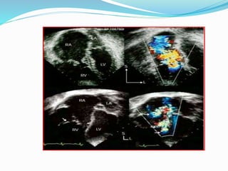
  • #46: these apical four-chamber images show two patients with ebstein’s malformation. the case illustrated in the upper panels shows a valve that is freely mobile (upper left panel) and colour flow mapping (upper right panel) revealed that there was only a single, central jet of regurgitation. this patient subsequently had a successful valve repair with only mild residual tricuspid regurgitation and no stenosis. the case shown in the lower panels displays a large muscular insertion to the middle of the anterosuperior leaflet (lower left) and multiple fenestrations and sites of regurgitation. the tethering and multiple origins of regurgitant flow dramatically decrease the chance for successful repair.