Enamel: Composition, Formation
and Structure Dr. Yumna
Lecturer
Dental Hygiene and Technology
Introduction
• Enamel is a protective covering for teeth
• Cells responsible for formation of enamel are
ameloblasts
• Enamel acquires a complex structural organization
• It possess high degree of mineralization
Composition
Physical Characteristics
• Enamel is the most highly mineralized extracellular
matrix
• It consists of approximately 96% mineral and 4%
organic material
• Inorganic content of enamel is a crystalline calcium
phosphate (hydroxyapatite)
Physical Characteristics
• Various ions strontium, magnessium, lead and
fluoride may also be incorporated into the crystals of
enamel
• High mineral content renders enamel extremely hard
• When crystals start dissolution, dental caries starts
Physical Characteristics
• Hardness of enamel is comparable to that of steel
• Enamel is brittle
• Dentine is resilient and it maintains integrity of
enamel
• Enamel is translucent
Physical Characteristics
• Enamel varies in color from light yellow to gray white
Structure of
Enamel
Structure
• Conventional demineralized sections of enamel only
have an empty space which was previously occupied
by mature enamel because the mineral has been
dissolved and traced organic material has been
washed away
Structure
• Fundamental organizational
units of enamel are the
– Rods (prisms)
– Interrod enamel
(interprismatic
substance)
Enamel composition and structure
Structure
• Enamel rod was first described as hexagonal and
prism like in cross section
• Enamel is built from closely packed and long, ribbon
like carbonatoapatite crystals
• Fully mature enamel crystals are no longer hexagonal
Enamel composition and structure
Amelogenesis
Amelogenesis
• It is a two step process
• Enamel first mineralizes to only 30%, later on crystals
grow wider
• Organic matter and water are lost and mineral is
added to reach full thickness of enamel
• At this stage greater than 96% mineral content is
found
Amelogenesis
• Ameloblasts secrete matrix proteins and are
responsible for creating and maintaining an
extracellular environment favorable to mineral
deposition
• Amelogenesis has been described in as many as six
phases
Amelogenesis
• Generally it is subdivided into three main functional
stages referred to as the
– Presecretory stage
– Secretory stage
– Maturation stage
Presecretory Stage
• Differentiating ameloblasts acquire their phenotype
• Change their polarity
• Develop extensive protein apparatus
• Secrete organic matrix of enamel
Secretory Stage
• Also called formative stage
• Ameloblasts elaborate and organize the entire
enamel thickness
• Results in the formation of highly ordered tissue
Maturation Stage
• Ameloblasts modulate
• Ameloblasts transport specific ions required for
concurrent accretion of enamel
• Ameloblasts are considered cells that carry out
multiple activities throughout their life cycle
Maturation Stage
• When differentiation of the ameloblasts occurs and
dentin starts forming, these cells are distanced from
the blood vessels
• Vascular supply is achieved by blood vessels
invaginating outer enamel epithelium
• There is loss of stellate reticulum
Light Microscopy of Amelogenesis
• At the late bell stage, most of light microscopic
features of amelogenesis can be seen
• In the region of cervical loop, low columnar cells in
inner enamel epithelium are identifiable
Enamel composition and structure
Light Microscopy of Amelogenesis
• An important event for the production of enamel is
the development of cytoplasmic extensions on
ameloblasts called Tome’s process
• Tome’s process give the junction between the
enamel and the ameloblast a saw-toothed
appearance
Light Microscopy of Amelogenesis
• As the inner enamel epithelium
is traced coronally in a crown
stage tooth germ, it cells
become tall and columnar
Enamel composition and structure
Light Microscopy of Amelogenesis
• Blood vessels invaginate deeply into
the cell, without disrupting the
basal lamina associated with the
outer aspect of enamel organ to
form a convoluted structure called
papillary layer
Light Microscopy of Amelogenesis
• Finally when enamel is fully mature, the ameloblast
layer and the adjacent papillary layer regress and
together constitute the reduced enamel epithelium
• At this stage ameloblasts stop modulating, reduce
their size and assume a cuboidal appearance
Enamel composition and structure
Light Microscopy of Amelogenesis
• As the tooth passes
through oral epithelium,
incisally reduced enamel
epithelium is destroyed
but remain cervically to
form the junctional
epithelium
Enamel composition and structure
Presecretory
Stage
Presecretory Stage
Morphogenetic Phase:
• During bell stage shape of the crown is determined
• Cells of inner enamel epithelium still can undergo
mitotic division
• They are cuboidal or low columnar cells
Presecretory Stage
Differentiation Phase:
• As the cells of inner enamel epithelium differentiate
into ameloblasts they elongate
• Ameloblasts become polarized
• These cells can no longer divide
Presecretory Stage
• Junctional complexes are present between
ameloblasts
• They play an important role in amelogenesis by
tightly holding together ameloblasts and pass on
required nutrients to them
Secretory
Stage
Secretory Stage
• Ameloblasts reflects their intense synthetic and
secretory activity
• The cell granules migrate to the distal extremity of
the cell that is into Tome’s process
Secretory Stage
• When enamel formation begins, Tomes’ process
comprises only a proximal portion
• The content of secretory granules is
• Secretion from the proximal part of the process and
from adjoining ameloblasts, results in the formation
of enamel pits
Maturation
Stage
Maturation Stage
• Amelogenesis is a slow developmental process
• It can take as long as 5 years to complete
• Maturation stage ameloblasts are referred to as post
secretory cells
Maturation Stage
• In this stage messenger RNA and protein signal for
– Amelogenin and
– Ameloblastin
• The two enamel proteins found in maturation
ameloblasts
Maturation Stage
Transitional Phase
• Ameloblasts undergo programmed cell death
(apoptosis)
• Approximately 25% cells die during transitional phase
• Other 25
% cells die as enamel maturation proceeds
Maturation Stage
• In embryogenesis, cells die at specific times during
development to permit orderly morphogenesis
• Two major ways by which cell death can occur are
– Necrosis (pathological)
– Apoptosis (physiological)
Maturation Stage
• Specialized proteinases (caspases) also inactivate
cellular survival pathways and activate factors that
promote death
Maturation Stage
Maturation Proper:
• Next principal activity of ameloblasts is the bulk
removal of water and organic material from the
enamel to allow introduction of additional inorganic
material
• The most dramatic activity is the modulation of cells
Maturation Stage
• They maintain the environment that allows accretion
of mineral content
• Maintain pH for functioning of the matrix degrading
enzymes
Enamel
Proteins
Enamel Proteins
• The organic matrix of enamel is made of non-
collagenous proteins only and made up of enamel
proteins and enzymes
• 90% of proteins are heterogenous group of low
molecular weight proteins known as amelogenins
• Remaining 10% are enamelins and ameloblastins
Enamel Proteins
• Amelogenins are hydrophobic proteins
• They have molecular weight between 5 and 45kDa
• Amelogenins regulate growth in thickness and width
of crystals
• Ameloblastins are concentrated near the cell surface
at sites where they are secreted
Enamel composition and structure
Structural and
Organizational
Features of
Enamel
Rod Interrelationships
• Rods tend to maintain in group
• Rods run in a perpendicular direction to the surface
of the dentin with a slight inclination towards the
cusp
Enamel composition and structure
Striae of Retzius
• They are identified using ground sections of calcified
teeth
• They are seen as a series of dark lines extending from
dentino-enamel junction towards tooth surface
• In cross sections they appear as concentric rings
Striae of Retzius
• The basis for their production is
still not clear
• A proposal suggests that they
reflect appositional or
incremental growth of the
enamel layer
Enamel composition and structure
Neonatal Line
• The neonatal line when present, is an enlarged stria
of Retzius that reflects changes occurring at birth
• Accentuated incremental lines also are produced by
systemic disturbances (e.g. fever) that affect
amelogenesis
Enamel composition and structure
Cross Striations
• Human enamel is known to form at a rate of 4µm per day
• Ground sections of enamel reveal periodic bands or cross
striations make up rods of enamel
• Alternating expansion and contraction of bands is
sometimes visible
• This pattern reflect diurnal rhythmicity in rod formation
Enamel composition and structure
Hunter Schreger Bands
• The bands of Hunter and Schreger are an optical
phenomenon produced by changes in direction
between adjacent groups of rods
• They are seen more clearly in longitudinal ground
sections
• Found in inner two thirds of enamel
• Appear as dark and light alternating zones
Enamel composition and structure
Gnarled Enamel
• Over the cusps of teeth
the rods appear twisted
around each other in a
seemingly complex
arrangement known as
gnarled enamel
Enamel composition and structure
Enamel Tufts and Lamellae
• They are like geological faults
• No known clinical significance
• Seen in transverse sections of enamel
•
Enamel Tufts
• Enamel tufts project from
dentino-enamel junction into
the enamel
• Contain greater concentration
of enamel proteins then rest of
enamel
• They occur developmentally
Enamel composition and structure
Enamel Lamellae
• They extend for varying depths from the surface of
enamel
• They contain linear, longitudinally oriented defects
filled with organic material
• Cracks in enamel can be sometimes mistaken for
lamellae but generally donot contain organic matter
Enamel Spindles
• Before enamel forms, some developing odontoblasts
processes extend into the ameloblast layer
• When enamel formation begins, become trapped to
form enamel spindles
• The shape and nature of DEJ prevent shearing of
enamel during function
Enamel composition and structure
Enamel Surface
• The striae of Retzius often extend from the
dentinoenamel junction to the outer surface of
enamel where they end in shallow furrows known as
perikymata
• Perikymeta runs in circumferentially horizontal lines
across the face of the crown
Enamel composition and structure
Enamel Surface
• Structure of enamel varies with age.
• In unerupted teeth enamel surface consists of a
structureless surface layer (finalenamel) that is lost
abruptly by abrasion, attrition and erosion in erupted
teeth
Enamel Surface
• As the tooth erupts it is covered with pellicle,
consisting of debris from enamel organ
• Salivary pellicle always reapperas on surface of teeth
shortly after mechanical polishing of teeth
• Dental plaque forms readily on teeth
Enamel composition and structure
Age Changes of Enamel
• With age enamel becomes
progressively worn in
regions of masticatory
attrition
• Wear facets increase in old
ages
Age Changes of Enamel
• Characteristics of aging include
– Discoloration
• Teeth darkens with age
– Reduced permeability
– Modifications in the surface layer
Age Changes of Enamel
• Darkening of teeth can be caused by addition of
organic matter
• It may also be caused by darkening of dentin layer
• Enamel becomes less permeable with age
• Young enamel acts as semipermeable membrane
Defects of
Amelogenesis
Defects of Amelogenesis
• Many conditions produce defects in enamel structure
• Defects occur because amelobalsts are cells sensitive
to changes in environment
Defects of Amelogenesis
• Defects in enamel can be caused by
– Febrile disease
– Nutrient deficiency
– Protein malformation in enamel
– Disarrangement of ameloblasts
– Maternal issues
– Fluoridation
Clinical
Implications
Fluoridation
• If fluoride is incorporated into the hydroxyapatite
crystals, the crystal becomes more resistant to acid
dissolution
• fluoride play role in caries prevention
• Presence of fluoride enhances chemical reactions
that lead to the precipitation of calcium phosphate
Enamel composition and structure
Acid Etching
• Use of fissure sealants, bonding of restorative
materials to enamel and cementing of orthodontic
brackets involve acid etching
• In first step etching removes plaque and other debris
along with a thin layer of enamel
Acid Etching
• Second step: it increases
porosity of the exposed
surface of enamel which
helps in proper bonding for
restoratives and adhesive
materials
Enamel composition and structure

More Related Content

PPT
A special tray is defined final
PPTX
02.dental caries
PPT
PPTX
Amelogenesis imperfecta
PPTX
Intro to dental material
PPTX
Enamel.pptx
PPTX
Life cycle of ameloblast
PPT
Introduction to dental materials
A special tray is defined final
02.dental caries
Amelogenesis imperfecta
Intro to dental material
Enamel.pptx
Life cycle of ameloblast
Introduction to dental materials

What's hot (20)

PPTX
Histology of Pulp
PPT
Enamel clinical aspect sagar hiwale
PPTX
age change in dental hard tissue
PPTX
ODP
Oral mucous membrane
PPTX
Tooth development
PPT
Tooth Dentin and dentinogenesis ppt
PPTX
HERTWIG’S EPITHELIAL ROOTH SHEATH
PPTX
Development ang growth of teeth
PPT
Cementum
PPTX
PPTX
Amelogenesis Slideshare.pptx
PPT
Dentine
PPTX
Age changes in enamel, dentin and pulp1.pptx
DOC
Dento enamel junction
PPTX
Theories of tooth eruption
PPTX
development and growth of teeth
PPTX
Hypomineralised structure of enamel
PPTX
Amelogenesis
Histology of Pulp
Enamel clinical aspect sagar hiwale
age change in dental hard tissue
Oral mucous membrane
Tooth development
Tooth Dentin and dentinogenesis ppt
HERTWIG’S EPITHELIAL ROOTH SHEATH
Development ang growth of teeth
Cementum
Amelogenesis Slideshare.pptx
Dentine
Age changes in enamel, dentin and pulp1.pptx
Dento enamel junction
Theories of tooth eruption
development and growth of teeth
Hypomineralised structure of enamel
Amelogenesis
Ad

Similar to Enamel composition and structure (20)

PDF
Amelogenesis.pdf
PPTX
Amelogenisis / dental implant courses by Indian dental academy
PPTX
Enamel & amelogenesis part 1
PPTX
The word amelogenesis is derived from two root words namely “Amelo” and “gene...
PPTX
Amelogenesis (enamel formation) oral biology anf histology
PPTX
Life cycle of ameloblast
PPTX
Amelogenesis Dr. Sherif Hassan
PPTX
Enamel _ Ashel _ up . pptx
PPTX
Amelogenesis
PPTX
Amelogenesis by dr.suriya visithiriyan
PPTX
AMELOGENESIS is the process of production of enamel.pptx
PPTX
ENAMEL AND DENTIN in paediatric dentistry
PPTX
Development of tooth
PPT
Tooth development on early stages of intra uterine life
PPTX
Enamel Seminar - Anu presented.pptx......
PPTX
3_2018_10_06!02_23_21_PM.pptx
PPTX
Enamel presentation. prepared by mohammed yahia
PPTX
EMBRYOLOGY OF THE DENTITION.pptx
Amelogenesis.pdf
Amelogenisis / dental implant courses by Indian dental academy
Enamel & amelogenesis part 1
The word amelogenesis is derived from two root words namely “Amelo” and “gene...
Amelogenesis (enamel formation) oral biology anf histology
Life cycle of ameloblast
Amelogenesis Dr. Sherif Hassan
Enamel _ Ashel _ up . pptx
Amelogenesis
Amelogenesis by dr.suriya visithiriyan
AMELOGENESIS is the process of production of enamel.pptx
ENAMEL AND DENTIN in paediatric dentistry
Development of tooth
Tooth development on early stages of intra uterine life
Enamel Seminar - Anu presented.pptx......
3_2018_10_06!02_23_21_PM.pptx
Enamel presentation. prepared by mohammed yahia
EMBRYOLOGY OF THE DENTITION.pptx
Ad

More from Dr. Yumna (7)

PPTX
Landmarks of maxilla
PPTX
Intro to dental terminology
PPT
Inlays and onlays
PPTX
Development of teeth
PPTX
Human skull (Bones of Jaw)
PPTX
Types of Teeth
PPTX
Types of crown & bridges
Landmarks of maxilla
Intro to dental terminology
Inlays and onlays
Development of teeth
Human skull (Bones of Jaw)
Types of Teeth
Types of crown & bridges

Recently uploaded (20)

PPTX
Chinmaya Tiranga Azadi Quiz (Class 7-8 )
PPTX
Share_Module_2_Power_conflict_and_negotiation.pptx
PDF
OBE - B.A.(HON'S) IN INTERIOR ARCHITECTURE -Ar.MOHIUDDIN.pdf
PDF
Hazard Identification & Risk Assessment .pdf
PPTX
B.Sc. DS Unit 2 Software Engineering.pptx
PDF
احياء السادس العلمي - الفصل الثالث (التكاثر) منهج متميزين/كلية بغداد/موهوبين
PDF
MBA _Common_ 2nd year Syllabus _2021-22_.pdf
PDF
1.3 FINAL REVISED K-10 PE and Health CG 2023 Grades 4-10 (1).pdf
PPTX
TNA_Presentation-1-Final(SAVE)) (1).pptx
PPTX
CHAPTER IV. MAN AND BIOSPHERE AND ITS TOTALITY.pptx
PDF
A GUIDE TO GENETICS FOR UNDERGRADUATE MEDICAL STUDENTS
PDF
advance database management system book.pdf
PDF
Trump Administration's workforce development strategy
PDF
David L Page_DCI Research Study Journey_how Methodology can inform one's prac...
PPTX
20th Century Theater, Methods, History.pptx
PPTX
202450812 BayCHI UCSC-SV 20250812 v17.pptx
PDF
ChatGPT for Dummies - Pam Baker Ccesa007.pdf
PDF
FORM 1 BIOLOGY MIND MAPS and their schemes
PDF
medical_surgical_nursing_10th_edition_ignatavicius_TEST_BANK_pdf.pdf
PDF
Empowerment Technology for Senior High School Guide
Chinmaya Tiranga Azadi Quiz (Class 7-8 )
Share_Module_2_Power_conflict_and_negotiation.pptx
OBE - B.A.(HON'S) IN INTERIOR ARCHITECTURE -Ar.MOHIUDDIN.pdf
Hazard Identification & Risk Assessment .pdf
B.Sc. DS Unit 2 Software Engineering.pptx
احياء السادس العلمي - الفصل الثالث (التكاثر) منهج متميزين/كلية بغداد/موهوبين
MBA _Common_ 2nd year Syllabus _2021-22_.pdf
1.3 FINAL REVISED K-10 PE and Health CG 2023 Grades 4-10 (1).pdf
TNA_Presentation-1-Final(SAVE)) (1).pptx
CHAPTER IV. MAN AND BIOSPHERE AND ITS TOTALITY.pptx
A GUIDE TO GENETICS FOR UNDERGRADUATE MEDICAL STUDENTS
advance database management system book.pdf
Trump Administration's workforce development strategy
David L Page_DCI Research Study Journey_how Methodology can inform one's prac...
20th Century Theater, Methods, History.pptx
202450812 BayCHI UCSC-SV 20250812 v17.pptx
ChatGPT for Dummies - Pam Baker Ccesa007.pdf
FORM 1 BIOLOGY MIND MAPS and their schemes
medical_surgical_nursing_10th_edition_ignatavicius_TEST_BANK_pdf.pdf
Empowerment Technology for Senior High School Guide

Enamel composition and structure

  • 1. Enamel: Composition, Formation and Structure Dr. Yumna Lecturer Dental Hygiene and Technology
  • 2. Introduction • Enamel is a protective covering for teeth • Cells responsible for formation of enamel are ameloblasts • Enamel acquires a complex structural organization • It possess high degree of mineralization
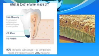
  • 4. Physical Characteristics • Enamel is the most highly mineralized extracellular matrix • It consists of approximately 96% mineral and 4% organic material • Inorganic content of enamel is a crystalline calcium phosphate (hydroxyapatite)
  • 5. Physical Characteristics • Various ions strontium, magnessium, lead and fluoride may also be incorporated into the crystals of enamel • High mineral content renders enamel extremely hard • When crystals start dissolution, dental caries starts
  • 6. Physical Characteristics • Hardness of enamel is comparable to that of steel • Enamel is brittle • Dentine is resilient and it maintains integrity of enamel • Enamel is translucent
  • 7. Physical Characteristics • Enamel varies in color from light yellow to gray white
  • 9. Structure • Conventional demineralized sections of enamel only have an empty space which was previously occupied by mature enamel because the mineral has been dissolved and traced organic material has been washed away
  • 10. Structure • Fundamental organizational units of enamel are the – Rods (prisms) – Interrod enamel (interprismatic substance)
  • 12. Structure • Enamel rod was first described as hexagonal and prism like in cross section • Enamel is built from closely packed and long, ribbon like carbonatoapatite crystals • Fully mature enamel crystals are no longer hexagonal
  • 15. Amelogenesis • It is a two step process • Enamel first mineralizes to only 30%, later on crystals grow wider • Organic matter and water are lost and mineral is added to reach full thickness of enamel • At this stage greater than 96% mineral content is found
  • 16. Amelogenesis • Ameloblasts secrete matrix proteins and are responsible for creating and maintaining an extracellular environment favorable to mineral deposition • Amelogenesis has been described in as many as six phases
  • 17. Amelogenesis • Generally it is subdivided into three main functional stages referred to as the – Presecretory stage – Secretory stage – Maturation stage
  • 18. Presecretory Stage • Differentiating ameloblasts acquire their phenotype • Change their polarity • Develop extensive protein apparatus • Secrete organic matrix of enamel
  • 19. Secretory Stage • Also called formative stage • Ameloblasts elaborate and organize the entire enamel thickness • Results in the formation of highly ordered tissue
  • 20. Maturation Stage • Ameloblasts modulate • Ameloblasts transport specific ions required for concurrent accretion of enamel • Ameloblasts are considered cells that carry out multiple activities throughout their life cycle
  • 21. Maturation Stage • When differentiation of the ameloblasts occurs and dentin starts forming, these cells are distanced from the blood vessels • Vascular supply is achieved by blood vessels invaginating outer enamel epithelium • There is loss of stellate reticulum
  • 22. Light Microscopy of Amelogenesis • At the late bell stage, most of light microscopic features of amelogenesis can be seen • In the region of cervical loop, low columnar cells in inner enamel epithelium are identifiable
  • 24. Light Microscopy of Amelogenesis • An important event for the production of enamel is the development of cytoplasmic extensions on ameloblasts called Tome’s process • Tome’s process give the junction between the enamel and the ameloblast a saw-toothed appearance
  • 25. Light Microscopy of Amelogenesis • As the inner enamel epithelium is traced coronally in a crown stage tooth germ, it cells become tall and columnar
  • 27. Light Microscopy of Amelogenesis • Blood vessels invaginate deeply into the cell, without disrupting the basal lamina associated with the outer aspect of enamel organ to form a convoluted structure called papillary layer
  • 28. Light Microscopy of Amelogenesis • Finally when enamel is fully mature, the ameloblast layer and the adjacent papillary layer regress and together constitute the reduced enamel epithelium • At this stage ameloblasts stop modulating, reduce their size and assume a cuboidal appearance
  • 30. Light Microscopy of Amelogenesis • As the tooth passes through oral epithelium, incisally reduced enamel epithelium is destroyed but remain cervically to form the junctional epithelium
  • 33. Presecretory Stage Morphogenetic Phase: • During bell stage shape of the crown is determined • Cells of inner enamel epithelium still can undergo mitotic division • They are cuboidal or low columnar cells
  • 34. Presecretory Stage Differentiation Phase: • As the cells of inner enamel epithelium differentiate into ameloblasts they elongate • Ameloblasts become polarized • These cells can no longer divide
  • 35. Presecretory Stage • Junctional complexes are present between ameloblasts • They play an important role in amelogenesis by tightly holding together ameloblasts and pass on required nutrients to them
  • 37. Secretory Stage • Ameloblasts reflects their intense synthetic and secretory activity • The cell granules migrate to the distal extremity of the cell that is into Tome’s process
  • 38. Secretory Stage • When enamel formation begins, Tomes’ process comprises only a proximal portion • The content of secretory granules is • Secretion from the proximal part of the process and from adjoining ameloblasts, results in the formation of enamel pits
  • 40. Maturation Stage • Amelogenesis is a slow developmental process • It can take as long as 5 years to complete • Maturation stage ameloblasts are referred to as post secretory cells
  • 41. Maturation Stage • In this stage messenger RNA and protein signal for – Amelogenin and – Ameloblastin • The two enamel proteins found in maturation ameloblasts
  • 42. Maturation Stage Transitional Phase • Ameloblasts undergo programmed cell death (apoptosis) • Approximately 25% cells die during transitional phase • Other 25 % cells die as enamel maturation proceeds
  • 43. Maturation Stage • In embryogenesis, cells die at specific times during development to permit orderly morphogenesis • Two major ways by which cell death can occur are – Necrosis (pathological) – Apoptosis (physiological)
  • 44. Maturation Stage • Specialized proteinases (caspases) also inactivate cellular survival pathways and activate factors that promote death
  • 45. Maturation Stage Maturation Proper: • Next principal activity of ameloblasts is the bulk removal of water and organic material from the enamel to allow introduction of additional inorganic material • The most dramatic activity is the modulation of cells
  • 46. Maturation Stage • They maintain the environment that allows accretion of mineral content • Maintain pH for functioning of the matrix degrading enzymes
  • 48. Enamel Proteins • The organic matrix of enamel is made of non- collagenous proteins only and made up of enamel proteins and enzymes • 90% of proteins are heterogenous group of low molecular weight proteins known as amelogenins • Remaining 10% are enamelins and ameloblastins
  • 49. Enamel Proteins • Amelogenins are hydrophobic proteins • They have molecular weight between 5 and 45kDa • Amelogenins regulate growth in thickness and width of crystals • Ameloblastins are concentrated near the cell surface at sites where they are secreted
  • 52. Rod Interrelationships • Rods tend to maintain in group • Rods run in a perpendicular direction to the surface of the dentin with a slight inclination towards the cusp
  • 54. Striae of Retzius • They are identified using ground sections of calcified teeth • They are seen as a series of dark lines extending from dentino-enamel junction towards tooth surface • In cross sections they appear as concentric rings
  • 55. Striae of Retzius • The basis for their production is still not clear • A proposal suggests that they reflect appositional or incremental growth of the enamel layer
  • 57. Neonatal Line • The neonatal line when present, is an enlarged stria of Retzius that reflects changes occurring at birth • Accentuated incremental lines also are produced by systemic disturbances (e.g. fever) that affect amelogenesis
  • 59. Cross Striations • Human enamel is known to form at a rate of 4µm per day • Ground sections of enamel reveal periodic bands or cross striations make up rods of enamel • Alternating expansion and contraction of bands is sometimes visible • This pattern reflect diurnal rhythmicity in rod formation
  • 61. Hunter Schreger Bands • The bands of Hunter and Schreger are an optical phenomenon produced by changes in direction between adjacent groups of rods • They are seen more clearly in longitudinal ground sections • Found in inner two thirds of enamel • Appear as dark and light alternating zones
  • 63. Gnarled Enamel • Over the cusps of teeth the rods appear twisted around each other in a seemingly complex arrangement known as gnarled enamel
  • 65. Enamel Tufts and Lamellae • They are like geological faults • No known clinical significance • Seen in transverse sections of enamel •
  • 66. Enamel Tufts • Enamel tufts project from dentino-enamel junction into the enamel • Contain greater concentration of enamel proteins then rest of enamel • They occur developmentally
  • 68. Enamel Lamellae • They extend for varying depths from the surface of enamel • They contain linear, longitudinally oriented defects filled with organic material • Cracks in enamel can be sometimes mistaken for lamellae but generally donot contain organic matter
  • 69. Enamel Spindles • Before enamel forms, some developing odontoblasts processes extend into the ameloblast layer • When enamel formation begins, become trapped to form enamel spindles • The shape and nature of DEJ prevent shearing of enamel during function
  • 71. Enamel Surface • The striae of Retzius often extend from the dentinoenamel junction to the outer surface of enamel where they end in shallow furrows known as perikymata • Perikymeta runs in circumferentially horizontal lines across the face of the crown
  • 73. Enamel Surface • Structure of enamel varies with age. • In unerupted teeth enamel surface consists of a structureless surface layer (finalenamel) that is lost abruptly by abrasion, attrition and erosion in erupted teeth
  • 74. Enamel Surface • As the tooth erupts it is covered with pellicle, consisting of debris from enamel organ • Salivary pellicle always reapperas on surface of teeth shortly after mechanical polishing of teeth • Dental plaque forms readily on teeth
  • 76. Age Changes of Enamel • With age enamel becomes progressively worn in regions of masticatory attrition • Wear facets increase in old ages
  • 77. Age Changes of Enamel • Characteristics of aging include – Discoloration • Teeth darkens with age – Reduced permeability – Modifications in the surface layer
  • 78. Age Changes of Enamel • Darkening of teeth can be caused by addition of organic matter • It may also be caused by darkening of dentin layer • Enamel becomes less permeable with age • Young enamel acts as semipermeable membrane
  • 80. Defects of Amelogenesis • Many conditions produce defects in enamel structure • Defects occur because amelobalsts are cells sensitive to changes in environment
  • 81. Defects of Amelogenesis • Defects in enamel can be caused by – Febrile disease – Nutrient deficiency – Protein malformation in enamel – Disarrangement of ameloblasts – Maternal issues – Fluoridation
  • 83. Fluoridation • If fluoride is incorporated into the hydroxyapatite crystals, the crystal becomes more resistant to acid dissolution • fluoride play role in caries prevention • Presence of fluoride enhances chemical reactions that lead to the precipitation of calcium phosphate
  • 86. Acid Etching • Use of fissure sealants, bonding of restorative materials to enamel and cementing of orthodontic brackets involve acid etching • In first step etching removes plaque and other debris along with a thin layer of enamel
  • 87. Acid Etching • Second step: it increases porosity of the exposed surface of enamel which helps in proper bonding for restoratives and adhesive materials