SlideShare a Scribd company logo
FRO.ppt
• The Knee Joint
 Structure
 Movements
 Biomechanics
• Manual Muscle Testing
 Hamstrings
 Quadriceps Femoris
• Floor reaction Orthosis
 Introduction
 Prerequisites
 Indications
 Design
 Fabrication
 Biomechanics
 Advantages
 Disadvantages
 Contraindications
FRO.ppt
• Knee is the largest and most
complex joint of the body. It is
a compound Synovial joint,
incorporating-
Two Condylar joint between the
condyles of Femur and Tibia.
One Saddle joint between the
Femur and Patella.
• The femoral condyles
articulate with tibial condyles
below and behind and with the
patella in front.
• Flexion/ Extension-
Flexion is the rolling of the
femoral condyles
posteriorly on the tibial
condyle.
Extension is the anterior
rolling of the femoral
condyles on the tibial
plateau, displacing the
femoral condyles back to a
neutral position.
• The main flexors of knee joint
are the Hamstrings. They
include:
Semimembranosus
Semitendinosus
Biceps femoris
• The main extensors of knee
joint are the Quadriceps
Femoris. It includes:
Rectus femoris
Vastus medialis
• Medial/ Lateral rotation-
• Medial/ Lateral rotation of the knee
joint are angular motions that are
named for the motion of the tibia on
the femur.
• These axial rotations of the knee joint
occur about a longitudinal axis that
runs through or close to the medial
tibial intercondylar tubercle.
• Consequently, the medial condyle
acts as a pivot point while the lateral
condyle moves through an arc of
motion, regardless of the direction of
rotation.
• As the tibia laterally rotates on the femur, the
medial tibial condyle moves only slightly
anteriorly on the relatively fixed medial femoral
condyle, whereas the lateral tibial condyle moves
a larger distance posteriorly on the relatively fixed
lateral femoral condyle.
• During tibial medial rotation, the medial tibial
condyle moves only slightly posteriorly, whereas
the lateral condyle moves anteriorly through a
larger arc of motion.
• The medial rotators of
the knee joint are:
Popliteus
Semimembranosus
Semitendinosus
• The lateral rotator of the
knee joint are:
Biceps femoris
• The longitudinal axis of femur is oblique, directed
inferiorly & medially from proximal to distal end. The
longitudinal axis of tibia is directed almost vertical.
• The femoral and tibial longitudinal axes normally form
an angle medially at the knee joint of 180°-185°.
• If the medial tibiofemoral angle is greater than 185°, an
abnormal condition called Genu Valgum (Knock Knees)
exists.
• If the medial tibiofemoral angle is 175° or less, an
abnormal condition called Genu Varum (Bow Legs) exist.
• Both active (muscular) and passive
(capsuloligamentous) forces contribute to the stability
of knee joint. These forces not only depend upon the
position of knee joint but also of the surrounding
tissues, magnitude & direction of applied forces and
availability of secondary restraints.
FRO.ppt
FRO.ppt
• Position of Patient: Prone with limbs
straight and toes hanging over the edge of
table.
• Position of Therapist: Standing next to
the limb to be tested. Hand giving
resistance is contoured around the
posterior surface of the leg just above the
ankle. Resistance is applied in the
direction of knee extension for Grades 5
and 4. the other hand is placed over the
hamstring tendons on the posterior thigh.
• Test: Patient flexes knee while maintaining
leg in neutral position.
• Position of Patient: Prone
with knee flexed to less than
90˚. Leg in internal rotation
(toes pointing towards
midline).
• Position of Therapist: Hand
giving resistance grasps the leg
at the ankle. Resistance is
applied in oblique direction
(down and out) towards knee
extension.
• Test: Patient flexes knee,
maintaining the leg in internal
rotation (heel towards
examiner, toes pointing
• Position of patient: Prone
with knee flexed to less than
90˚. Leg is in external
rotation (toes pointing
laterally).
• Position of Therapist:
Therapist resists knee flexion
at the ankle using a downward
and inward force.
• Test: Patient flexes knee,
maintaining leg in external
rotation (heel away from
• Grade 5 (Normal) for all
three tests: Resistance will
be maximal and the end knee
flexion position (approx 90˚)
cannot be broken.
• Grade 4 (Good) for all
three tests: End knee
flexion position is held
against strong to moderate
resistance.
• Grade 3 (Fair) for all
three tests: Holds end range
• Position of patient: Side-lying with
test limb (uppermost limb) supported by
examiner. Lower limb flexed for stability.
• Position of Therapist: Standing
behind the patient at knee level. One arm
is used to cradle thigh, providing hand
support at the medial side of thigh. Other
hand supports the leg at the ankle just
above the malleolus.
• Test: Patient flexes knee through
available range of motion.
• Instructions to the patient: “Bend
your knee.”
Grade 2 (Poor): Completes available
• Position of Patient: Prone. Limbs are
straight with toes extending over end of
table. Knee is partially flexed and
supported at ankle by examiner.
• Position of Therapist: Standing next to
test limb at knee level. One hand supports
the flexed limb at the ankle. The opposite
hand palpates both medial and lateral
hamstring tendons just above posterior
knee.
• Test: Patient attempts to flex the knee.
• Instructions to Patient: “Try to bend your knee.”
• Grade 1 (Trace): Tendons become prominent, but
no visible movement occurs.
• Grade 2 (Zero): No palpable contraction of the
muscles and tendons do not stand out.
• Position of Patient: Short sitting. Place the wedge or
pad under the distal thigh to maintain the femur in
horizontal position. Hands rest on the table on either side
of the body for stability, or may grasp the table edge. The
patient should be allowed to lean backward to relieve
hamstring muscle tension. Do not allow the patient to
hyperextend the knee because this may lock it into
position.
• Position of the Therapist: Standing on the side of limb
to be tested. The hand giving resistance is contoured over
the anterior surface of the distal leg just above the ankle.
For Grades 5 &4, resistance is applied in a downward
direction in the direction of knee flexion.
• Test: Patient extends knee through available ROM but not
beyond 0˚
• Instructions to Patient: “Straighten your knee. Hold it!
Don’t let me bend it.”
• Grade 5 (Normal):
Holds and position
against maximal
resistance.
• Grade 4 (Good):
Holds end position
against strong to
moderate resistance.
• Grade 3 (Fair):
Completes available
range and holds
• Position of Patient: Side-
lying with test limb uppermost.
Lowermost limb may be flexed
for stability. Limb to be tested is
held in about 90˚of knee
flexion. The hip should be in full
extension.
• Position of Therapist:
Standing behind patient at knee
level. One arm cradles the test
limb around the thigh with the
hand supporting the underside
of the knee. The other hand
holds the leg just above the
• Test: Patient extends knee through the available
ROM. The therapist supporting the limb provides
neither assistance nor resistance to the patient’s
voluntary movement.
• Instructions to Patient: “Straighten your knee.”
• Grade 2 (Poor): Completes available ROM.
• Position of Patient: Supine.
• Position of Therapist:
Standing next to the limb to be
tested at knee level. Hand used
for palpation should be on the
quadriceps tendon just above the
knee with the tendon “held”
gently between the thumb and
fingers. The examiner may also
want to palpate the patellar
tendon with 2 or 4 fingers just
below the knee.
• Test: Patient attempts to
Instructions to Patient: “Push the back of
your knee down to the table:” or “tighten your
kneecap”
Grade 1 (Trace): Contractile activity can be
palpated in muscle through the tendon. No
joint movement occurs.
Grade 0 (Zero): No palpable contractile
activity.
FRO.ppt
• It is a device that harnesses the Ground Reaction
Force as primary source of sagittal plane stability for
knee joint during stance.
• It relies on foot planter flexion-knee extension
couple where a fixed, slightly planter flexed ankle
creates an extension moment at knee.
• Plantar flexion angle, length and rigidity of toe-plate
helps to determine the magnitude of knee extension
moment.
• Anterior shell and medio-lateral
trimlines are padded at proximal
pre-patellar areas so that the extra
extension force being transmitted
to knee is more tolerable.
• FRO can be fabricated as
Single solid unit (May be
difficult to don).
Solid ankle AFO with an
additional anterior shell that is
snugly strapped in place
(Anterior shell can be removed
and the orthosis can be used as
traditional AFO).
Absence of knee or hip flexion contracture exceeding
10˚.
Careful determination of the angle to which the ankle
is set within the orthosis.
Presence of some trunk balance and/or ability to use
auxiliary walking aids in the event of diminished trunk
balance.
Presence of minimum of Grade 3 quadriceps strength.
Whether or not the orthosis is U/L or B/L.
• Post polio paralysis
• Cerebral palsy (FRAFOs in children with spastic diplegic CP who exhibit a moderate or
severe knee flexion (crouch gait) with excessive ankle dorsiflexion motion during the stance
phase. FRAFO use showed significant kinematic gait improvements including a reduction of
abnormal ankle dorsiflexion and knee flexion motion).
• Brain injury
• Spina bifida
• Spinal cord injury
 In these cases, FRO functions to maintain affected joints in proper alignment , to accentuate
knee extension in midstance and compensate for weak or absent gastrocnemius-soleus
muscles.
• Poliomyelitis often called
polio or infantile paralysis, is
an acute, viral, infectious
disease spread from person to
person, primarily via the
fecal-oral route.
• It generally affects lowerlimbs
and causes partial or full
paralysis.
• The main muscle of the knee
joint that is paralysed is the
quadriceps.
Quadriceps paralysis
Loss of Knee extension power
Patient will press the knee by
hand to extend it
Hand-to-Knee gait
• The conventional remedy for paralysed quadriceps is a long leg calliper made out of mild steel,
leather and wood.
• This not only weighs excessively but the long lever arm rigidises the limb and adds to the
difficulties.
• All the joints provided in the side bars are not congruent with the axis of movements of
anatomical joints.
• Apart from this, its fabrication is labour-intensive and time-consuming.
• When a polio patient with paralysed quadriceps, comes initial contact at heel it causes the
weight line to pass posterior to the knee. Here quadriceps action is needed to prevent buckling
but this action is absent in polio affected limb.
• To compensate for this, FRO is prescribed.
• Spina bifida is a
developmental congenital
disorderr caused by the
incomplete closing of the
embryonic neural tube.
• Patients with
myelomeningocele tend to
stand in a crouched
position (flexed hips,
knees, and ankles) due to
lower-extremity weakness.
• In this crouched position, the ground-reaction forces generally act posterior
to the knee center, encouraging further knee flexion.
• A floor-reaction ankle-foot orthosis (FRAFO) may be prescribed with the
goal of shifting the ground-reaction force more anterior to the knee center,
providing a knee-extension moment and increasing knee extension in stance.
• Compound this problem with knee-flexion contractures, and the patient's
ability to achieve an efficient gait pattern is severely decreased.
• The loss of knee extension due to the contractures result in a shortening of
step length seen at terminal swing.
• The inability to appropriately extend the knee in mid and terminal stance
increases the force on the quadriceps.
• Cerebral palsy (CP) is an umbrella
term encompassing a group of
non-progressive , non-contagious
motor conditions that cause
physical disability in human
development, chiefly in the
various areas of body movement
• In patients with cerebral palsy,
labored gait associated with
excessive knee flexion in stance
phase is presented with significant
• Crouch gait commences with overactivity of
the hamstrings, which increases knee flexion
and thus requires large increase in quadriceps
force to stabilize the knee.
• The FRO involves harnessing the floor
reaction and directing it to the anterior aspect
of the distal shank, thus stabilizing the joint in
extension.
• One piece: encloses the back of
lower calf, shin and bottom of the
foot.
• Two piece: same as one piece but
has a removable anterior panel.
• The design of a FRAFO included a full
toe plate, rigid ankle, and an anterior
tibial shell section. The combination
of these three components allow
the plantarflexion-knee
extension couple (PF/KE) to
occur, causing a knee-extension
moment. This knee-extension couple
helps to support weak quadriceps and
plantarflexor muscles.
FRO.ppt
• The patient is seated on a casting chair and all the bony prominences are
identified & marked with an indelible pencil.
• A latex rubber tube is run anteriorly over the dorsum of the foot along the tibial
crest to the level of neck of fibula. This allows easy and safe removal of the
cast.
• A length of stockinette is wrapped to act as a cast separator and the shank is
then wrapped with bandage to the level of fibular neck.
• The foot is then placed on a footboard in neutral subtalar position with slight
planterflexion to accommodate the heel height of patient’s shoe.
• Care must be taken to position the foot in the correct amount of toe-out and to
maintain the shank in proper orientation to mid-sagittal alignment.
• When the ankle-foot section has hardened, the knee is
extended to approx. 15˚ of flexion and wrapped with
the bandage to the level of proximal border of patella.
• The patellar tendon is then outlined as a reaction
point from floor for knee extension by compressing
with thumb and forefinger pressing in the same
manner as with PTB orthosis casting.
• Since weight bearing is not a consideration, the
popliteal area and contour of the posterior calf need
not be disturbed.
• The negative cast is removed and filled in for modification.
• Modification follows standard procedures with plaster build ups
over all bony prominences for pressure relief.
• The foot plate is then modified for support of the medial and
lateral longitudinal arches.
• In addition, a slightly more aggressive modification is made
under the sustentalaculum tali ( eminence on the medial surface
of calcaneum for the attachment of spring ligament) and
transverse metatarsal arch to provide a stable base for calcaneum
in a slight planterflexion position and comfortable distal reaction
point under metatarsal heads.
• A sheet of COPP is drape vacuum formed over the positive model
from posterior to anterior, creating an anterior seam.
• In the area of patellar tendon, the seam is pinched together tightly
to endure good bonding at this point.
• The orthosis is trimmed as follows:
Proximal trimline: Anteriorly, it should extend just above
the patellar tendon. Posteriorly, it should extend till the level
of maximum calf.
Distal trimline: It extends to the end of the toes.
Medial trimline: It encompasses the medial malleolus and
lies just anterior to it.
FRO.ppt
• The ankle joint is kept in 5 to 7 degrees of plantar flexion in order to
shift the line of floor reaction.
• By keeping ankle in plantar flexion, the resultant of floor reaction
passes posterior to the ankle joint and anterior to the knee joint
generating a posteriorly directed moment which tends to push the
knee in extension thus controlling knee flexion allowing the patient
to remove his or her hand.
• The brace acta as a First class lever and the planterflexed attitude at
the ankle utilizes the fulcrum establised by the distal trimline of the
footplate.
FRO.ppt
FRO.ppt
• Besides stabilizing the ankle and subtalar joints, it assists the knee
extension capabilities of the patient with fair quadriceps strength
and therefore, helps prevent the knee joint from buckling.
• The ankle in FRO is almost in neutral position, so that patient can
begin gait cycle with heel strike and descend stairs more easily.
• Gait is more physiological because it provides opportunity for
quadriceps to work as the patient walks.
• Provides patient good security and gives him a sense of freedom.
• Light weight.
• Device provides total-contact, thus preventing pressure sores over
bony prominences.
• Durable.
• Compact.
• FRO may have a negative impact on balance reactions.
If the patient do not have effective postural control, an
assistive device may be required.
• Fabrication is time-consuming.
• Lack of adjustibility in the degree of ankle equinus &
consequently the amount of knee extension moment
control force.
• Long term use can lead to Genu recurvatum.
• Inapproriate for the patients with Genu recurvatum.
• Not suitable in case of structural instability of knee
joint.
• Difficult for the patients with fixed flexion contracture
of more than 10˚.(In this case, FRO will resist
dorsiflexion and knee flexion but does not create a
true extension moment.)
• If used in bilateral cases, tip-toe type of gait will be
achieved which is very uncomfortable for the patients
and hard to maintain the balance, hence
contraindicated in bilateral cases.
• It was observed that by using FRO in patients with
cerebral plasy and spina bifida, crouch gait was greatly
reduced or eliminated.
• When the pre requisites and specifications were met,
FRO proved to be more successful than its
predecessor, the KAFO.
• Following the careful determination of candidacy in
the patients, the floor reaction orthosis can provide
more stability and minimize componentary, weight
and bulk.
FRO.ppt

More Related Content

PPTX
knee biomechanics.pptx
PPTX
Hand splinting in common orthopedic & neurological condition 1
PPTX
prehension.pptx
PDF
Shoulder anatomy__biomechanics__pathomechanics
PPTX
BIOMECHANICS OF HIP JOINT
PPTX
Evolution of hip disarticulation prosthesis
PPT
Active movement resisted exercises
PPTX
Biomechanics of knee complex 1
knee biomechanics.pptx
Hand splinting in common orthopedic & neurological condition 1
prehension.pptx
Shoulder anatomy__biomechanics__pathomechanics
BIOMECHANICS OF HIP JOINT
Evolution of hip disarticulation prosthesis
Active movement resisted exercises
Biomechanics of knee complex 1

What's hot (20)

PPTX
Biomechanics of cane, crutch and walker
PDF
Hip joint biomechanics and pathomechanics
PPTX
Pelvic tilt
PPTX
Maitland concept.pptx.....................
PPTX
Biomechanics of knee joint
PPTX
Biomechanics of Wrist and Hand Complex- Dr Gurjant Singh (PT)
PPTX
Biomechanics of Gait, Posture and ADL
PPT
Foot biomechanics
PPTX
WALKING AIDS by Dr. Mumux
PPTX
Biomechanics of knee complex 8 patellofemoral joint
KEY
Joint biomechanics
PPTX
mobility aids.pptx
PPT
A to z of exercise stability ball
PPTX
CERVICAL BIOMECHANICS.pptx
PPT
Gait analysis and.ppt by ramachandra
PPTX
Biomechanics of hip complex 2
PPTX
Range Of Motion Assessment
PPTX
Biomechanics of hip complex 4
PPTX
muscle re_education by chintu.pptx
PPTX
Cervical spine gonio
Biomechanics of cane, crutch and walker
Hip joint biomechanics and pathomechanics
Pelvic tilt
Maitland concept.pptx.....................
Biomechanics of knee joint
Biomechanics of Wrist and Hand Complex- Dr Gurjant Singh (PT)
Biomechanics of Gait, Posture and ADL
Foot biomechanics
WALKING AIDS by Dr. Mumux
Biomechanics of knee complex 8 patellofemoral joint
Joint biomechanics
mobility aids.pptx
A to z of exercise stability ball
CERVICAL BIOMECHANICS.pptx
Gait analysis and.ppt by ramachandra
Biomechanics of hip complex 2
Range Of Motion Assessment
Biomechanics of hip complex 4
muscle re_education by chintu.pptx
Cervical spine gonio
Ad

Similar to FRO.ppt (20)

PDF
MMT of Knee.pdf
PPTX
Knee Joint Assessment Tests and Mobilizations
PPTX
4.Meniscal Injury Assessment & Management (1).pptx
PPTX
derived positions.pptx
PPTX
The Elbow Joint 1_a93e492f7cfcdba2d5a0fde7c8fecec5(1).pptx
PPTX
The Elbow Joint 1_a93e492f7cfcdba2d5a0fde7c8fecec5.pptx
PPTX
The Elbow Joint 1_a93e492f7cfcdba2d5a0fde7c8fecec5(2).pptx
PPTX
Lecture 9 ttt an d also different and .pptx
PPTX
Mmt f0r hip
PPTX
musculoskeletal_system_examination[1] (1) (3).pptx
PPTX
Seminar rotator cuff
PPTX
complete hip joint msk.pptx dpt SIPMR bapu
PPTX
Tightness test for lower quadrent muscle
PPTX
Club foot
PPTX
this is a description of spinal injuries in sports players and this includes ...
PPTX
MMT Neck & Scapula.pptx
PPTX
ASSISTIVE DEVICES in Nursing Proceduress
PPTX
Dislocation of hip
PPTX
Dislocation of hip
PDF
Manualmusletesting 211 240
MMT of Knee.pdf
Knee Joint Assessment Tests and Mobilizations
4.Meniscal Injury Assessment & Management (1).pptx
derived positions.pptx
The Elbow Joint 1_a93e492f7cfcdba2d5a0fde7c8fecec5(1).pptx
The Elbow Joint 1_a93e492f7cfcdba2d5a0fde7c8fecec5.pptx
The Elbow Joint 1_a93e492f7cfcdba2d5a0fde7c8fecec5(2).pptx
Lecture 9 ttt an d also different and .pptx
Mmt f0r hip
musculoskeletal_system_examination[1] (1) (3).pptx
Seminar rotator cuff
complete hip joint msk.pptx dpt SIPMR bapu
Tightness test for lower quadrent muscle
Club foot
this is a description of spinal injuries in sports players and this includes ...
MMT Neck & Scapula.pptx
ASSISTIVE DEVICES in Nursing Proceduress
Dislocation of hip
Dislocation of hip
Manualmusletesting 211 240
Ad

Recently uploaded (20)

PPTX
1 General Principles of Radiotherapy.pptx
PPT
Management of Acute Kidney Injury at LAUTECH
PPT
OPIOID ANALGESICS AND THEIR IMPLICATIONS
PPTX
CEREBROVASCULAR DISORDER.POWERPOINT PRESENTATIONx
DOCX
NEET PG 2025 | Pharmacology Recall: 20 High-Yield Questions Simplified
PPTX
15.MENINGITIS AND ENCEPHALITIS-elias.pptx
PPTX
Imaging of parasitic D. Case Discussions.pptx
PPTX
Uterus anatomy embryology, and clinical aspects
PPTX
SKIN Anatomy and physiology and associated diseases
PPTX
Slider: TOC sampling methods for cleaning validation
PPT
Obstructive sleep apnea in orthodontics treatment
PPT
1b - INTRODUCTION TO EPIDEMIOLOGY (comm med).ppt
PPTX
CME 2 Acute Chest Pain preentation for education
PPT
Breast Cancer management for medicsl student.ppt
PPTX
surgery guide for USMLE step 2-part 1.pptx
PPT
ASRH Presentation for students and teachers 2770633.ppt
PPTX
Note on Abortion.pptx for the student note
PPTX
post stroke aphasia rehabilitation physician
PDF
Medical Evidence in the Criminal Justice Delivery System in.pdf
PPTX
Electromyography (EMG) in Physiotherapy: Principles, Procedure & Clinical App...
1 General Principles of Radiotherapy.pptx
Management of Acute Kidney Injury at LAUTECH
OPIOID ANALGESICS AND THEIR IMPLICATIONS
CEREBROVASCULAR DISORDER.POWERPOINT PRESENTATIONx
NEET PG 2025 | Pharmacology Recall: 20 High-Yield Questions Simplified
15.MENINGITIS AND ENCEPHALITIS-elias.pptx
Imaging of parasitic D. Case Discussions.pptx
Uterus anatomy embryology, and clinical aspects
SKIN Anatomy and physiology and associated diseases
Slider: TOC sampling methods for cleaning validation
Obstructive sleep apnea in orthodontics treatment
1b - INTRODUCTION TO EPIDEMIOLOGY (comm med).ppt
CME 2 Acute Chest Pain preentation for education
Breast Cancer management for medicsl student.ppt
surgery guide for USMLE step 2-part 1.pptx
ASRH Presentation for students and teachers 2770633.ppt
Note on Abortion.pptx for the student note
post stroke aphasia rehabilitation physician
Medical Evidence in the Criminal Justice Delivery System in.pdf
Electromyography (EMG) in Physiotherapy: Principles, Procedure & Clinical App...

FRO.ppt

  • 2. • The Knee Joint  Structure  Movements  Biomechanics • Manual Muscle Testing  Hamstrings  Quadriceps Femoris • Floor reaction Orthosis  Introduction  Prerequisites  Indications  Design  Fabrication  Biomechanics  Advantages  Disadvantages  Contraindications
  • 4. • Knee is the largest and most complex joint of the body. It is a compound Synovial joint, incorporating- Two Condylar joint between the condyles of Femur and Tibia. One Saddle joint between the Femur and Patella. • The femoral condyles articulate with tibial condyles below and behind and with the patella in front.
  • 5. • Flexion/ Extension- Flexion is the rolling of the femoral condyles posteriorly on the tibial condyle. Extension is the anterior rolling of the femoral condyles on the tibial plateau, displacing the femoral condyles back to a neutral position.
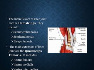
  • 6. • The main flexors of knee joint are the Hamstrings. They include: Semimembranosus Semitendinosus Biceps femoris • The main extensors of knee joint are the Quadriceps Femoris. It includes: Rectus femoris Vastus medialis
  • 7. • Medial/ Lateral rotation- • Medial/ Lateral rotation of the knee joint are angular motions that are named for the motion of the tibia on the femur. • These axial rotations of the knee joint occur about a longitudinal axis that runs through or close to the medial tibial intercondylar tubercle. • Consequently, the medial condyle acts as a pivot point while the lateral condyle moves through an arc of motion, regardless of the direction of rotation.
  • 8. • As the tibia laterally rotates on the femur, the medial tibial condyle moves only slightly anteriorly on the relatively fixed medial femoral condyle, whereas the lateral tibial condyle moves a larger distance posteriorly on the relatively fixed lateral femoral condyle. • During tibial medial rotation, the medial tibial condyle moves only slightly posteriorly, whereas the lateral condyle moves anteriorly through a larger arc of motion.
  • 9. • The medial rotators of the knee joint are: Popliteus Semimembranosus Semitendinosus • The lateral rotator of the knee joint are: Biceps femoris
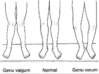
  • 10. • The longitudinal axis of femur is oblique, directed inferiorly & medially from proximal to distal end. The longitudinal axis of tibia is directed almost vertical. • The femoral and tibial longitudinal axes normally form an angle medially at the knee joint of 180°-185°. • If the medial tibiofemoral angle is greater than 185°, an abnormal condition called Genu Valgum (Knock Knees) exists. • If the medial tibiofemoral angle is 175° or less, an abnormal condition called Genu Varum (Bow Legs) exist.
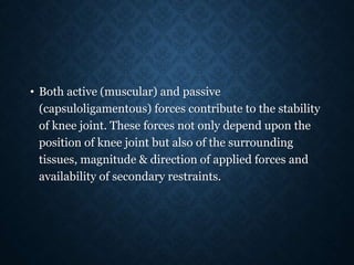
  • 11. • Both active (muscular) and passive (capsuloligamentous) forces contribute to the stability of knee joint. These forces not only depend upon the position of knee joint but also of the surrounding tissues, magnitude & direction of applied forces and availability of secondary restraints.
  • 14. • Position of Patient: Prone with limbs straight and toes hanging over the edge of table. • Position of Therapist: Standing next to the limb to be tested. Hand giving resistance is contoured around the posterior surface of the leg just above the ankle. Resistance is applied in the direction of knee extension for Grades 5 and 4. the other hand is placed over the hamstring tendons on the posterior thigh. • Test: Patient flexes knee while maintaining leg in neutral position.
  • 15. • Position of Patient: Prone with knee flexed to less than 90˚. Leg in internal rotation (toes pointing towards midline). • Position of Therapist: Hand giving resistance grasps the leg at the ankle. Resistance is applied in oblique direction (down and out) towards knee extension. • Test: Patient flexes knee, maintaining the leg in internal rotation (heel towards examiner, toes pointing
  • 16. • Position of patient: Prone with knee flexed to less than 90˚. Leg is in external rotation (toes pointing laterally). • Position of Therapist: Therapist resists knee flexion at the ankle using a downward and inward force. • Test: Patient flexes knee, maintaining leg in external rotation (heel away from
  • 17. • Grade 5 (Normal) for all three tests: Resistance will be maximal and the end knee flexion position (approx 90˚) cannot be broken. • Grade 4 (Good) for all three tests: End knee flexion position is held against strong to moderate resistance. • Grade 3 (Fair) for all three tests: Holds end range
  • 18. • Position of patient: Side-lying with test limb (uppermost limb) supported by examiner. Lower limb flexed for stability. • Position of Therapist: Standing behind the patient at knee level. One arm is used to cradle thigh, providing hand support at the medial side of thigh. Other hand supports the leg at the ankle just above the malleolus. • Test: Patient flexes knee through available range of motion. • Instructions to the patient: “Bend your knee.” Grade 2 (Poor): Completes available
  • 19. • Position of Patient: Prone. Limbs are straight with toes extending over end of table. Knee is partially flexed and supported at ankle by examiner. • Position of Therapist: Standing next to test limb at knee level. One hand supports the flexed limb at the ankle. The opposite hand palpates both medial and lateral hamstring tendons just above posterior knee. • Test: Patient attempts to flex the knee.
  • 20. • Instructions to Patient: “Try to bend your knee.” • Grade 1 (Trace): Tendons become prominent, but no visible movement occurs. • Grade 2 (Zero): No palpable contraction of the muscles and tendons do not stand out.
  • 21. • Position of Patient: Short sitting. Place the wedge or pad under the distal thigh to maintain the femur in horizontal position. Hands rest on the table on either side of the body for stability, or may grasp the table edge. The patient should be allowed to lean backward to relieve hamstring muscle tension. Do not allow the patient to hyperextend the knee because this may lock it into position.
  • 22. • Position of the Therapist: Standing on the side of limb to be tested. The hand giving resistance is contoured over the anterior surface of the distal leg just above the ankle. For Grades 5 &4, resistance is applied in a downward direction in the direction of knee flexion. • Test: Patient extends knee through available ROM but not beyond 0˚ • Instructions to Patient: “Straighten your knee. Hold it! Don’t let me bend it.”
  • 23. • Grade 5 (Normal): Holds and position against maximal resistance. • Grade 4 (Good): Holds end position against strong to moderate resistance. • Grade 3 (Fair): Completes available range and holds
  • 24. • Position of Patient: Side- lying with test limb uppermost. Lowermost limb may be flexed for stability. Limb to be tested is held in about 90˚of knee flexion. The hip should be in full extension. • Position of Therapist: Standing behind patient at knee level. One arm cradles the test limb around the thigh with the hand supporting the underside of the knee. The other hand holds the leg just above the
  • 25. • Test: Patient extends knee through the available ROM. The therapist supporting the limb provides neither assistance nor resistance to the patient’s voluntary movement. • Instructions to Patient: “Straighten your knee.” • Grade 2 (Poor): Completes available ROM.
  • 26. • Position of Patient: Supine. • Position of Therapist: Standing next to the limb to be tested at knee level. Hand used for palpation should be on the quadriceps tendon just above the knee with the tendon “held” gently between the thumb and fingers. The examiner may also want to palpate the patellar tendon with 2 or 4 fingers just below the knee. • Test: Patient attempts to
  • 27. Instructions to Patient: “Push the back of your knee down to the table:” or “tighten your kneecap” Grade 1 (Trace): Contractile activity can be palpated in muscle through the tendon. No joint movement occurs. Grade 0 (Zero): No palpable contractile activity.
  • 29. • It is a device that harnesses the Ground Reaction Force as primary source of sagittal plane stability for knee joint during stance. • It relies on foot planter flexion-knee extension couple where a fixed, slightly planter flexed ankle creates an extension moment at knee. • Plantar flexion angle, length and rigidity of toe-plate helps to determine the magnitude of knee extension moment.
  • 30. • Anterior shell and medio-lateral trimlines are padded at proximal pre-patellar areas so that the extra extension force being transmitted to knee is more tolerable. • FRO can be fabricated as Single solid unit (May be difficult to don). Solid ankle AFO with an additional anterior shell that is snugly strapped in place (Anterior shell can be removed and the orthosis can be used as traditional AFO).
  • 31. Absence of knee or hip flexion contracture exceeding 10˚. Careful determination of the angle to which the ankle is set within the orthosis. Presence of some trunk balance and/or ability to use auxiliary walking aids in the event of diminished trunk balance. Presence of minimum of Grade 3 quadriceps strength. Whether or not the orthosis is U/L or B/L.
  • 32. • Post polio paralysis • Cerebral palsy (FRAFOs in children with spastic diplegic CP who exhibit a moderate or severe knee flexion (crouch gait) with excessive ankle dorsiflexion motion during the stance phase. FRAFO use showed significant kinematic gait improvements including a reduction of abnormal ankle dorsiflexion and knee flexion motion). • Brain injury • Spina bifida • Spinal cord injury  In these cases, FRO functions to maintain affected joints in proper alignment , to accentuate knee extension in midstance and compensate for weak or absent gastrocnemius-soleus muscles.
  • 33. • Poliomyelitis often called polio or infantile paralysis, is an acute, viral, infectious disease spread from person to person, primarily via the fecal-oral route. • It generally affects lowerlimbs and causes partial or full paralysis. • The main muscle of the knee joint that is paralysed is the quadriceps.
  • 34. Quadriceps paralysis Loss of Knee extension power Patient will press the knee by hand to extend it Hand-to-Knee gait
  • 35. • The conventional remedy for paralysed quadriceps is a long leg calliper made out of mild steel, leather and wood. • This not only weighs excessively but the long lever arm rigidises the limb and adds to the difficulties. • All the joints provided in the side bars are not congruent with the axis of movements of anatomical joints. • Apart from this, its fabrication is labour-intensive and time-consuming. • When a polio patient with paralysed quadriceps, comes initial contact at heel it causes the weight line to pass posterior to the knee. Here quadriceps action is needed to prevent buckling but this action is absent in polio affected limb. • To compensate for this, FRO is prescribed.
  • 36. • Spina bifida is a developmental congenital disorderr caused by the incomplete closing of the embryonic neural tube. • Patients with myelomeningocele tend to stand in a crouched position (flexed hips, knees, and ankles) due to lower-extremity weakness.
  • 37. • In this crouched position, the ground-reaction forces generally act posterior to the knee center, encouraging further knee flexion. • A floor-reaction ankle-foot orthosis (FRAFO) may be prescribed with the goal of shifting the ground-reaction force more anterior to the knee center, providing a knee-extension moment and increasing knee extension in stance. • Compound this problem with knee-flexion contractures, and the patient's ability to achieve an efficient gait pattern is severely decreased. • The loss of knee extension due to the contractures result in a shortening of step length seen at terminal swing. • The inability to appropriately extend the knee in mid and terminal stance increases the force on the quadriceps.
  • 38. • Cerebral palsy (CP) is an umbrella term encompassing a group of non-progressive , non-contagious motor conditions that cause physical disability in human development, chiefly in the various areas of body movement • In patients with cerebral palsy, labored gait associated with excessive knee flexion in stance phase is presented with significant
  • 39. • Crouch gait commences with overactivity of the hamstrings, which increases knee flexion and thus requires large increase in quadriceps force to stabilize the knee. • The FRO involves harnessing the floor reaction and directing it to the anterior aspect of the distal shank, thus stabilizing the joint in extension.
  • 40. • One piece: encloses the back of lower calf, shin and bottom of the foot. • Two piece: same as one piece but has a removable anterior panel. • The design of a FRAFO included a full toe plate, rigid ankle, and an anterior tibial shell section. The combination of these three components allow the plantarflexion-knee extension couple (PF/KE) to occur, causing a knee-extension moment. This knee-extension couple helps to support weak quadriceps and plantarflexor muscles.
  • 42. • The patient is seated on a casting chair and all the bony prominences are identified & marked with an indelible pencil. • A latex rubber tube is run anteriorly over the dorsum of the foot along the tibial crest to the level of neck of fibula. This allows easy and safe removal of the cast. • A length of stockinette is wrapped to act as a cast separator and the shank is then wrapped with bandage to the level of fibular neck. • The foot is then placed on a footboard in neutral subtalar position with slight planterflexion to accommodate the heel height of patient’s shoe. • Care must be taken to position the foot in the correct amount of toe-out and to maintain the shank in proper orientation to mid-sagittal alignment.
  • 43. • When the ankle-foot section has hardened, the knee is extended to approx. 15˚ of flexion and wrapped with the bandage to the level of proximal border of patella. • The patellar tendon is then outlined as a reaction point from floor for knee extension by compressing with thumb and forefinger pressing in the same manner as with PTB orthosis casting. • Since weight bearing is not a consideration, the popliteal area and contour of the posterior calf need not be disturbed.
  • 44. • The negative cast is removed and filled in for modification. • Modification follows standard procedures with plaster build ups over all bony prominences for pressure relief. • The foot plate is then modified for support of the medial and lateral longitudinal arches. • In addition, a slightly more aggressive modification is made under the sustentalaculum tali ( eminence on the medial surface of calcaneum for the attachment of spring ligament) and transverse metatarsal arch to provide a stable base for calcaneum in a slight planterflexion position and comfortable distal reaction point under metatarsal heads.
  • 45. • A sheet of COPP is drape vacuum formed over the positive model from posterior to anterior, creating an anterior seam. • In the area of patellar tendon, the seam is pinched together tightly to endure good bonding at this point. • The orthosis is trimmed as follows: Proximal trimline: Anteriorly, it should extend just above the patellar tendon. Posteriorly, it should extend till the level of maximum calf. Distal trimline: It extends to the end of the toes. Medial trimline: It encompasses the medial malleolus and lies just anterior to it.
  • 47. • The ankle joint is kept in 5 to 7 degrees of plantar flexion in order to shift the line of floor reaction. • By keeping ankle in plantar flexion, the resultant of floor reaction passes posterior to the ankle joint and anterior to the knee joint generating a posteriorly directed moment which tends to push the knee in extension thus controlling knee flexion allowing the patient to remove his or her hand. • The brace acta as a First class lever and the planterflexed attitude at the ankle utilizes the fulcrum establised by the distal trimline of the footplate.
  • 50. • Besides stabilizing the ankle and subtalar joints, it assists the knee extension capabilities of the patient with fair quadriceps strength and therefore, helps prevent the knee joint from buckling. • The ankle in FRO is almost in neutral position, so that patient can begin gait cycle with heel strike and descend stairs more easily. • Gait is more physiological because it provides opportunity for quadriceps to work as the patient walks. • Provides patient good security and gives him a sense of freedom. • Light weight. • Device provides total-contact, thus preventing pressure sores over bony prominences. • Durable. • Compact.
  • 51. • FRO may have a negative impact on balance reactions. If the patient do not have effective postural control, an assistive device may be required. • Fabrication is time-consuming. • Lack of adjustibility in the degree of ankle equinus & consequently the amount of knee extension moment control force. • Long term use can lead to Genu recurvatum.
  • 52. • Inapproriate for the patients with Genu recurvatum. • Not suitable in case of structural instability of knee joint. • Difficult for the patients with fixed flexion contracture of more than 10˚.(In this case, FRO will resist dorsiflexion and knee flexion but does not create a true extension moment.) • If used in bilateral cases, tip-toe type of gait will be achieved which is very uncomfortable for the patients and hard to maintain the balance, hence contraindicated in bilateral cases.
  • 53. • It was observed that by using FRO in patients with cerebral plasy and spina bifida, crouch gait was greatly reduced or eliminated. • When the pre requisites and specifications were met, FRO proved to be more successful than its predecessor, the KAFO. • Following the careful determination of candidacy in the patients, the floor reaction orthosis can provide more stability and minimize componentary, weight and bulk.