Myelodysplastic syndromes
MDS/MPN
By
Azza Mostafa Elkady
Assistant lecturer of clinical pathology
Assuit Universty
A.R.E
MYELODYSPLASTIC
SYNDROMES
Introduction
The myelodysplastic syndromes are a
heterogeneous group of clonal stem cell
disorders characterized by:
• Cytopenias due to impaired blood cell
production,
• Hypercellular and dysplastic bone marrow,
• Increased risk of leukaemic transformation*
They each exhibit various
morphological abnormalities of the blood
and bone marrow that are indicative of
defective haemopoiesis in one or more
lineages.
Other names
• Dysmyelopoietic syndrome
• Preleukemic syndrome
• Smoldering acute leukemia
• Oligoblastic leukemia
• Sub acute myelogenous leukemia
History
• The first description of MDS was in 1900 by
Leube who used the term leukanamie to describe
a patient with severe megaloblastic anaemia that
progressed to acute leukaemia.
• In the 1930s, the term ‘ refractory anaemia ’ was
coined to refer to a group of patients with a
macrocytic anaemia that was unresponsive to
iron or other dietary haematinics.
• In the 1950s, it was appreciated that AML in the
elderly was often preceded by a pre leukaemic
state characterized by peripheral blood cytopenias
and increased numbers of blasts in the marrow.
• However, the term ‘ pre -leukaemia ’ fell
away in the 1970s when it became apparent that
many patients never developed leukaemia but died
instead of complications arising directly from the
cytopenias.
• In the 1980s, the term ‘ myelodysplasia ’ ,
or more properly myelodysplastic
syndromes to reflect the heterogeneity of
the disease, became more widely used.
Incidence
• A disease of the elderly (seventh decade)
• Incidence : 3 – 30 /100,000*
• Increasing number of therapy related MDS
• Males more likely to be diagnosed with MDS than
females by a ratio of 1.4 : 1.
Etiology
Primary Unknown …but
There is some evidence to suggest that
polymorphic variation in certain genes may
increase susceptibility to MDS, particularly
where the role of the encoded protein* is to
counter environmental insults to the cell.
Secondary
– Possible etiologies: Virus, Benzene, cigarette (2 fold risk),
Fanconi anemia.
– therapy-related (15%)
Chemotherapy (alkylating agents)*
Radiation Therapy
prolonged immunosuppressive therapy
autologous transplantation for lymphoma.
By comparison with primary MDS, these cases
of therapy -related MDS are associated with a
higher incidence of trilineage dysplasia, genetic
abnormalities, evolution to AML and poor
response to treatment
Pathogenesis
1.MDS : a stem cell disorder:
• The presence of trilineage dysplasia and cytogenetic
abnormalities provides evidence for a multipotent
stem/progenitor cell origin.
• However, whether this is a haemopoietic stem cell
or a myeloid progenitor cell, or a lineage -committed
cell in cases of unilineage dysplasia, is not entirely
clear.
Mds&mds mpn
• Thus, in 5q – syndrome, the cytogenetic
abnormality can be found in both CD34 + CD38 −
myeloid progenitors and CD34 + CD19 + pro - B
cells, indicative of a true haemopoietic stem cell
origin,
• while in patients with trisomy 8 the cytogenetic
abnormality is often absent from the CD34 +
CD38 − fraction suggesting that there might be a
different initiating event occurring within the
haemopoietic stem cell.
2.Immunological a bnormalities in MDS:
This is particularly apparent in cases of
hypoplastic (10%) MDS that share a number of
features in common with aplastic anaemia,
• Both appear to be characterized by immune
response triggered by abnormal haemopoietic
stem cells.
• Both characterized by a clonal expansion of T cells,
Sometimes, the expanded T cells become
neoplastic,resulting eventually in the diagnosis of T
- cell large granular lymphocytic leukaemia.
• clinical presentation with macrocytosis and varying
levels of dyserythropoiesis.
• hypocellular MDS, like aplastic anaemia can
respond well to immunosuppressive therapy.
Role of immune system in the pathogenesis of
MDS supported by the higher incidence of
autoimmune disease in these patients.
3.Apoptosis in MDS
The presence of cytopenias despite a typically
hypercellular bone marrow ????
• For those patients undergoing leukaemic
transformation,the cytopenias due to maturation
block of the malignant cells
• However, in cases that lack an excess of blasts, the
cytopenias are a reflection of the ineffective
haemopoiesis that is a hallmark of the disease.
The mechanism appears to be one of increased apoptosis
of haemopoietic precursors in the marrow.
• Apoptosis is more prominent in early MDS, as RA and
RARS, than in advanced MDS with excess myeloblasts
• This finding is corroborated by flow cytometry analysis of
MDS marrow samples to measure relative levels of
apoptosis (by annexin V) versus proliferation (by Ki67)
that demonstrates a shift from apoptosis to proliferation as
the disease progresses.
Classification
FAB -1982
WHO- 2001
FAB classification
• In 1982 The FAB group classified MDS according to
Morphology and the % of myeloblasts in the BM and
PB
• These included
– Refractory anaemia (RA)
– Refractory anaemia with ringed sideroblasts (RARS)
– Refractory anaemia with excess blast in marrow (RAEB)
– Refractory anaemia with excess blast in transformation
(RAEB-t)
-CMML
FAB classification
Bone marrow
Blood
Subtype
Dysplasia
< 5% blasts
As for RA
and > 15%
Ringed
sideroblasts
< 1% blasts
< 1% blasts
Refractory anaemia (RA)
Refractory anaemia
with ringed sideroblasts
(RARS)
Refractory anaemia with
excess blasts
< 5% blasts Dysplasia
5– 19% blasts
Refractory anaemia with
excess blasts in
transformation (RAEBt)
< 5% blasts Dysplasia
20– 29% blasts
or
Auer rods
Chronic myelomonocytic
Leukaemia (CMML)
> 1 × 10 9 /L
monocytes
Dysplasia
< 30% blasts
Mds&mds mpn
WHO classification(2001)
– Myelodysplastic Syndromes
• RA
• RARS
• RCMD & RCMD-RS
• RAEB-1 & RAEB-2
• MDS Unclassified
• MDS del(5q)
– Myelodysplastic/Myeloproliferative Diseases
• CMML
• Atypical CML
• Juvenile CMML
• MDS/MPD, unclassified
The revised WHO
classification of MDS (2008)
Subtype Blood Bone marrow
1. Refractory cytopenias with
unilineage dysplasia (RCUD)
Refractory anaemia (RA)
Refractory neutropenia (RN)
Refractory thrombocytopenia
(RT)
Unicytopenia
or
Bicytopenia
No or rare
blasts (1%)
Dysplasia in >
10% of cells of
one myeloid
lineage only
< 5% blasts
< 15% ring
sideroblasts
2.(RARS)
3.(RCMD)
Anaemia
No blasts
Cytopenia
No or rare blasts1%
No Auer rods
1 ×10 9 /L monocytes
Erythroid dysplasia only
>15% ring sideroblasts
5% blasts.
Dysplasia in > 10% of
cells in two or more
myeloid lineages
< 5% blasts
± 15% ring sideroblasts
4. (RAEB -1)
5.(RAEB -2)
Cytopenia(s)
< 5% blasts
No Auer rods
1 ×10 9 /L
monocytes
Cytopenia(s)
5– 19%
blasts
or Auer rods
1 ×10 9 /L
monocytes
Unilineage or multilineage
dysplasia
5– 9% blasts
No Auer rods
Unilineage or multilineage
dysplasia
10– 19% blasts
± Auer rods
6.(MDS-U) Cytopenia(s)
< 1% blasts
Dysplasia in < 10% of cells in
one or more myeloid lineage
Cytogenetic abnormality
supportive of diagnosis
< 5% blasts
7. MDS
associated
with isolated
del(5q)
Anaemia
± thrombo-
Cytosis
< 1% blasts
Prominent megakaryocytes
with hypolobated nuclei
Isolated del(5q) cytogenetic
abnormality
No Auer rods
SUMMARY OF FEATURES OF
WHO CATEGORIES
Type PB Blasts BM Blasts RS Mono Dysplasia
(%) (%)
RA 0 <5 <15 <1x10⁹/L E only
RARS 0 <5 >15 <1x10⁹/L E only
RCMD <5 <5 <15 <1x10⁹/L >10% in >2
lineages
RCMD-RS <5 <5 >15 <1x10⁹/L >10% in >2
lineages
RAEB 1 <5 5-9 var. <1x10⁹/L E, G, Mega
RAEB 2 5-19 10-19 var. <1x10⁹/L E, G, Meg
MAJOR CHANGES IN WHO
CLASSIFICATION OF MDS
• Blast count for a diagnosis of AML is reduced from
30% to 20%
• RAEB-T is eliminated
• Refractory cytopenia with multilineage dysplasia
(RCMD) is added
• 5q- syndrome is added
• CMML is incorporated into a bridging MDS-MPS
classification
5q- syndrome
• Myelodysplastic syndrome associated with
isolated del (5q) chromosome
• < 5% Blasts in marrow and blood
• Predominantly middle-aged to older women
• Severe Refractory Anemia (Macrocytic)
• Hypercellular marrow with abnormal
megakaryocytes.
Diagnostic
findings in 5q– syndrome
(a) chromosomal abnormality
(b) Hypolobated megakaryocytes with occasional
blasts.
5q- syndrome
5q- syndrome
Myelodysplastic syndrome,
unclassifiable
Definition:
• Is a subtype of MDS which initially lacks
findings appropriate for classification into any
other MDS category.
• Three possible situations which qualify
pateints for inclusion in this category.
1. Patients with the findings of (RCUD) or
(RCMD) but with 1% blasts in the PB qualify
for MDS-U.
2.Cases of MDS with Unilineage dysplasia which
are associated with pancytopenia. In contrast. the
RCUD category only allows for a single
cytopenia or bi-cytoperna .
3. Patients with persistent cytopenia(s) with 1% or
fewer blasts in the blood and fewer than 5% in
the BM, unequivocal dysplasia in less than 10%
of the cells in one or more myeloid lineages. and
who have cytogenetic abnormalities considered
as presumptive evidence of MDS.
• MDS-U patients should be carefully
followed for evidence of evolution to a
more specific MDS type.
Diagnosis
Clinical features:
• Approximately 20% of cases of MDS are
detected incidentally
• Majority of the remainder present with
symptoms and signs of bone marrow
failure,
(notably fatigue due to anaemia in up to 80%
and infections or bleeding in up to 20%).
• Features of lymphadenopathy,
splenomegaly and hepatomegaly are rarely
found.
• There is association between MDS and
several rare disorders that seem to have an
immunological basis, including neutrophilic
dermatosis (Sweet syndrome), pyoderma
gangrenosum and cutaneous vasculitis.
Lab features: 1. Blood count
• Anaemia is the predominant in most patients at presentation,
Occurring as pancytopenia in 30 – 50% OR
As bicytopenia in 20 – 30%.
Isolated neutropenia or thrombocytopenia is rarer, in 5 – 10%.
• Occasionally, the blood count is normal and the diagnosis is
suggested by abnormal parameters that reflect aberrant
morphology, such as RDW that canitself become a useful
marker of MDS.
2.Peripheral blood morphology
RBC
While non- specific, but very helpful
• Oval macrocytosis (typical)
• Reticulocyte count low
• Hypochromia (rare; in acquired HbH disease).
• Dimorphic cells in sideroblastic anaemia
(minority are hypochromic microcytic cells;
Pappenheimer bodies by iron stain, basophilic
stippling
• Megaloblastoid erythroblasts (rare)
A). Peripheral blood (dimorphic red cells with a population
of macrocytes and a population of hypochromic microcytes
• http://www.haematologica.org/content/96/6/
789/F2.large.jpg
• Peripheral blood from a patient with RAEB, ring
sideroblasts and acquired haemoglobin H, showing a
grossly dimorphic picture.
WBC
• Hypogranular
• Agranular neutrophils highly specific for
MDS.
• Hypolobated neutrophils (psedo pelger-
Huet= Acquired ) classically bilobed ( like
spectacles) or even non-lobed (like dumb -
bell) pathognomonic.
Peripheral blood smear with a circulating blast and a
pseudo Pelger-Huet cell
• Monocytosis is present, in CMML and monocytes
(often morphologically abnormal. While often
reduced in MDS,
• basophils and eosinophils might also be raised in
the proliferative overlap syndromes.
• Type 1( non granular) Type 2 ( granular) blasts may
be found in all categories but if in significant
numbers are more usually indicative of RAEB.
Platelets
• Hypogranular platelets
• Giant platelets
3.Bone marrow Aspiration
• Bone marrow is hypercellular in majority of
patients,
• But can be normocellular
• in 10 – 20% of cases, hypocellular.
Dyserythropoiesis
• Nuclear budding
• Inter-nuclear bridging
• Karyorrhexis
• Binuclearity & Multinuclearity
• Megaloblastoid maturation
• Ringed sideroblast (iron staining should
performed)
• Vacuolation ( in sideroblastic anaemia)
Dyserythropoiesis
Dyserythropoiesis
Dyserythropoiesis
PERINUCLEAR
SIDEROTIC GRANULES
Perls stain from a patient with RARS showing ring
sideroblasts
Dysgranulopoiesis
Quite difficult to appreciate
• Nuclear hypolobulation (pseudo-Pelger
Heut)
• Hypogranularity
• Hypersegmentation
• prominent basophilic and eosinophilic
differentiation
• increased numbers of blasts may be present
Erythroid
&Meyloid dysplasia
Dysgranulopoiesis
Dysgranulopoiesis
Dysmegakaryocytopoiesis
• Hypolobulated micro-megakaryocyte
• Non-lobulated nuclei in megakaryocyte of
all sizes
• Multiple, widely separated nuclei
Megakaryocyte
dysplasia
Megakaryocyte dysplasia
with widely separatednuclei
Megakaryocyte
dysplasia
Micromegakaryocyte
Mds&mds mpn
4.Bone marrow Biopsy
• Assessment of cellularity
• Abnormal clustering of megakaryocytes (
micromegakaryocytes) that can be more easily
detected by immunohistochemical staining.
• BM fibrosis (Reticulin can increased).
• ALIP (immature cells present in centre of marrow)
- tends to correlate with the blast percentage
- may signify propensity to leuk. Trans.
Abnormal localization of
immature precursors
ALIP
ALIP stained
for neutrophil elastase
5.Cytogenetic abnormalities
• Cytogenetic analysis of marrow samples
plays an important role in the evaluation of
MDS with regard to establishing clonality
and determining prognosis.
• Clonal abnormalities are observed in
approximately 50% of primary MDS cases
and in up to 90% of cases of secondary TR-
MDS
Cytogenetic
abnormalities in MDS
Abnormality Primary Therapy -
related
MDS (%) MDS (%)
Complex karyotype 15 – 20 80 – 90
del(5q)/monosomy 5 15– 20 30– 40
del(7q)/monosomy 7 10– 15 40– 50
Trisomy 8 10– 15 10– 15
del(20q) 5– 10 –
del(17p) < 5 –
del(13q) < 5 –
del(11q) < 5 –
Recurrent chromosomal abnormalities that
provide presumptive evidence of primary MDS
Abnormality
-7 or (7q)
-5 or (5q)
i(17q) or t(17p)
-13 or (13q)
(11q)
(12p) or t(12p)
(9q)
idic(X)(q13)
t(11;16)(q23;p13.3)
t(3;21)(q26.2;q22.1)
t(1;3)(p36.3;q21.2)
t(2;11)(p21;q23)
inv(3)(q21q26.2)
t(6;9)(p23;q34)
New practical application of 8 color flow cytometry 11-12th
Feb 2014
Monosomy 7
• is the second most common chromosomal
abnormality.
• lower median age compared with deletions
of 5q.
• severe refractory cytopenias, and tendency
to life - threatening infections.
• Monosomy7 confers a poor prognosis
Cytogenetics and prognosis
• Good risk
– Normal, isoloted 5q-, isolated 20q-, -Y
• Poor risk
– Complex changes (> 3 abnormalities)
– Chromosome 7 abnormalities
• Intermediate risk
– All other changes
6.Recurrent mutated genes in MDS
Gene function Abnormal gene
Cell surface receptor KIT , FMS , PDGFRB , GCSFR ,
MPL , FLT3
Signal transduction NRAS , JAK2
Transcription factor A ML1 , G ATA - 1 , P U.1 ,
C EBPA ,TP53
Epigenetic factor M LL , A TRX
Protein degradation CBL
Unknown function TET2
Known molecular
abnormalities in MDS
Gene Type of anomaly Incidence
(%)
RAS
(N or K)
Point mutation
(codon 12, 13 or 61)
10-30%
P53 Point mutation or
deletion of other allele
5
FMS
(encodes M-CSF
receptor)
Point mutation
(codon 969 or rarely 301)
5-10
6.Other i nvestigations
• Immunophenotyping does not play a major role
-low side - scatter,
- reduced expression of normal myeloid markers,
-and aberrant patterns of expression of other markers.
-CD34 expression, and to a lesser degree CD117,
often correlates with the blast percentage,
while coexpression of CD7 is significant for
conferring a worse prognosis.
Analysis of the (mature) myeloid lineage in
normal bone
New practical application of 8 color flow cytometry 11-12th
Feb 2014
CD13-CD11b staining of maturing neutrophils
New practical application of 8 color flow cytometry 11-12th
Feb 2014
Lineage infidelity markers on immature
myeloid cells
In approximately
40% of patients:
lineage
infidelity marker
expression
detectable on
myeloid
progenitor cells
New practical application of 8 color flow cytometry 11-12th
Flow Cytometric myeloid dysplasia:
myeloid blasts granulocytes
(maturing cells)
monocytes
increased percentage decreased myeloid/lymphoid ratio
(<1)
decreased/increased number as
compared to lymphocytes
abnormal granularity* abnormal granularity* abnormal granularity*
abnormal expression of CD45 abnormal expression of CD45 abnormal expression of CD45
abnormal expression of CD34 abnormal CD11b/CD13 pattern abnormal expression of CD14
abnormal expression of CD117 abnormal CD16/CD13 pattern abnormal CD11b/HLA-DR
pattern
abnormal expression of CD13 abnormal expression of CD15 abnormal expression of CD13
abnormal expression of CD33 abnormal expression of CD33 abnormal expression of CD33
abnormal expression of HLA-
DR
expression of HLA-DR abnormal expression of CD36
expression of CD11b expression of CD34 abnormal expression of HLA-DR
expression of CD15 asynchronous shift to the left expression of CD34
expression of lineage infidelity
markers CD5, CD7, CD19 or
CD56
expression of lineage infidelity
markers CD5, CD7, CD19
expression of lineage infidelity
markers CD5, CD7, CD19
Flow Cytometric Scoring System
(FCSS)
score definition
0 no FC aberrancies in either subpopulation analyzed
1 - a single aberrancy in either granulocytes or monocytes
2  a single aberrancy in both granulocytes and monocytes or …
 two or three aberrancies in either granulocytes and monocytes
or …
 expression of CD34 or lineage infidelity markers on either
granulocytes or monocytes
3 - four or more aberrancies in either granulocytes and monocytes
4 - two or three aberrancies in both granulocytes and monocytes
New practical application of 8 color flow cytometry 11-12th
Flow Cytometric Scoring System
(FCSS)
score definition
+ 1  decreased myeloid/lymphoid ratio (<1)
 normal percentage of myeloid blasts (<5%) with flow
cytometric aberrancies
+ 2 - increased percentage of abnormal myeloid blasts (5-10%)
+ 3 - increased percentage of abnormal myeloid blasts (11-20%)
+ 4 - increased percentage of abnormal myeloid blasts (>20%)
New practical application of 8 color flow cytometry 11-12th
Total flow score: 0-1: normal-mild 2-3: moderate 4-9:
severe
Conclusions: Clinical significance of
FC in MDS
• FC is instrumental in the diagnosis or exclusion of
MDS
• Aberrancies on CD34+ cells and
myelomonocytic cells by FC is correlated to
transfusion dependency and overall survival
• FCSS identifies patients at risk for shortened OS
within the IPSS-r low risk and good
cytogenetic subgroups
New practical application of 8 color flow cytometry 11-12th
Feb 2014
• Granulocyte function tests to demonstrate defective
phagocytosis, cell killing and motility.
• Platelet function tests : reduced aggregation, prolonged
bleeding time.
• Haemoglobin electrophoresis, or HPLC, to detect HbH
(raised in acquired HbH disease) and HbF (raised in
juvenile myelomonocytic leukaemia).
• Ferrokinetics to assess erythropoiesis → In effective
erythropoiesis
• Autoantibodies (found in up to 50% of CMML patients).
• Serum protein electrophoresis to assess immunoglobulins
and detect paraprotein.(↑ polyclonal, ↓ Igs, paraprotein)
• Lymphocyte populations to detect altered numbers of T –
cell subsets and natural killer cells.
Diagnostic tool Diagnostic value Priority
Peripheral blood smear
• Evaluation of dysplasia in one or more cell lines Mandatory
• Enumeration of blasts
Bone marrow aspirate
• Evaluation of dysplasia in one or more myeloid cell lines Mandatory
• Enumeration of blasts
• Enumeration of ring sideroblasts
Bone marrow biopsy • Assessment of cellularity, CD34+ cells, and fibrosis Mandatory
Cytogenetic analysis
• Detection of acquired clonal chromosomal abnormalities that
can allow a conclusive diagnosis and also prognostic
assessment
Mandatory
FISH
• Detection of targeted chromosomal abnormalities in
interphase nuclei following failure of standard Gbanding Recommended
Flow cytometry
immunophenotyping
• Detection of abnormalities in erythroid, immature myeloid,
maturing granulocytes, monocytes, immature and mature
lymphoid compartments
Recommended
SNP-array
• Detection of chromosomal defects at a high resolution in
combination with metaphase cytogenetics
Suggested (likely to
become a diagnostic tool
in the near future)
Mutation analysis of candidate
genes*
• Detection of somatic mutations that can allow a conclusive
diagnosis and also reliable prognostic evaluation
Suggested (likely to
become a diagnostic tool
in
Diagnostic approach to MDS 2012
Minimal diagnostic criteria in
MDS
A. Prerequisite Criteria
• constant cytopenia in one or more cell
lineages
• exclusion of all other hematopoietic or non-
hematopoietic disorders
B. MDS-related (Decisive) Criteria
• dysplasia in > 10% of all cells in one of the
lineages, or > 15% ring sideroblasts (iron
stain)
• 5–19% blast cells in bone marrow smears
• typical chromosomal abnormality
(karyotyping or FISH)
C. Co-criteria
abnormal phenotype of bone marrow cells by flow
cytometry
molecular signs of a monoclonal cell population
o HUMARA assay, gene profiling, point
mutation analysis
o markedly and persistently reduced colony-
formation (CFU-assay)
DIFFERENTIAL
DIAGNOSIS OF MDS
Non-neoplastic causes of myelodysplasia
• Megaloblastic anemia
• Toxic agents, i.e., heavy metals, acute alcohol
intoxication
• Drug effects - primarily anti-neoplastic
• Congenital dyserythropoietic anemia
• Chronic infectious disease
• Acquired immunodeficiency syndrome (AIDS)
•A.A
•NH
Neoplastic Diseases
• Chronic myeloproliferative disease
• Acute myeloid leukemia
Marrow blast percentage:
• < 5 0
• 5-10 0.5
• 11-20 1.5
• 21-30 2.0
Cytogentic fetures
• Good prognosis 0
(–Y, 5q- , 20q-)
• Intermediate prognosis 0.5
(+8, miscellaneous singleabnormality, double abnormalities)
• Poor prognosis 1.0
(abnor. 7, complex- >3 abnor.)
Cytopenias
• None or one type 0
• 2 or 3 type 0.5
Myelodysplastic syndromes
IPSS risk-based classification system
Cytopenias defined as
Haemoglobin < 10 g/dL,
Neutrophils < 1.8 × 10 9 /L
Platelets < 100 × 10 9 /L.
Mds&mds mpn
Mds&mds mpn
Emerging new prognostic
variables in MDS: 2012
• Bone marrow fibrosis
• Co-morbidity score
• New karyotyping subgroups
(conventional/FISH/flow-FISH)
• Newly identified mutations (TET2, EZH2, TP53,
RUNX1, NRAS, ASXL1, ETV6, GNAS, SF3B1,
and others)
• IPSS-revise
• Flow cytometric aberrancies (FCSS)
Manegment
Myelodysplasia of
childhood
Myelodysplastic syndrome (MDS) is very
uncommen in children, accounting for less than
5% of all haematopoietic neoplasms in patients
less than 14 years of age
• Childhood MDS is recognized as an entity of its
own in the current version of the WHO classifi-
cation but excludes JMML, which is grouped
within the MDS/MPD category.
• MDS associated with Down syndrome, which
previously accounted for up to 25% of
paediatric MDS, is now grouped within a new
entity of Down syndrome - related myeloid
leukaemia.
• Many of the morphologic
,immonophenotypic and genetic features
observed in MDS in adults are also seen in
childhood forms of the disease but there are
some significant differences reported
• For example, the subtypes refractory
anaemia with ring sideroblaets and MDS
associated with isolated del (5q)
chromosomal abnormality are exceedingly
rare in children .
Isolated anaemia, which is the major presenting
rnamtestatton of refractory anaemia (RA) in
adults, is uncommen in children, who are more
likely to present with neutropenia and
thrombocytopenia.
In addition, hypocellularity of the BM is more
commonly observed in childhood MDS than in
older patients.
• In contrast to adult MDS, there are no
available studies that have investigated the
prognostic significance of distinguishing
RAEB-1 and RAEB-2 in children, but it is
recommended that this distinction be made
for future investigation.
• Children with RAEB generally have
relatively stable PB counts for weeks or
months .
• The term ‘ refractory cytopenia of
childhood ’ is reserved for cases of MDS
associated with persistent cytopenia and less
than 5% blasts in the marrow and less than
2% blasts in peripheral blood.
Mds&mds mpn
Mds&mds mpn
• About 75% of children with RCC show
considerable hypocellularity of the bone
marrow, making it difficult to differentiate
from congenital bone marrow failure
syndromes that can lead to secondary
myelodysplasia in affected children.
• These congenital syndromes include
disorders such as Fanconi anaemia,
dyskeratosis congenita, Schwachman –
Diamond syndrome, amegakaryocytic
thrombocytopenia, and pancytopenia with
radioulnar synostosis.
Mds&mds mpn
Myelodysplastic/
Myeloproliferative Neoplasms
• Chronic myelomonocytic leukaemia (CMML)
• Atypical chronic myeloid leukaemia (aCML)
• Juvenile myelomonocytic leukaemia (JMML)
• Myelodysplastic myeloproliferative neoplasm.
Unclassifiable (MDS/MPN, U.)
The WHO classification established a
new diagnostic entity for those diseases that
share features characteristic of both the
myelodysplastic syndromes and the
myeloproliferative neoplasms
.
CMML
CMML
• CMML constitutes 20 – 30% of cases of MDS.
• The hallmark feature is peripheral blood monocytosis
accompanied by morphological dysplasia of other
lineages.
• It has a male predominance and a median age of
presentation of about 70 years (only 10% of CMML
cases occur in individuals less than 60 years)
• The etiology is largely unknown, therapy - related cases
of CMML are very rare.
Clinical features
• Approximately half of patients have
splenomegaly, hepatomegaly, at diagnosis.
• Individuals with high monocyte counts may
develop a maculopapular skin infiltration,
gum infiltration, and monocytic pleural and
pericardial effusions.
• Lymphadenopathy is uncommon but when
it occurs it may signal a more acute phase
with infiltration of lymph nodes by
myeloblasts.
• Weight loss, fevers and night sweats may
occur in symptomatic patients
laboratory features
• The majority of patients present with a
leucocytosis.
• That is often accompanied by mild anaemia and
thrombocytopenia.
(Reactive causes due to infection, notably TB,
must be excluded)
• The monocyte count must be greater than 1 ×
1000 /cmm and is usually in the range 2 – 5 ×
1000 /cmm, but may exceed 80 × 1000 /cmm.
• The monocytes generally appear mature but
may have agranular cytoplasm and/or abnormal
nuclear lobulation (abnormal monocytes)*.
• Evidence of dysgranulopoiesis is commonly
present, including hypogranular and
hypolobated neutrophils that are sometimes
difficult to distinguish from dysplastic
monocytes
(A)-WBC is elevated with minimal dysplasia in the neutrophilis.
(B)- A normal WBC with absolute monocytosis , nuetropenia and
dysgranulopoiesis.
• Mild basophilia is sometimes present.
Eosinophils are usually normal or slightly
increased in number, but in some cases
eosinophilia may be striking*.
• CMML with eosinophilia may be diagnosed
when the criteria of CMML are present*.
but in addition the eosinophil count in the
PB is ≥1500 /cmm.
• The BM is generally hypercellular, usually
with striking granulocytic proliferation and
invariably monocytic proliferation that can be
distinguished using cytochemical studies.
• Typical features of dysplasia can be
identified in all three lineages in over 80% of
patients
• The blast cells + promonocytes should account
for  5% of peripheral blood leucocytes &
and  10% of nucleated marrow cells to give a
diagnosis of CMML - 1.
• If the blast/promonocyte count is  this but 
20% in either the PB or BM, or if Auer rods
are present, the diagnosis is CMML – 2
• CMML-2 has worse prognosis and higher
risk of transformation to AML, which is
diagnosed when the blast/promonocyte
count  20%.
CMML
Peripheral blood
Marrow aspirate from a patient with CMML showing
monocytes, promonocytes and blast cells.
The folded nucluei and delicate nuclear chromatin can be
appreciated among granulocytes.
Mds&mds mpn
• Lysozyme used in conjunction with cyto-
chemistry for CAE can also facilate the
identifica tion of monocytic cells,which are
lysozyme-positive but negative for CAE, in
contrast with the granulocytrc precursor
cells, which are positive for both.
The monocytic component stained with naphthol-ASD-
chloroacetate estrase raection combined with alpha naphthyl
butytrate estrase (monocyte brown, nutrophil blue, dual staining
cells have mixture of brown & blue.
• Imunohistochemistry on tissue sections for
the identification of monocytic cells is
relatively insensitive as compared with
cytochemistry or flow cytometry.
• The most reliable markes ars are CD 68 and
CD l63
BM biobsy section stained with CD 163, note the postivity of the
scattered monocytic cells as well as the strong positivity in the
macrophage in the stain.
Immunophenotyping may be helpful for
identifying myelomonocytic populations
• The PB and BM cells usually express CD33 and
CD13, with variable expressionof CD14, CD68
and CD64.
• but can also give prognostic information such as
reduced CD14 expression indicating monocytic
immaturity, aberrant expression of CD2 and
CD56, and the CD34 positive cell percentage.
Cytogenetic analysis is important for confirming
clonality, although abnormalities are only found
in 30 – 40% of cases,
• Up to 40% of patients have point mutations
of RAS genes.
• Hypermethylation of the CDKN2B gene
(which encodes the tumour suppressor
p15INK4b), resulting in reduced
expression,in about 50% of patients..
• Recently, the CBL gene, has been
implicated in progressive CMML
• Exclude certain chromosomal
translocations, by cytogenetic and (PCR),
that are indicative of alternative diagnoses.
1. BCR – ABL1 rearrangement (CML).
2.(eosinophilia - related disorders) associated
with abnormalities of PDGFRA , PDGFRB
and FGFR1.
3. Mutations of the JAK2 gene
WHO diagnostic craitria of CMML
Atypical chronic myeloid
leukaemia
• Atypical chronic myeloid leukaemia is a
poorly defined entity.
• Importantly, it is negative for the BCR –
ABL1 fusion gene of typical chronic
myeloid leukaemia
• aCML principally involving the neutrophil
lineage.
• Myeloid precursors often comprise more
than 10% of cells but blasts are rarely more
than 5%.
• Dysgranulopoiesis is prominent but
multilineage dysplasia is also seen.
• Patients usually present with anaemia
and/or thrombocytopenia that accompany
the leucocytosis.
• Splenomegaly is common and often causes
symptoms because of its massive
enlargement.
Mds&mds mpn
• Cytogenetic abnormalities can be identified
in 80% of cases although is none specific.
• The outlook is generally poorer for atypical
chronic myeloid leukaemia than for
CMML, with a median survival of less than
20 months. .
Mds&mds mpn
JMML
• JMML is a clonal haemopoietic disorder of
childhood characterized principally by
proliferation of the granulocytic and mono
cytic lineages.
• comprising approximately 2 – 3% of all
childhood leukaemias but 40% of childhood
MDS.
• The majority of cases occur in children
under 3 years and twice as commonly in
boys than girls.
• There are associations with neurofi
bromatosis type 1 and Noonan syndrome
due to germline mutations in the NF1 and
PTPN11 or KRAS genes respectively
• There is marked in vitro hypersensitivity of
myeloid progenitors to (granulocyte/macrophage
colony - stimulating factor) that is a hallmark
feature of JMML and suggestive of defective
RAS – MAP kinase signalling that is often
attributable to RAS mutations.
• Monosomy 7 is the most common chromosomal
abnormality, found in 25% of cases.
• Clinically, most patients present with
constitutional symptoms or evidence of
infection
• and are found to have marked
hepatosplenomegaly
• Lymphoid and tonsillar enlargement is also
common.
• Typically, there is a leucocytosis
comprising neutrophils,
• myeloid precursors and monocytes, with
blasts constituting less than 5% of cells. The
marrow is hypercellular and dysplastic
features are minimal.
Mds&mds mpn
• A marked increase in the synthesis of HbF is a
recurrent finding that has poor prognostic
implications.
• The prognosis of JMML is variable with a
median survival of 1 year. It is usually rapidly
fatal without treatment, causing organ failure,
especially respiratory failure due to leukaemic
infiltration, while blast transformation occurs
infrequently.
Mds&mds mpn
MDS/MPN, U.
Mds&mds mpn
Refractory anaemia
with
ring sideroblasts and
thrombocytosis
• The precise nature of (RARS - T) is a
controversial and unresolved issue.
• These patients meet the criteria for RARS but
also have persistently elevated platelet counts
over 450 × 10 9 /L.
• The majority (50 – 60% of cases) carry the
V617F mutation of the JAK2 gene that is more
commonly associated with myeloproliferative
disorders.
• Whether RARS - T represents a JAK2 -
positive myeloproliferative neoplasm with
acquired dysplastic features or,
• Conversely, a form of MDS with an acquired
proliferative mutation remains a question ?
• For this reason, RARS - T exists as a
provisional entity in the current version of the
WHO classification.
Mds&mds mpn
Mds&mds mpn

More Related Content

PPT
Mds csbrp
PPTX
Leukemoid and lekoerythroblastic reaction
PPTX
Classification and diagnostic approach to fnac of mediastinal
PPTX
Myelodysplastic syndromes
PPTX
Acute myeloid leukemia
PPTX
Chronic Myeloid Leukemia
PPTX
Acute myeloid leukemia
PPT
Acute myeloid leukemia
Mds csbrp
Leukemoid and lekoerythroblastic reaction
Classification and diagnostic approach to fnac of mediastinal
Myelodysplastic syndromes
Acute myeloid leukemia
Chronic Myeloid Leukemia
Acute myeloid leukemia

What's hot (20)

PPTX
Myelodysplastic syndrome (MDS)
PPTX
Myelodysplastic Syndrome
PPTX
Bone marrow failure syndromes.ppt
PPTX
Myelodysplastic syndrome according to WHO 2016
PPTX
PPTX
Myelodysplastic Syndrome
PDF
Myelodysplastic Syndromes (MDS)
PPTX
Minimal Residual Disease in leukaemia andhematological malignancies
PPTX
PPTX
Approach to hemolytic anemia
PPTX
Myelodysplastic syndrome
PDF
PPTX
Acute myeloid leukemia
PDF
Myeloprolmiferative Neoplasms (2021)
PPTX
C3 glomerulopathy
PPTX
Multiple Myeloma
PPTX
MOLECULAR PROFILING OF OSTEOARTICULAR NEOPLASM.pptx
PPTX
Approach to Hemolytic Anemia
PPT
Hematological manifestations of hiv
PPTX
leukemoid reaction and leukemia
Myelodysplastic syndrome (MDS)
Myelodysplastic Syndrome
Bone marrow failure syndromes.ppt
Myelodysplastic syndrome according to WHO 2016
Myelodysplastic Syndrome
Myelodysplastic Syndromes (MDS)
Minimal Residual Disease in leukaemia andhematological malignancies
Approach to hemolytic anemia
Myelodysplastic syndrome
Acute myeloid leukemia
Myeloprolmiferative Neoplasms (2021)
C3 glomerulopathy
Multiple Myeloma
MOLECULAR PROFILING OF OSTEOARTICULAR NEOPLASM.pptx
Approach to Hemolytic Anemia
Hematological manifestations of hiv
leukemoid reaction and leukemia
Ad

Similar to Mds&mds mpn (20)

PPT
Haematopathology: An introduction to the various myeloid cell neoplasms
PPT
MYELODYSPLASTICSYNDROMES.ppt
PPTX
The 2016 revision to the WHO classification of myelodysplastic syndromes
PPT
Medicine 5th year, 5th lecture/part two (Dr. Abdulla Sharief)
PPTX
MDS.pptx
PPTX
Myelodysplastic syndromes
PPTX
myelodysplastic syndrome
PPTX
Myelodysplasticsyndromes
PPTX
MDS 1.pptx
PPTX
MYELODYSPLASTIC SYNDROME-3.pptx
PPT
myelodysplastic_syndromes.ppt
PPT
Myelodysplastic syndrome by dr narmada
PPTX
Myelodysplastic syndrome
PPTX
Myelodysplastic Syndrome
PPTX
AML ALL HL NHL.pptx
PPTX
myeloid malignancy overview
PDF
AML ASH SAP 6th edition
PPTX
Myelodysplastic Syndromes
PPTX
Acute myeloid leukemia.pptx Acute myeloid leukemia.pptx
Haematopathology: An introduction to the various myeloid cell neoplasms
MYELODYSPLASTICSYNDROMES.ppt
The 2016 revision to the WHO classification of myelodysplastic syndromes
Medicine 5th year, 5th lecture/part two (Dr. Abdulla Sharief)
MDS.pptx
Myelodysplastic syndromes
myelodysplastic syndrome
Myelodysplasticsyndromes
MDS 1.pptx
MYELODYSPLASTIC SYNDROME-3.pptx
myelodysplastic_syndromes.ppt
Myelodysplastic syndrome by dr narmada
Myelodysplastic syndrome
Myelodysplastic Syndrome
AML ALL HL NHL.pptx
myeloid malignancy overview
AML ASH SAP 6th edition
Myelodysplastic Syndromes
Acute myeloid leukemia.pptx Acute myeloid leukemia.pptx
Ad

Recently uploaded (20)

PPTX
Genetics and health: study of genes and their roles in inheritance
PPTX
Biostatistics Lecture Notes_Dadason.pptx
PPTX
Approch to weakness &paralysis pateint.pptx
PPT
fiscal planning in nursing and administration
PDF
Cranial nerve palsies (I-XII) - AMBOSS.pdf
PPT
ANTI-HYPERTENSIVE PHARMACOLOGY Department.ppt
PPSX
Man & Medicine power point presentation for the first year MBBS students
PPTX
المحاضرة الثالثة Urosurgery (Inflammation).pptx
PPTX
Bacteriology and purification of water supply
DOCX
ORGAN SYSTEM DISORDERS Zoology Class Ass
PDF
heliotherapy- types and advantages procedure
PPTX
Communicating with the FDA During an Inspection -August 26, 2025 - GMP.pptx
PDF
neonatology-for-nurses.pdfggghjjkkkkkkjhhg
PPTX
ANTI BIOTICS. SULPHONAMIDES,QUINOLONES.pptx
PPTX
Phamacology Presentation (Anti cance drugs).pptx
PPTX
Approach to Abdominal trauma Gemme(COMMENT).pptx
PPTX
Peripheral Arterial Diseases PAD-WPS Office.pptx
PDF
Diabetes mellitus - AMBOSS.pdf
PPTX
PLANNING in nursing administration study
PPTX
GAIT IN HUMAN AMD PATHOLOGICAL GAIT ...............
Genetics and health: study of genes and their roles in inheritance
Biostatistics Lecture Notes_Dadason.pptx
Approch to weakness &paralysis pateint.pptx
fiscal planning in nursing and administration
Cranial nerve palsies (I-XII) - AMBOSS.pdf
ANTI-HYPERTENSIVE PHARMACOLOGY Department.ppt
Man & Medicine power point presentation for the first year MBBS students
المحاضرة الثالثة Urosurgery (Inflammation).pptx
Bacteriology and purification of water supply
ORGAN SYSTEM DISORDERS Zoology Class Ass
heliotherapy- types and advantages procedure
Communicating with the FDA During an Inspection -August 26, 2025 - GMP.pptx
neonatology-for-nurses.pdfggghjjkkkkkkjhhg
ANTI BIOTICS. SULPHONAMIDES,QUINOLONES.pptx
Phamacology Presentation (Anti cance drugs).pptx
Approach to Abdominal trauma Gemme(COMMENT).pptx
Peripheral Arterial Diseases PAD-WPS Office.pptx
Diabetes mellitus - AMBOSS.pdf
PLANNING in nursing administration study
GAIT IN HUMAN AMD PATHOLOGICAL GAIT ...............

Mds&mds mpn

  • 1. Myelodysplastic syndromes MDS/MPN By Azza Mostafa Elkady Assistant lecturer of clinical pathology Assuit Universty A.R.E
  • 3. Introduction The myelodysplastic syndromes are a heterogeneous group of clonal stem cell disorders characterized by: • Cytopenias due to impaired blood cell production, • Hypercellular and dysplastic bone marrow, • Increased risk of leukaemic transformation*
  • 4. They each exhibit various morphological abnormalities of the blood and bone marrow that are indicative of defective haemopoiesis in one or more lineages.
  • 5. Other names • Dysmyelopoietic syndrome • Preleukemic syndrome • Smoldering acute leukemia • Oligoblastic leukemia • Sub acute myelogenous leukemia
  • 6. History • The first description of MDS was in 1900 by Leube who used the term leukanamie to describe a patient with severe megaloblastic anaemia that progressed to acute leukaemia. • In the 1930s, the term ‘ refractory anaemia ’ was coined to refer to a group of patients with a macrocytic anaemia that was unresponsive to iron or other dietary haematinics.
  • 7. • In the 1950s, it was appreciated that AML in the elderly was often preceded by a pre leukaemic state characterized by peripheral blood cytopenias and increased numbers of blasts in the marrow. • However, the term ‘ pre -leukaemia ’ fell away in the 1970s when it became apparent that many patients never developed leukaemia but died instead of complications arising directly from the cytopenias.
  • 8. • In the 1980s, the term ‘ myelodysplasia ’ , or more properly myelodysplastic syndromes to reflect the heterogeneity of the disease, became more widely used.
  • 9. Incidence • A disease of the elderly (seventh decade) • Incidence : 3 – 30 /100,000* • Increasing number of therapy related MDS • Males more likely to be diagnosed with MDS than females by a ratio of 1.4 : 1.
  • 10. Etiology Primary Unknown …but There is some evidence to suggest that polymorphic variation in certain genes may increase susceptibility to MDS, particularly where the role of the encoded protein* is to counter environmental insults to the cell.
  • 11. Secondary – Possible etiologies: Virus, Benzene, cigarette (2 fold risk), Fanconi anemia. – therapy-related (15%) Chemotherapy (alkylating agents)* Radiation Therapy prolonged immunosuppressive therapy autologous transplantation for lymphoma.
  • 12. By comparison with primary MDS, these cases of therapy -related MDS are associated with a higher incidence of trilineage dysplasia, genetic abnormalities, evolution to AML and poor response to treatment
  • 13. Pathogenesis 1.MDS : a stem cell disorder: • The presence of trilineage dysplasia and cytogenetic abnormalities provides evidence for a multipotent stem/progenitor cell origin. • However, whether this is a haemopoietic stem cell or a myeloid progenitor cell, or a lineage -committed cell in cases of unilineage dysplasia, is not entirely clear.
  • 15. • Thus, in 5q – syndrome, the cytogenetic abnormality can be found in both CD34 + CD38 − myeloid progenitors and CD34 + CD19 + pro - B cells, indicative of a true haemopoietic stem cell origin, • while in patients with trisomy 8 the cytogenetic abnormality is often absent from the CD34 + CD38 − fraction suggesting that there might be a different initiating event occurring within the haemopoietic stem cell.
  • 16. 2.Immunological a bnormalities in MDS: This is particularly apparent in cases of hypoplastic (10%) MDS that share a number of features in common with aplastic anaemia, • Both appear to be characterized by immune response triggered by abnormal haemopoietic stem cells.
  • 17. • Both characterized by a clonal expansion of T cells, Sometimes, the expanded T cells become neoplastic,resulting eventually in the diagnosis of T - cell large granular lymphocytic leukaemia. • clinical presentation with macrocytosis and varying levels of dyserythropoiesis.
  • 18. • hypocellular MDS, like aplastic anaemia can respond well to immunosuppressive therapy. Role of immune system in the pathogenesis of MDS supported by the higher incidence of autoimmune disease in these patients.
  • 19. 3.Apoptosis in MDS The presence of cytopenias despite a typically hypercellular bone marrow ???? • For those patients undergoing leukaemic transformation,the cytopenias due to maturation block of the malignant cells • However, in cases that lack an excess of blasts, the cytopenias are a reflection of the ineffective haemopoiesis that is a hallmark of the disease.
  • 20. The mechanism appears to be one of increased apoptosis of haemopoietic precursors in the marrow. • Apoptosis is more prominent in early MDS, as RA and RARS, than in advanced MDS with excess myeloblasts • This finding is corroborated by flow cytometry analysis of MDS marrow samples to measure relative levels of apoptosis (by annexin V) versus proliferation (by Ki67) that demonstrates a shift from apoptosis to proliferation as the disease progresses.
  • 22. FAB classification • In 1982 The FAB group classified MDS according to Morphology and the % of myeloblasts in the BM and PB • These included – Refractory anaemia (RA) – Refractory anaemia with ringed sideroblasts (RARS) – Refractory anaemia with excess blast in marrow (RAEB) – Refractory anaemia with excess blast in transformation (RAEB-t) -CMML
  • 23. FAB classification Bone marrow Blood Subtype Dysplasia < 5% blasts As for RA and > 15% Ringed sideroblasts < 1% blasts < 1% blasts Refractory anaemia (RA) Refractory anaemia with ringed sideroblasts (RARS)
  • 24. Refractory anaemia with excess blasts < 5% blasts Dysplasia 5– 19% blasts Refractory anaemia with excess blasts in transformation (RAEBt) < 5% blasts Dysplasia 20– 29% blasts or Auer rods Chronic myelomonocytic Leukaemia (CMML) > 1 × 10 9 /L monocytes Dysplasia < 30% blasts
  • 26. WHO classification(2001) – Myelodysplastic Syndromes • RA • RARS • RCMD & RCMD-RS • RAEB-1 & RAEB-2 • MDS Unclassified • MDS del(5q) – Myelodysplastic/Myeloproliferative Diseases • CMML • Atypical CML • Juvenile CMML • MDS/MPD, unclassified
  • 27. The revised WHO classification of MDS (2008) Subtype Blood Bone marrow 1. Refractory cytopenias with unilineage dysplasia (RCUD) Refractory anaemia (RA) Refractory neutropenia (RN) Refractory thrombocytopenia (RT) Unicytopenia or Bicytopenia No or rare blasts (1%) Dysplasia in > 10% of cells of one myeloid lineage only < 5% blasts < 15% ring sideroblasts
  • 28. 2.(RARS) 3.(RCMD) Anaemia No blasts Cytopenia No or rare blasts1% No Auer rods 1 ×10 9 /L monocytes Erythroid dysplasia only >15% ring sideroblasts 5% blasts. Dysplasia in > 10% of cells in two or more myeloid lineages < 5% blasts ± 15% ring sideroblasts
  • 29. 4. (RAEB -1) 5.(RAEB -2) Cytopenia(s) < 5% blasts No Auer rods 1 ×10 9 /L monocytes Cytopenia(s) 5– 19% blasts or Auer rods 1 ×10 9 /L monocytes Unilineage or multilineage dysplasia 5– 9% blasts No Auer rods Unilineage or multilineage dysplasia 10– 19% blasts ± Auer rods
  • 30. 6.(MDS-U) Cytopenia(s) < 1% blasts Dysplasia in < 10% of cells in one or more myeloid lineage Cytogenetic abnormality supportive of diagnosis < 5% blasts
  • 31. 7. MDS associated with isolated del(5q) Anaemia ± thrombo- Cytosis < 1% blasts Prominent megakaryocytes with hypolobated nuclei Isolated del(5q) cytogenetic abnormality No Auer rods
  • 32. SUMMARY OF FEATURES OF WHO CATEGORIES Type PB Blasts BM Blasts RS Mono Dysplasia (%) (%) RA 0 <5 <15 <1x10⁹/L E only RARS 0 <5 >15 <1x10⁹/L E only RCMD <5 <5 <15 <1x10⁹/L >10% in >2 lineages RCMD-RS <5 <5 >15 <1x10⁹/L >10% in >2 lineages RAEB 1 <5 5-9 var. <1x10⁹/L E, G, Mega RAEB 2 5-19 10-19 var. <1x10⁹/L E, G, Meg
  • 33. MAJOR CHANGES IN WHO CLASSIFICATION OF MDS • Blast count for a diagnosis of AML is reduced from 30% to 20% • RAEB-T is eliminated • Refractory cytopenia with multilineage dysplasia (RCMD) is added • 5q- syndrome is added • CMML is incorporated into a bridging MDS-MPS classification
  • 34. 5q- syndrome • Myelodysplastic syndrome associated with isolated del (5q) chromosome • < 5% Blasts in marrow and blood • Predominantly middle-aged to older women • Severe Refractory Anemia (Macrocytic) • Hypercellular marrow with abnormal megakaryocytes.
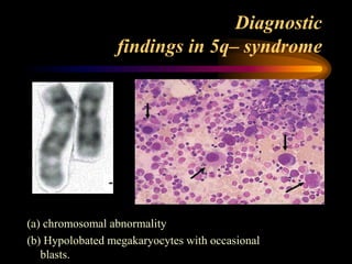
  • 35. Diagnostic findings in 5q– syndrome (a) chromosomal abnormality (b) Hypolobated megakaryocytes with occasional blasts.
  • 38. Myelodysplastic syndrome, unclassifiable Definition: • Is a subtype of MDS which initially lacks findings appropriate for classification into any other MDS category. • Three possible situations which qualify pateints for inclusion in this category.
  • 39. 1. Patients with the findings of (RCUD) or (RCMD) but with 1% blasts in the PB qualify for MDS-U. 2.Cases of MDS with Unilineage dysplasia which are associated with pancytopenia. In contrast. the RCUD category only allows for a single cytopenia or bi-cytoperna .
  • 40. 3. Patients with persistent cytopenia(s) with 1% or fewer blasts in the blood and fewer than 5% in the BM, unequivocal dysplasia in less than 10% of the cells in one or more myeloid lineages. and who have cytogenetic abnormalities considered as presumptive evidence of MDS.
  • 41. • MDS-U patients should be carefully followed for evidence of evolution to a more specific MDS type.
  • 42. Diagnosis Clinical features: • Approximately 20% of cases of MDS are detected incidentally • Majority of the remainder present with symptoms and signs of bone marrow failure, (notably fatigue due to anaemia in up to 80% and infections or bleeding in up to 20%).
  • 43. • Features of lymphadenopathy, splenomegaly and hepatomegaly are rarely found. • There is association between MDS and several rare disorders that seem to have an immunological basis, including neutrophilic dermatosis (Sweet syndrome), pyoderma gangrenosum and cutaneous vasculitis.
  • 44. Lab features: 1. Blood count • Anaemia is the predominant in most patients at presentation, Occurring as pancytopenia in 30 – 50% OR As bicytopenia in 20 – 30%. Isolated neutropenia or thrombocytopenia is rarer, in 5 – 10%. • Occasionally, the blood count is normal and the diagnosis is suggested by abnormal parameters that reflect aberrant morphology, such as RDW that canitself become a useful marker of MDS.
  • 45. 2.Peripheral blood morphology RBC While non- specific, but very helpful • Oval macrocytosis (typical) • Reticulocyte count low • Hypochromia (rare; in acquired HbH disease). • Dimorphic cells in sideroblastic anaemia (minority are hypochromic microcytic cells; Pappenheimer bodies by iron stain, basophilic stippling • Megaloblastoid erythroblasts (rare)
  • 46. A). Peripheral blood (dimorphic red cells with a population of macrocytes and a population of hypochromic microcytes • http://www.haematologica.org/content/96/6/ 789/F2.large.jpg
  • 47. • Peripheral blood from a patient with RAEB, ring sideroblasts and acquired haemoglobin H, showing a grossly dimorphic picture.
  • 48. WBC • Hypogranular • Agranular neutrophils highly specific for MDS. • Hypolobated neutrophils (psedo pelger- Huet= Acquired ) classically bilobed ( like spectacles) or even non-lobed (like dumb - bell) pathognomonic.
  • 49. Peripheral blood smear with a circulating blast and a pseudo Pelger-Huet cell
  • 50. • Monocytosis is present, in CMML and monocytes (often morphologically abnormal. While often reduced in MDS, • basophils and eosinophils might also be raised in the proliferative overlap syndromes. • Type 1( non granular) Type 2 ( granular) blasts may be found in all categories but if in significant numbers are more usually indicative of RAEB.
  • 52. 3.Bone marrow Aspiration • Bone marrow is hypercellular in majority of patients, • But can be normocellular • in 10 – 20% of cases, hypocellular.
  • 53. Dyserythropoiesis • Nuclear budding • Inter-nuclear bridging • Karyorrhexis • Binuclearity & Multinuclearity • Megaloblastoid maturation • Ringed sideroblast (iron staining should performed) • Vacuolation ( in sideroblastic anaemia)
  • 58. Perls stain from a patient with RARS showing ring sideroblasts
  • 59. Dysgranulopoiesis Quite difficult to appreciate • Nuclear hypolobulation (pseudo-Pelger Heut) • Hypogranularity • Hypersegmentation • prominent basophilic and eosinophilic differentiation • increased numbers of blasts may be present
  • 63. Dysmegakaryocytopoiesis • Hypolobulated micro-megakaryocyte • Non-lobulated nuclei in megakaryocyte of all sizes • Multiple, widely separated nuclei
  • 69. 4.Bone marrow Biopsy • Assessment of cellularity • Abnormal clustering of megakaryocytes ( micromegakaryocytes) that can be more easily detected by immunohistochemical staining. • BM fibrosis (Reticulin can increased). • ALIP (immature cells present in centre of marrow) - tends to correlate with the blast percentage - may signify propensity to leuk. Trans.
  • 71. ALIP
  • 73. 5.Cytogenetic abnormalities • Cytogenetic analysis of marrow samples plays an important role in the evaluation of MDS with regard to establishing clonality and determining prognosis. • Clonal abnormalities are observed in approximately 50% of primary MDS cases and in up to 90% of cases of secondary TR- MDS
  • 74. Cytogenetic abnormalities in MDS Abnormality Primary Therapy - related MDS (%) MDS (%) Complex karyotype 15 – 20 80 – 90 del(5q)/monosomy 5 15– 20 30– 40 del(7q)/monosomy 7 10– 15 40– 50 Trisomy 8 10– 15 10– 15 del(20q) 5– 10 – del(17p) < 5 – del(13q) < 5 – del(11q) < 5 –
  • 75. Recurrent chromosomal abnormalities that provide presumptive evidence of primary MDS Abnormality -7 or (7q) -5 or (5q) i(17q) or t(17p) -13 or (13q) (11q) (12p) or t(12p) (9q) idic(X)(q13) t(11;16)(q23;p13.3) t(3;21)(q26.2;q22.1) t(1;3)(p36.3;q21.2) t(2;11)(p21;q23) inv(3)(q21q26.2) t(6;9)(p23;q34) New practical application of 8 color flow cytometry 11-12th Feb 2014
  • 76. Monosomy 7 • is the second most common chromosomal abnormality. • lower median age compared with deletions of 5q. • severe refractory cytopenias, and tendency to life - threatening infections. • Monosomy7 confers a poor prognosis
  • 77. Cytogenetics and prognosis • Good risk – Normal, isoloted 5q-, isolated 20q-, -Y • Poor risk – Complex changes (> 3 abnormalities) – Chromosome 7 abnormalities • Intermediate risk – All other changes
  • 78. 6.Recurrent mutated genes in MDS Gene function Abnormal gene Cell surface receptor KIT , FMS , PDGFRB , GCSFR , MPL , FLT3 Signal transduction NRAS , JAK2 Transcription factor A ML1 , G ATA - 1 , P U.1 , C EBPA ,TP53 Epigenetic factor M LL , A TRX Protein degradation CBL Unknown function TET2
  • 79. Known molecular abnormalities in MDS Gene Type of anomaly Incidence (%) RAS (N or K) Point mutation (codon 12, 13 or 61) 10-30% P53 Point mutation or deletion of other allele 5 FMS (encodes M-CSF receptor) Point mutation (codon 969 or rarely 301) 5-10
  • 80. 6.Other i nvestigations • Immunophenotyping does not play a major role -low side - scatter, - reduced expression of normal myeloid markers, -and aberrant patterns of expression of other markers. -CD34 expression, and to a lesser degree CD117, often correlates with the blast percentage, while coexpression of CD7 is significant for conferring a worse prognosis.
  • 81. Analysis of the (mature) myeloid lineage in normal bone New practical application of 8 color flow cytometry 11-12th Feb 2014
  • 82. CD13-CD11b staining of maturing neutrophils New practical application of 8 color flow cytometry 11-12th Feb 2014
  • 83. Lineage infidelity markers on immature myeloid cells In approximately 40% of patients: lineage infidelity marker expression detectable on myeloid progenitor cells New practical application of 8 color flow cytometry 11-12th
  • 84. Flow Cytometric myeloid dysplasia: myeloid blasts granulocytes (maturing cells) monocytes increased percentage decreased myeloid/lymphoid ratio (<1) decreased/increased number as compared to lymphocytes abnormal granularity* abnormal granularity* abnormal granularity* abnormal expression of CD45 abnormal expression of CD45 abnormal expression of CD45 abnormal expression of CD34 abnormal CD11b/CD13 pattern abnormal expression of CD14 abnormal expression of CD117 abnormal CD16/CD13 pattern abnormal CD11b/HLA-DR pattern abnormal expression of CD13 abnormal expression of CD15 abnormal expression of CD13 abnormal expression of CD33 abnormal expression of CD33 abnormal expression of CD33 abnormal expression of HLA- DR expression of HLA-DR abnormal expression of CD36 expression of CD11b expression of CD34 abnormal expression of HLA-DR expression of CD15 asynchronous shift to the left expression of CD34 expression of lineage infidelity markers CD5, CD7, CD19 or CD56 expression of lineage infidelity markers CD5, CD7, CD19 expression of lineage infidelity markers CD5, CD7, CD19
  • 85. Flow Cytometric Scoring System (FCSS) score definition 0 no FC aberrancies in either subpopulation analyzed 1 - a single aberrancy in either granulocytes or monocytes 2  a single aberrancy in both granulocytes and monocytes or …  two or three aberrancies in either granulocytes and monocytes or …  expression of CD34 or lineage infidelity markers on either granulocytes or monocytes 3 - four or more aberrancies in either granulocytes and monocytes 4 - two or three aberrancies in both granulocytes and monocytes New practical application of 8 color flow cytometry 11-12th
  • 86. Flow Cytometric Scoring System (FCSS) score definition + 1  decreased myeloid/lymphoid ratio (<1)  normal percentage of myeloid blasts (<5%) with flow cytometric aberrancies + 2 - increased percentage of abnormal myeloid blasts (5-10%) + 3 - increased percentage of abnormal myeloid blasts (11-20%) + 4 - increased percentage of abnormal myeloid blasts (>20%) New practical application of 8 color flow cytometry 11-12th Total flow score: 0-1: normal-mild 2-3: moderate 4-9: severe
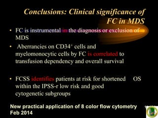
  • 87. Conclusions: Clinical significance of FC in MDS • FC is instrumental in the diagnosis or exclusion of MDS • Aberrancies on CD34+ cells and myelomonocytic cells by FC is correlated to transfusion dependency and overall survival • FCSS identifies patients at risk for shortened OS within the IPSS-r low risk and good cytogenetic subgroups New practical application of 8 color flow cytometry 11-12th Feb 2014
  • 88. • Granulocyte function tests to demonstrate defective phagocytosis, cell killing and motility. • Platelet function tests : reduced aggregation, prolonged bleeding time. • Haemoglobin electrophoresis, or HPLC, to detect HbH (raised in acquired HbH disease) and HbF (raised in juvenile myelomonocytic leukaemia). • Ferrokinetics to assess erythropoiesis → In effective erythropoiesis • Autoantibodies (found in up to 50% of CMML patients). • Serum protein electrophoresis to assess immunoglobulins and detect paraprotein.(↑ polyclonal, ↓ Igs, paraprotein) • Lymphocyte populations to detect altered numbers of T – cell subsets and natural killer cells.
  • 89. Diagnostic tool Diagnostic value Priority Peripheral blood smear • Evaluation of dysplasia in one or more cell lines Mandatory • Enumeration of blasts Bone marrow aspirate • Evaluation of dysplasia in one or more myeloid cell lines Mandatory • Enumeration of blasts • Enumeration of ring sideroblasts Bone marrow biopsy • Assessment of cellularity, CD34+ cells, and fibrosis Mandatory Cytogenetic analysis • Detection of acquired clonal chromosomal abnormalities that can allow a conclusive diagnosis and also prognostic assessment Mandatory FISH • Detection of targeted chromosomal abnormalities in interphase nuclei following failure of standard Gbanding Recommended Flow cytometry immunophenotyping • Detection of abnormalities in erythroid, immature myeloid, maturing granulocytes, monocytes, immature and mature lymphoid compartments Recommended SNP-array • Detection of chromosomal defects at a high resolution in combination with metaphase cytogenetics Suggested (likely to become a diagnostic tool in the near future) Mutation analysis of candidate genes* • Detection of somatic mutations that can allow a conclusive diagnosis and also reliable prognostic evaluation Suggested (likely to become a diagnostic tool in Diagnostic approach to MDS 2012
  • 90. Minimal diagnostic criteria in MDS A. Prerequisite Criteria • constant cytopenia in one or more cell lineages • exclusion of all other hematopoietic or non- hematopoietic disorders
  • 91. B. MDS-related (Decisive) Criteria • dysplasia in > 10% of all cells in one of the lineages, or > 15% ring sideroblasts (iron stain) • 5–19% blast cells in bone marrow smears • typical chromosomal abnormality (karyotyping or FISH)
  • 92. C. Co-criteria abnormal phenotype of bone marrow cells by flow cytometry molecular signs of a monoclonal cell population o HUMARA assay, gene profiling, point mutation analysis o markedly and persistently reduced colony- formation (CFU-assay)
  • 93. DIFFERENTIAL DIAGNOSIS OF MDS Non-neoplastic causes of myelodysplasia • Megaloblastic anemia • Toxic agents, i.e., heavy metals, acute alcohol intoxication • Drug effects - primarily anti-neoplastic • Congenital dyserythropoietic anemia • Chronic infectious disease • Acquired immunodeficiency syndrome (AIDS) •A.A •NH
  • 94. Neoplastic Diseases • Chronic myeloproliferative disease • Acute myeloid leukemia
  • 95. Marrow blast percentage: • < 5 0 • 5-10 0.5 • 11-20 1.5 • 21-30 2.0 Cytogentic fetures • Good prognosis 0 (–Y, 5q- , 20q-) • Intermediate prognosis 0.5 (+8, miscellaneous singleabnormality, double abnormalities) • Poor prognosis 1.0 (abnor. 7, complex- >3 abnor.) Cytopenias • None or one type 0 • 2 or 3 type 0.5 Myelodysplastic syndromes IPSS risk-based classification system
  • 96. Cytopenias defined as Haemoglobin < 10 g/dL, Neutrophils < 1.8 × 10 9 /L Platelets < 100 × 10 9 /L.
  • 99. Emerging new prognostic variables in MDS: 2012 • Bone marrow fibrosis • Co-morbidity score • New karyotyping subgroups (conventional/FISH/flow-FISH) • Newly identified mutations (TET2, EZH2, TP53, RUNX1, NRAS, ASXL1, ETV6, GNAS, SF3B1, and others) • IPSS-revise • Flow cytometric aberrancies (FCSS)
  • 102. Myelodysplastic syndrome (MDS) is very uncommen in children, accounting for less than 5% of all haematopoietic neoplasms in patients less than 14 years of age
  • 103. • Childhood MDS is recognized as an entity of its own in the current version of the WHO classifi- cation but excludes JMML, which is grouped within the MDS/MPD category. • MDS associated with Down syndrome, which previously accounted for up to 25% of paediatric MDS, is now grouped within a new entity of Down syndrome - related myeloid leukaemia.
  • 104. • Many of the morphologic ,immonophenotypic and genetic features observed in MDS in adults are also seen in childhood forms of the disease but there are some significant differences reported
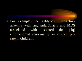
  • 105. • For example, the subtypes refractory anaemia with ring sideroblaets and MDS associated with isolated del (5q) chromosomal abnormality are exceedingly rare in children .
  • 106. Isolated anaemia, which is the major presenting rnamtestatton of refractory anaemia (RA) in adults, is uncommen in children, who are more likely to present with neutropenia and thrombocytopenia. In addition, hypocellularity of the BM is more commonly observed in childhood MDS than in older patients.
  • 107. • In contrast to adult MDS, there are no available studies that have investigated the prognostic significance of distinguishing RAEB-1 and RAEB-2 in children, but it is recommended that this distinction be made for future investigation. • Children with RAEB generally have relatively stable PB counts for weeks or months .
  • 108. • The term ‘ refractory cytopenia of childhood ’ is reserved for cases of MDS associated with persistent cytopenia and less than 5% blasts in the marrow and less than 2% blasts in peripheral blood.
  • 111. • About 75% of children with RCC show considerable hypocellularity of the bone marrow, making it difficult to differentiate from congenital bone marrow failure syndromes that can lead to secondary myelodysplasia in affected children.
  • 112. • These congenital syndromes include disorders such as Fanconi anaemia, dyskeratosis congenita, Schwachman – Diamond syndrome, amegakaryocytic thrombocytopenia, and pancytopenia with radioulnar synostosis.
  • 114. Myelodysplastic/ Myeloproliferative Neoplasms • Chronic myelomonocytic leukaemia (CMML) • Atypical chronic myeloid leukaemia (aCML) • Juvenile myelomonocytic leukaemia (JMML) • Myelodysplastic myeloproliferative neoplasm. Unclassifiable (MDS/MPN, U.)
  • 115. The WHO classification established a new diagnostic entity for those diseases that share features characteristic of both the myelodysplastic syndromes and the myeloproliferative neoplasms .
  • 116. CMML
  • 117. CMML • CMML constitutes 20 – 30% of cases of MDS. • The hallmark feature is peripheral blood monocytosis accompanied by morphological dysplasia of other lineages. • It has a male predominance and a median age of presentation of about 70 years (only 10% of CMML cases occur in individuals less than 60 years) • The etiology is largely unknown, therapy - related cases of CMML are very rare.
  • 118. Clinical features • Approximately half of patients have splenomegaly, hepatomegaly, at diagnosis. • Individuals with high monocyte counts may develop a maculopapular skin infiltration, gum infiltration, and monocytic pleural and pericardial effusions.
  • 119. • Lymphadenopathy is uncommon but when it occurs it may signal a more acute phase with infiltration of lymph nodes by myeloblasts. • Weight loss, fevers and night sweats may occur in symptomatic patients
  • 120. laboratory features • The majority of patients present with a leucocytosis. • That is often accompanied by mild anaemia and thrombocytopenia. (Reactive causes due to infection, notably TB, must be excluded) • The monocyte count must be greater than 1 × 1000 /cmm and is usually in the range 2 – 5 × 1000 /cmm, but may exceed 80 × 1000 /cmm.
  • 121. • The monocytes generally appear mature but may have agranular cytoplasm and/or abnormal nuclear lobulation (abnormal monocytes)*. • Evidence of dysgranulopoiesis is commonly present, including hypogranular and hypolobated neutrophils that are sometimes difficult to distinguish from dysplastic monocytes
  • 122. (A)-WBC is elevated with minimal dysplasia in the neutrophilis. (B)- A normal WBC with absolute monocytosis , nuetropenia and dysgranulopoiesis.
  • 123. • Mild basophilia is sometimes present. Eosinophils are usually normal or slightly increased in number, but in some cases eosinophilia may be striking*. • CMML with eosinophilia may be diagnosed when the criteria of CMML are present*. but in addition the eosinophil count in the PB is ≥1500 /cmm.
  • 124. • The BM is generally hypercellular, usually with striking granulocytic proliferation and invariably monocytic proliferation that can be distinguished using cytochemical studies. • Typical features of dysplasia can be identified in all three lineages in over 80% of patients
  • 125. • The blast cells + promonocytes should account for  5% of peripheral blood leucocytes & and  10% of nucleated marrow cells to give a diagnosis of CMML - 1. • If the blast/promonocyte count is  this but  20% in either the PB or BM, or if Auer rods are present, the diagnosis is CMML – 2
  • 126. • CMML-2 has worse prognosis and higher risk of transformation to AML, which is diagnosed when the blast/promonocyte count  20%.
  • 128. Marrow aspirate from a patient with CMML showing monocytes, promonocytes and blast cells.
  • 129. The folded nucluei and delicate nuclear chromatin can be appreciated among granulocytes.
  • 131. • Lysozyme used in conjunction with cyto- chemistry for CAE can also facilate the identifica tion of monocytic cells,which are lysozyme-positive but negative for CAE, in contrast with the granulocytrc precursor cells, which are positive for both.
  • 132. The monocytic component stained with naphthol-ASD- chloroacetate estrase raection combined with alpha naphthyl butytrate estrase (monocyte brown, nutrophil blue, dual staining cells have mixture of brown & blue.
  • 133. • Imunohistochemistry on tissue sections for the identification of monocytic cells is relatively insensitive as compared with cytochemistry or flow cytometry. • The most reliable markes ars are CD 68 and CD l63
  • 134. BM biobsy section stained with CD 163, note the postivity of the scattered monocytic cells as well as the strong positivity in the macrophage in the stain.
  • 135. Immunophenotyping may be helpful for identifying myelomonocytic populations • The PB and BM cells usually express CD33 and CD13, with variable expressionof CD14, CD68 and CD64. • but can also give prognostic information such as reduced CD14 expression indicating monocytic immaturity, aberrant expression of CD2 and CD56, and the CD34 positive cell percentage.
  • 136. Cytogenetic analysis is important for confirming clonality, although abnormalities are only found in 30 – 40% of cases,
  • 137. • Up to 40% of patients have point mutations of RAS genes. • Hypermethylation of the CDKN2B gene (which encodes the tumour suppressor p15INK4b), resulting in reduced expression,in about 50% of patients.. • Recently, the CBL gene, has been implicated in progressive CMML
  • 138. • Exclude certain chromosomal translocations, by cytogenetic and (PCR), that are indicative of alternative diagnoses. 1. BCR – ABL1 rearrangement (CML). 2.(eosinophilia - related disorders) associated with abnormalities of PDGFRA , PDGFRB and FGFR1. 3. Mutations of the JAK2 gene
  • 141. • Atypical chronic myeloid leukaemia is a poorly defined entity. • Importantly, it is negative for the BCR – ABL1 fusion gene of typical chronic myeloid leukaemia • aCML principally involving the neutrophil lineage.
  • 142. • Myeloid precursors often comprise more than 10% of cells but blasts are rarely more than 5%. • Dysgranulopoiesis is prominent but multilineage dysplasia is also seen.
  • 143. • Patients usually present with anaemia and/or thrombocytopenia that accompany the leucocytosis. • Splenomegaly is common and often causes symptoms because of its massive enlargement.
  • 145. • Cytogenetic abnormalities can be identified in 80% of cases although is none specific. • The outlook is generally poorer for atypical chronic myeloid leukaemia than for CMML, with a median survival of less than 20 months. .
  • 147. JMML
  • 148. • JMML is a clonal haemopoietic disorder of childhood characterized principally by proliferation of the granulocytic and mono cytic lineages. • comprising approximately 2 – 3% of all childhood leukaemias but 40% of childhood MDS.
  • 149. • The majority of cases occur in children under 3 years and twice as commonly in boys than girls. • There are associations with neurofi bromatosis type 1 and Noonan syndrome due to germline mutations in the NF1 and PTPN11 or KRAS genes respectively
  • 150. • There is marked in vitro hypersensitivity of myeloid progenitors to (granulocyte/macrophage colony - stimulating factor) that is a hallmark feature of JMML and suggestive of defective RAS – MAP kinase signalling that is often attributable to RAS mutations. • Monosomy 7 is the most common chromosomal abnormality, found in 25% of cases.
  • 151. • Clinically, most patients present with constitutional symptoms or evidence of infection • and are found to have marked hepatosplenomegaly • Lymphoid and tonsillar enlargement is also common.
  • 152. • Typically, there is a leucocytosis comprising neutrophils, • myeloid precursors and monocytes, with blasts constituting less than 5% of cells. The marrow is hypercellular and dysplastic features are minimal.
  • 154. • A marked increase in the synthesis of HbF is a recurrent finding that has poor prognostic implications. • The prognosis of JMML is variable with a median survival of 1 year. It is usually rapidly fatal without treatment, causing organ failure, especially respiratory failure due to leukaemic infiltration, while blast transformation occurs infrequently.
  • 159. • The precise nature of (RARS - T) is a controversial and unresolved issue. • These patients meet the criteria for RARS but also have persistently elevated platelet counts over 450 × 10 9 /L. • The majority (50 – 60% of cases) carry the V617F mutation of the JAK2 gene that is more commonly associated with myeloproliferative disorders.
  • 160. • Whether RARS - T represents a JAK2 - positive myeloproliferative neoplasm with acquired dysplastic features or, • Conversely, a form of MDS with an acquired proliferative mutation remains a question ? • For this reason, RARS - T exists as a provisional entity in the current version of the WHO classification.

Editor's Notes

  • #4: approximately 40% of patients will transform to AML
  • #10: MDS in adults is 3 – 4 per 100 000. However, this rate increases markedly with age, exceeding 30 per 100 000 for individuals over the age of 80 years.
  • #11: enzymes for the metabolism of carcinogens, proteins involved with oxidative stress,repair damaged DNA.
  • #12: alkylating agents,as cyclophosphamide, and topoisomerase II inhibitors,as etoposide, &amp;antimetabolite drugs
  • #121: however, the WBCs normal or slightly decreased with variable neutropenia and the disease resembles MDS., nearly one half of patients it is increased due not only to monocytosis but also to neutrophilia
  • #122: But in comparison to promonocytes (and monoblasts). Have denser chromatin, nuclear convolutions and folds, and a more greyish cytoplasm
  • #124: Patients in this category may have complications related to the degranulation the eosinophils. These &amp;quot;hypereosinophilic &amp;quot; cases of CMML may
  • #140: Blasts include myeloblasts, monoblasts and promonoblast.