SlideShare a Scribd company logo
MICROARRAY
MOHAMMAD SHAHAN
DNA Microarray
 DNA microarrays are microscope slides that are
printed with thousands of tiny spots in defined
positions, with each spot containing a known DNA
sequence or gene.
 These slides are referred to as gene chips or DNA
chips.
 DNA microarrays are solid supports, usually of glass
or silicon, upon which DNA is attached in an
organized pre-determined grid fashion.
 Each spot of DNA, called a probe, represents a
single gene.
 A microarray is a laboratory tool used to detect the
expression of thousands of genes at the same time.
Microarray
A basic protocol for a DNA microarray is as follows:
Isolate and purify mRNA from samples of
interest. Since we are interested in comparing
gene expression, one sample usually serves as
control, and another sample would be the
experiment (healthy vs. disease, etc)
Reverse transcribe and label the mRNA. In order
to detect the transcripts by hybridization, they need
to be labeled, and because starting material maybe
limited, an amplification step is also used.
Hybridize the labeled target to the microarray. This step
involves placing labeled cDNAs onto a DNA microarray where
it will hybridize to their synthetic complementary DNA probes
attached on the microarray. A series of washes are used to
remove non-bound sequences.
Scan the microarray and quantitate the signal. The
fluorescent tags on bound cDNA are excited by a laser
and the fluorescently labeled target sequences that bind to
a probe generate a signal. The total strength of the signal
depends upon the amount of target sample binding to the
probes present on that spot
Microarray
cDNA slide
fabrication
mRNA
preparation,
fluorescence
dye labeling,
gene
hybridization
robotic
spotting
green and red
fluorophores
excitation by
lasers
imaging using
optics
slide scanning
analog to
digital
conversion
using
image storage
and archiving.
GENRAL cDNA
MICROARRAY
WORKFLOW
Parameters affecting fabrication of microarrays.
Spot density and array geometry.
Probe density is defined as number of probes located on a given surface while
hybridised density is defined as the number of target located on a given surface
Hybridisation efficiency is defined as the ratio between hybridised and probe
density
Classification of microarray based on the
mode of preparation
1. The spotted array on glass: spotted arrays are arrays made on
poly-lysine coated glass microscope slides. This provides binding of
high-density DNA by using slotted pins. It allows fluorescent labeling
of the sample.
2. Self-assembled arrays: these are fiber optic arrays made by the
deposition of DNA synthesized on small polystyrene beads. The
beads are deposited on the etched ends of the array. Different DNA
can be synthesized on different beads and applying a mixture of
beads to the fiber optic cable will make a randomly assembled array.
3. In-situ synthesized arrays: these arrays are made by chemical
synthesis on a solid substrate. In the chemical synthesis, photolabile
protecting groups are combined with photolithography to perform the
action. These arrays are used in expression analysis, genotyping,
and sequencing. Affymetrix for instance produces microarrays
routinely with millions of probes on 1.28 cm2 surface. There is
almost no space between the spots and spotsizes are below 10 mm.
Microarray
Microarray
(A) Unmodified DNA
is randomly
immobilised to
surfaces meaning
that some DNA
strands can
participate in
hybridisation (I) while
other cannot (II).
(B) Immobilisation of
DNA using end
modifications (I) can
also result in intra
chain bonds (II).
(C) Molecular organisation of end modified probes
directly immobilised to the active groups on the solid
support (I) or displaced from the surface using a
linker (II) or a dendrimeric linker (III).
Immobilisation
of DNA to
surfaces.
Microarray
Microarray
 A grid alignment (also known as addressing or spot finding or gridding) is
one of the processing steps in microarray image analysis that registers a set
of unevenly spaced, parallel, and perpendicular lines (a template) with the
image content representing a two-dimensional (2D) array of spots.
 Grid alignment methods: Manual grid alignment, semiautomated grid
alignment and fully automated grid alignment.
Grid alignment
Foreground and background detection.
The outcome of grid alignment is an approximation of spot locations. The
next task is to identify pixels that belong to foreground (signal) of
expected spot shape and to background. This task involves image
segmentation and clustering.
Image segmentation is associated with the problem of partitioning an
image into spatially contiguous regions with similar properties (e.g., color
or texture)
image clustering refers to the problem of partitioning an image into
sets of pixels with similar properties (e.g., intensity, color, or texture) but
not necessarily connected.
(1) circular signal area outside of allowed location
and radius deviations,
(2) small signal with respect to background
(3) disconnected signal areas
(4) inconsistent intensity probability distributions.
 quality assurance of the microaray image is
the process of elimination of the grid cells with
unreliable microarray information. This is done
by running the screening algorithm which performs
four types of quality assurance.
Quality assurance
Normalization of microarray data is aimed to correct for
the systematic measurement errors and bias in the
observed data. The errors and bias may be introduced by
several factors such as difference in probe labeling,
concentration of target DNA/ RNA sequence, efficiency of
hybridization, instrumental noise due to scanners or
printers etc.
Classification of microarray based on the
types of probes used
1. DNA microarrays: DNA microarray is also known as gene chip, DNA chip,
or biochip. It either measures DNA or uses DNA as a part of its detection
system. There are four different types of DNA microarrays:
 cDNA microarrays
 oligo DNA microarrays –
 In this type, the spotted probes contains of short, chemically synthesized
sequences, 20-25 mers/gene.
 specificity is high and cross hybridization can be inhibited.
 BAC microarrays - A BAC clone chip is a DNA microarray whose probe is
a template amplified by PCR. The template is a genome region
incorporated into a comprehensive BAC (bacterial artificial chromosome)
clone which various research institutions used in decoding the genome
sequence of various organisms.
 SNP microarrays. - SNP array is a type of DNA microarray which is used
to detect polymorphisms within a population
Protein
Microarrays
Analytical
microarrays
Functional
microarrays
Reverse
phase
microarrays
Analytical microarrays
 Analytical microarrays are typically used to profile a
complex mixture of proteins in order to measure binding
affinities, specificities, and protein expression levels of
the proteins in the mixture.
 Antibody microarrays are the most common analytical
microarray
 In this technique, a library of antibodies, aptamers, or
affibodies is arrayed on a glass microscope slide. The
array is then probed with a protein solution.
Microarray
Functional Microarrays
 functional protein arrays are composed of
arrays containing full-length functional
proteins or protein domains.
 These protein chips are used to study the
biochemical activities of an entire proteome
in a single experiment.
 They are used to study numerous protein
interactions, such as protein–protein,
protein–DNA, protein–RNA, protein–
phospholipid, and protein–small molecule
interactions
Microarray
 In RPA, cells are isolated from
various tissues of interest and are
lysed. The lysate is arrayed onto a
nitrocellulose slide using a contact
pin microarrayer.
 The slides are then probed with
antibodies against the target protein
of interest, and the antibodies are
typically detected with
chemiluminescent, fluorescent, or
colorimetric assays
 Several such multiprotein samples
are spotted on the microarray, which
is then probed with a single target
molecule.
Reverse Phase Microarrays
An ideal surface for protein microarray fabrication has to be capable of
immobilizing proteins and preserving their three‐dimensional (3‐D) conformation.
Microarray
3. Tissue microarrays: tissue microarray paraffin blocks that are formed by
separating cylindrical tissue cores from various donors and embedding it into
a single microarray. This is mainly used in pathology.
4. Cellular microarrays: they are also called transfection microarrays or living-
cell-microarrays, and are used for screening large-scale chemical and
genomic libraries and systematically investigating the local cellular
microenvironment.
5. Chemical compound microarrays: this is used for drug screening and drug
discovery. This microarray has the capacity to identify and evaluate small
molecules and so it is more useful than the other technologies used in the
pharmaceutical industry.
6. Carbohydrate arrays: they are also called glycoarrays. Carbohydrate arrays
are used in screening proteomes that are carbohydrate binding. They can also
be utilized in calculating protein binding affinities and automization of solid-
support synthesis for glycans.
7. Phenotype microarrays: phenotype microarrays or PMs are mainly used in
drug development. They quantitatively measure thousands of cellular
phenotypes all at once. It is also used in functional genomics and toxicological
testing.
Microarray

More Related Content

PPTX
DNA microarray
PPTX
DNA Microarray introdution and application
PPT
Microarray Analysis
PDF
DNA Microarray notes.pdf
PPTX
Dna chip
PPTX
DNA microarray ppt
PPTX
Applications of microarray
PPTX
Microarray technology, biochip, DNA chip
DNA microarray
DNA Microarray introdution and application
Microarray Analysis
DNA Microarray notes.pdf
Dna chip
DNA microarray ppt
Applications of microarray
Microarray technology, biochip, DNA chip

What's hot (20)

PPTX
lambda cloning vector
PPTX
M13 phage
PPTX
Introduction to Microarray in Gene Expression studies
PPTX
Third Generation Sequencing
PPTX
DNA Sequencing- Sanger's Method
PPTX
Next generation sequencing
PPTX
Lectut btn-202-ppt-l23. labeling techniques for nucleic acids
PPTX
cohesive and blunt end ligation
PDF
Basic information of s1 nuclease
PPTX
Genome mapping
PDF
DNA Microarray notes.pdf
PPT
Analysis of gene expression
PDF
10 safety guidelines for recombinant dna research
PPT
CDNA Library preparation. ppt for Jamil sir
PDF
Animal viral vector
PPTX
Pyrosequencing
PPTX
shotgun sequncing
PDF
DNA Ligation
DOCX
Yeast artificial chromosomes
PPT
DNA sequencing- Maxam- Gilbert sequencing
lambda cloning vector
M13 phage
Introduction to Microarray in Gene Expression studies
Third Generation Sequencing
DNA Sequencing- Sanger's Method
Next generation sequencing
Lectut btn-202-ppt-l23. labeling techniques for nucleic acids
cohesive and blunt end ligation
Basic information of s1 nuclease
Genome mapping
DNA Microarray notes.pdf
Analysis of gene expression
10 safety guidelines for recombinant dna research
CDNA Library preparation. ppt for Jamil sir
Animal viral vector
Pyrosequencing
shotgun sequncing
DNA Ligation
Yeast artificial chromosomes
DNA sequencing- Maxam- Gilbert sequencing
Ad

Similar to Microarray (20)

PPTX
DNA Microarray.pptx, Protein Microarray.pptx
PPT
A DNA microarray (also commonly known as gene or genome chip
PPTX
microarrary
PPTX
Power Point lecture slides on DNA microarray
PPTX
dna microarray .pptx
PPTX
Miarray:A new Technology For Life Science
PPTX
DNA MICROARRAY
PPTX
DNA microarray
PPT
A comprehensive study of microarray
PPTX
Toxicogenomics: microarray
PDF
Microarray @ujjwal sirohi
PPTX
DNA MICROARRAY TECHNOLOGY FOR PRINCIPLE OF DRUG DISCOVERY
PPTX
MICROARRAY
PDF
DNA microarray.pdfghghvjjsjsjdhdhdhddhdhdjdhd
PPTX
Nucleic acid microarray.pptx
PPTX
Microarray: A high-throughput tool used to analyze gene expression
PPTX
Nucleic acid microarray
PPTX
Microarray technique
PPTX
Microarray by dr.prabhash
PPTX
Microarray-Technology.pptx
DNA Microarray.pptx, Protein Microarray.pptx
A DNA microarray (also commonly known as gene or genome chip
microarrary
Power Point lecture slides on DNA microarray
dna microarray .pptx
Miarray:A new Technology For Life Science
DNA MICROARRAY
DNA microarray
A comprehensive study of microarray
Toxicogenomics: microarray
Microarray @ujjwal sirohi
DNA MICROARRAY TECHNOLOGY FOR PRINCIPLE OF DRUG DISCOVERY
MICROARRAY
DNA microarray.pdfghghvjjsjsjdhdhdhddhdhdjdhd
Nucleic acid microarray.pptx
Microarray: A high-throughput tool used to analyze gene expression
Nucleic acid microarray
Microarray technique
Microarray by dr.prabhash
Microarray-Technology.pptx
Ad

More from MOHAMMADSHAHAN2 (11)

PPTX
Xylometazoline
PPTX
Plumicort
PPTX
PPTX
Kogenate fs
PPTX
PPTX
PPTX
PPTX
Epogen copy
PPTX
Cetrizine
PPTX
Alteplase
PPTX
Cefpodoxime
Xylometazoline
Plumicort
Kogenate fs
Epogen copy
Cetrizine
Alteplase
Cefpodoxime

Recently uploaded (20)

PPTX
INTRODUCTION TO EVS | Concept of sustainability
PPTX
Protein & Amino Acid Structures Levels of protein structure (primary, seconda...
PPTX
Production technology of seed spices,,,,
PDF
SEHH2274 Organic Chemistry Notes 1 Structure and Bonding.pdf
PDF
Unveiling a 36 billion solar mass black hole at the centre of the Cosmic Hors...
PPTX
2. Earth - The Living Planet earth and life
PDF
Crime Scene Investigation: A Guide for Law Enforcement (2013 Update)
PPTX
7. General Toxicologyfor clinical phrmacy.pptx
PPTX
ECG_Course_Presentation د.محمد صقران ppt
PDF
IFIT3 RNA-binding activity primores influenza A viruz infection and translati...
PPT
protein biochemistry.ppt for university classes
PPTX
Taita Taveta Laboratory Technician Workshop Presentation.pptx
PPTX
Vitamins & Minerals: Complete Guide to Functions, Food Sources, Deficiency Si...
PPTX
cpcsea ppt.pptxssssssssssssssjjdjdndndddd
PPT
The World of Physical Science, • Labs: Safety Simulation, Measurement Practice
PDF
HPLC-PPT.docx high performance liquid chromatography
PPTX
SCIENCE10 Q1 5 WK8 Evidence Supporting Plate Movement.pptx
PPTX
microscope-Lecturecjchchchchcuvuvhc.pptx
PDF
The scientific heritage No 166 (166) (2025)
PPT
POSITIONING IN OPERATION THEATRE ROOM.ppt
INTRODUCTION TO EVS | Concept of sustainability
Protein & Amino Acid Structures Levels of protein structure (primary, seconda...
Production technology of seed spices,,,,
SEHH2274 Organic Chemistry Notes 1 Structure and Bonding.pdf
Unveiling a 36 billion solar mass black hole at the centre of the Cosmic Hors...
2. Earth - The Living Planet earth and life
Crime Scene Investigation: A Guide for Law Enforcement (2013 Update)
7. General Toxicologyfor clinical phrmacy.pptx
ECG_Course_Presentation د.محمد صقران ppt
IFIT3 RNA-binding activity primores influenza A viruz infection and translati...
protein biochemistry.ppt for university classes
Taita Taveta Laboratory Technician Workshop Presentation.pptx
Vitamins & Minerals: Complete Guide to Functions, Food Sources, Deficiency Si...
cpcsea ppt.pptxssssssssssssssjjdjdndndddd
The World of Physical Science, • Labs: Safety Simulation, Measurement Practice
HPLC-PPT.docx high performance liquid chromatography
SCIENCE10 Q1 5 WK8 Evidence Supporting Plate Movement.pptx
microscope-Lecturecjchchchchcuvuvhc.pptx
The scientific heritage No 166 (166) (2025)
POSITIONING IN OPERATION THEATRE ROOM.ppt

Microarray

  • 2. DNA Microarray  DNA microarrays are microscope slides that are printed with thousands of tiny spots in defined positions, with each spot containing a known DNA sequence or gene.  These slides are referred to as gene chips or DNA chips.  DNA microarrays are solid supports, usually of glass or silicon, upon which DNA is attached in an organized pre-determined grid fashion.  Each spot of DNA, called a probe, represents a single gene.  A microarray is a laboratory tool used to detect the expression of thousands of genes at the same time.
  • 4. A basic protocol for a DNA microarray is as follows: Isolate and purify mRNA from samples of interest. Since we are interested in comparing gene expression, one sample usually serves as control, and another sample would be the experiment (healthy vs. disease, etc) Reverse transcribe and label the mRNA. In order to detect the transcripts by hybridization, they need to be labeled, and because starting material maybe limited, an amplification step is also used. Hybridize the labeled target to the microarray. This step involves placing labeled cDNAs onto a DNA microarray where it will hybridize to their synthetic complementary DNA probes attached on the microarray. A series of washes are used to remove non-bound sequences. Scan the microarray and quantitate the signal. The fluorescent tags on bound cDNA are excited by a laser and the fluorescently labeled target sequences that bind to a probe generate a signal. The total strength of the signal depends upon the amount of target sample binding to the probes present on that spot
  • 6. cDNA slide fabrication mRNA preparation, fluorescence dye labeling, gene hybridization robotic spotting green and red fluorophores excitation by lasers imaging using optics slide scanning analog to digital conversion using image storage and archiving. GENRAL cDNA MICROARRAY WORKFLOW
  • 7. Parameters affecting fabrication of microarrays. Spot density and array geometry. Probe density is defined as number of probes located on a given surface while hybridised density is defined as the number of target located on a given surface Hybridisation efficiency is defined as the ratio between hybridised and probe density
  • 8. Classification of microarray based on the mode of preparation 1. The spotted array on glass: spotted arrays are arrays made on poly-lysine coated glass microscope slides. This provides binding of high-density DNA by using slotted pins. It allows fluorescent labeling of the sample. 2. Self-assembled arrays: these are fiber optic arrays made by the deposition of DNA synthesized on small polystyrene beads. The beads are deposited on the etched ends of the array. Different DNA can be synthesized on different beads and applying a mixture of beads to the fiber optic cable will make a randomly assembled array. 3. In-situ synthesized arrays: these arrays are made by chemical synthesis on a solid substrate. In the chemical synthesis, photolabile protecting groups are combined with photolithography to perform the action. These arrays are used in expression analysis, genotyping, and sequencing. Affymetrix for instance produces microarrays routinely with millions of probes on 1.28 cm2 surface. There is almost no space between the spots and spotsizes are below 10 mm.
  • 11. (A) Unmodified DNA is randomly immobilised to surfaces meaning that some DNA strands can participate in hybridisation (I) while other cannot (II). (B) Immobilisation of DNA using end modifications (I) can also result in intra chain bonds (II). (C) Molecular organisation of end modified probes directly immobilised to the active groups on the solid support (I) or displaced from the surface using a linker (II) or a dendrimeric linker (III). Immobilisation of DNA to surfaces.
  • 14.  A grid alignment (also known as addressing or spot finding or gridding) is one of the processing steps in microarray image analysis that registers a set of unevenly spaced, parallel, and perpendicular lines (a template) with the image content representing a two-dimensional (2D) array of spots.  Grid alignment methods: Manual grid alignment, semiautomated grid alignment and fully automated grid alignment. Grid alignment
  • 15. Foreground and background detection. The outcome of grid alignment is an approximation of spot locations. The next task is to identify pixels that belong to foreground (signal) of expected spot shape and to background. This task involves image segmentation and clustering. Image segmentation is associated with the problem of partitioning an image into spatially contiguous regions with similar properties (e.g., color or texture) image clustering refers to the problem of partitioning an image into sets of pixels with similar properties (e.g., intensity, color, or texture) but not necessarily connected.
  • 16. (1) circular signal area outside of allowed location and radius deviations, (2) small signal with respect to background (3) disconnected signal areas (4) inconsistent intensity probability distributions.  quality assurance of the microaray image is the process of elimination of the grid cells with unreliable microarray information. This is done by running the screening algorithm which performs four types of quality assurance. Quality assurance Normalization of microarray data is aimed to correct for the systematic measurement errors and bias in the observed data. The errors and bias may be introduced by several factors such as difference in probe labeling, concentration of target DNA/ RNA sequence, efficiency of hybridization, instrumental noise due to scanners or printers etc.
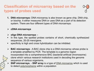
  • 17. Classification of microarray based on the types of probes used 1. DNA microarrays: DNA microarray is also known as gene chip, DNA chip, or biochip. It either measures DNA or uses DNA as a part of its detection system. There are four different types of DNA microarrays:  cDNA microarrays  oligo DNA microarrays –  In this type, the spotted probes contains of short, chemically synthesized sequences, 20-25 mers/gene.  specificity is high and cross hybridization can be inhibited.  BAC microarrays - A BAC clone chip is a DNA microarray whose probe is a template amplified by PCR. The template is a genome region incorporated into a comprehensive BAC (bacterial artificial chromosome) clone which various research institutions used in decoding the genome sequence of various organisms.  SNP microarrays. - SNP array is a type of DNA microarray which is used to detect polymorphisms within a population
  • 19. Analytical microarrays  Analytical microarrays are typically used to profile a complex mixture of proteins in order to measure binding affinities, specificities, and protein expression levels of the proteins in the mixture.  Antibody microarrays are the most common analytical microarray  In this technique, a library of antibodies, aptamers, or affibodies is arrayed on a glass microscope slide. The array is then probed with a protein solution.
  • 21. Functional Microarrays  functional protein arrays are composed of arrays containing full-length functional proteins or protein domains.  These protein chips are used to study the biochemical activities of an entire proteome in a single experiment.  They are used to study numerous protein interactions, such as protein–protein, protein–DNA, protein–RNA, protein– phospholipid, and protein–small molecule interactions
  • 23.  In RPA, cells are isolated from various tissues of interest and are lysed. The lysate is arrayed onto a nitrocellulose slide using a contact pin microarrayer.  The slides are then probed with antibodies against the target protein of interest, and the antibodies are typically detected with chemiluminescent, fluorescent, or colorimetric assays  Several such multiprotein samples are spotted on the microarray, which is then probed with a single target molecule. Reverse Phase Microarrays
  • 25. An ideal surface for protein microarray fabrication has to be capable of immobilizing proteins and preserving their three‐dimensional (3‐D) conformation.
  • 27. 3. Tissue microarrays: tissue microarray paraffin blocks that are formed by separating cylindrical tissue cores from various donors and embedding it into a single microarray. This is mainly used in pathology. 4. Cellular microarrays: they are also called transfection microarrays or living- cell-microarrays, and are used for screening large-scale chemical and genomic libraries and systematically investigating the local cellular microenvironment. 5. Chemical compound microarrays: this is used for drug screening and drug discovery. This microarray has the capacity to identify and evaluate small molecules and so it is more useful than the other technologies used in the pharmaceutical industry. 6. Carbohydrate arrays: they are also called glycoarrays. Carbohydrate arrays are used in screening proteomes that are carbohydrate binding. They can also be utilized in calculating protein binding affinities and automization of solid- support synthesis for glycans. 7. Phenotype microarrays: phenotype microarrays or PMs are mainly used in drug development. They quantitatively measure thousands of cellular phenotypes all at once. It is also used in functional genomics and toxicological testing.