SlideShare a Scribd company logo
Neuroanatomy
Lecture.4
Midbrain
GrossAppearanceGrossAppearance
of Midbrain:of Midbrain:
• connectstheponsand
cerebellum with the
forebrain.
• Itslong axisascends
through theopening in the
tentorium cerebelli.
• Themidbrain istraversed
by anarrow channel, the
cerebral aqueduct,
which isfilled with
cerebrospinal fluid
posterior
surface
1. Four colliculi
Thesearerounded
eminencesthat are
divided by avertical and
atransversegrooveinto :
• Superior colliculi :
arecentersfor visual
reflexes
• Inferior colliculi :
arelower auditory
centers.
2. Trochlear nerves:
emergeIn the
midlinebelow the
inferior colliculi,
(Theseareslender cranial
nervesthat wind around
thelateral aspect of the
midbrain to enter the
lateral wall of the
cavernoussinus).
• On thelateral aspect of
themidbrain,
3. Superior brachium
passesfrom thesuperior
colliculusto thelateral
geniculatebody and the
optic tract.
4. Inferior brachium
connectstheinferior
colliculusto themedial
geniculatebody.
Anterior aspect
1. thereisadeep depression
in themidline, called :
Interpeduncular fossa,
2. Thisdepression is
bounded on either sideby
the:
Cruscerebri.
Many small blood vessels
perforatethefloor of the
interpeduncular fossa, and
thisregion istermed the:
Posterior
perforated
substance
3. Theocculomotor nerve
emergesfrom agroove
on themedial sideof
thecruscerebri and
passesforward in the
lateral wall of the
cavernoussinus.
Arterial supply:
issupplied by:
1. Posterior cerebral
2. Superior cerebellar
3. Basilar arteries.
Venousdrainage:
into thebasal or great
cerebral veins
Internal StructureOf
Midbrain
Level Inferior colliculi
Cavity Cerebral aqueduct
Nuclei
Inferior colliculus, Substantianigra,
Trochlear nucleus, Mesencephalic
nuclei of cranial nerveV
MotorTracts
Corticospinal and corticonuclear tracts,
Temporopontine, Frontopontine,
Medial longitudinal fasciculus,
Sensory Tracts
Lateral, trigeminal, spinal, and medial
lemnisci; decussation of superior
cerebellar peduncles
Nuclie:
4. Mesencephalic nuclei of
cranial nerveV
1. Inferior colliculus,
2. Trochlear
nucleus,
3. Substantia
nigra,
Motor Tracts:
1. Temporo-
pontine
2. Corticospinal &
corticonuclear
3. Frontopontine,
4. Medial longitudinal
fasciculus
Sensory tracts
1. Lemnisci (Lmn.)
Lateral Lmn.
Trigeminal Lmn
Spinal Lmn.
Medial Lmn.
2. Decussation of2. Decussation of
superior cerebellarsuperior cerebellar
pedunclespeduncles
Cerebral aqueduct
Level Superior colliculi
Cavity Cerebral aqueduct
Nuclei
Superior colliculus, substantianigra,
Oculomotor nucleus, Edinger-Westphal
nucleus, red nucleus, Mesencephalic
nucleusof cranial nerveV
MotorTracts
Corticospinal and corticonuclear tracts,
temporopontine, frontopontine, medial
longitudinal fasciculus, decussation of
rubrospinal tract
Sensory Tracts Trigeminal, spinal, and medial lemnisci
Cerebral aqueduct
Nuclie:
1. Superior colliculus,
2. Mesencephalic nucleusof
cranial nerveV
2. Oculomotor nucleus,
3. Edinger-Westphal nucleus,
4. Red nucleus
5. Substantianigra,
Motor Tracts:
1. Temporo-
pontine
2. Corticospinal &
corticonuclear
3. Frontopontine,
5. Medial longitudinal
fasciculus
4. Decussation of rubrospinal tract
Sensory tracts
Lemnisci (Lmn.)
Trigeminal Lmn
Spinal Lmn.
Medial Lmn.
Clinical Notes
Clinical Significance of the Midbrain
• Themidbrain formstheupper end of thenarrow stalk of the
brain or brainstem. Asit ascendsout of theposterior cranial
fossathrough therelatively small rigid opening in the
tentorium cerebelli, it isvulnerableto traumatic injury.
• It possessestwo important cranial nervenuclei (oculomotor
and trochlear), reflex centers(thecolliculi), and thered
nucleusand substantianigra, which greatly influencemotor
function, and themidbrain servesasaconduit for many
important ascending and descending tracts.
• Asin other partsof thebrainstem, it isasitefor tumors,
hemorrhage, or infarcts that will produceawidevariety of
symptomsand signs.
1. Trauma to the Midbrain
• asudden lateral movement of thehead could result
in thecerebral pedunclesimpinging against the
sharp rigid freeedgeof thetentorium cerebelli.
• Sudden movementsof thehead resulting from
traumacausedifferent regionsof thebrain to move
at different velocities relativeto oneanother. For
example, thelargeanatomical unit, theforebrain,
may moveat adifferent velocity from theremainder
of thebrain, such asthecerebellum. Thiswill result
in themidbrain being bent, stretched, twisted, or
torn.
• Involvement of the oculomotornucleus will
produceipsilateral paralysis of thelevator
palpebraesuperioris; thesuperior, inferior, and
medial recti muscles; and theinferior oblique
muscle.
• Malfunction of the parasympathetic nucleus
of the oculomotornerve producesadilated
pupil that isinsensitiveto light and doesnot
constrict on accommodation.
• Involvement of the trochlearnucleus will
producecontralateral paralysis of thesuperior
obliquemuscleof theeyeball.
2. Blockage of the Cerebral Aqueduct
Thecavity of themidbrain, thecerebral
aqueduct, isoneof thenarrower
partsof theventricular system.
• In congenital hydrocephalus,
thecerebral aqueduct may be
blocked or replaced by numerous
small tubular passagesthat are
insufficient for thenormal flow of cerebrospinal fluid.
• When thecerebral aqueduct isblocked, theaccumulating
cerebrospinal fluid within thethird and lateral ventricles
produceslesionsin themidbrain.
• Thepresenceof theoculomotor and trochlear nervenuclei,
together with theimportant descending corticospinal and
corticonuclear tracts, will providesymptomsand signsthat
arehelpful in accurately localizing alesion in thebrainstem.
3. VascularLesions of the Midbrain
A. Weber Syndrome
• which iscommonly produced by
occlusion of abranch of theposterior
cerebral artery that suppliesthe
midbrain, resultsin thenecrosis
of brain tissueinvolving
oculomotor nerveand thecruscerebri.
• Thereisipsilateral ophthalmoplegiaand contralateral
paralysisof thelower part of theface, thetongue, and the
arm and leg. Theeyeball isdeviated laterally becauseof
theparalysisof themedial rectusmuscle; thereisdrooping
(ptosis) of theupper lid, and thepupil isdilated and fixed
to light and accommodation.
B. Benedikt Syndrome
• issimilar to Weber
syndrome, but thenecrosis
involvesthemedial
lemniscusand red nucleus,
• producing contralateral
hemianesthesiaand
involuntary movementsof
thelimbsof theopposite
side.
Cerebellum
DefinitionDefinition:
• The trilobed structure of the brain, lying posterior 
to the pons and medulla oblongata and inferior to 
occipital lobes of the cerebral hemispheres, thusit
liesin theposterior cranial fossa. 
• Responsible for the regulation and coordination of
 complex voluntary muscular movementsand the
maintainenceof postureand balance
GrossAppearanceof theCerebellum
• situated in theposterior cranial fossa
• covered superiorly by the tentorium cerebelli
• liesposterior to thefourth ventricle, thepons, and
themedullaoblongata
• issomewhat ovoid in shapeand constricted in its
median part.
It consistsof:
1. two cerebellar hemi-
spheres
2. Vermis: joining both hemi-
spheres.
Connected to posterior
aspect of thebrainstem by
threesymmetrical bundles
of nervefiberscalled the:
1.Superior cerebellar
peduncle
2.Middlecerebellar
peduncle
3.inferior cerebellar
peduncle
Thecerebellum is
divided into three
main lobes:
1. Anteriorlobe :
may beseen on the
superior surfaceof
thecerebellum and is
separated from the
middlelobeby awide
V-shaped fissure
called theprimary
fissure.
2. Middlelobe:
(sometimescalled the
posterior lobe), which is
thelargest part of the
cerebellum, issituated
between theprimary and
posterolateral fissures.
• Flocculonodular lobe:
• issituated posterior to
theposterolateral
fissure.
• Formed by two flocculi
and thenodule
Inferior veiw
Superior veiw
Tonsils
• Are roughly spherical
lobuleson theinferior
aspect of posterior lobe.
• Thetonsil may bedisplaced
down through theforamen
magnum in conditionsof
severeraised intracranial
pressureor in congenital
malformations
• horizontal fissure:
that isfound along the
margin of thecerebellum
separatesthesuperior
from theinferior surfaces.
The vermis
• consistsof ;
A. Superior part
B. Inferior part
• SuperiorVermis
liesbetween superior
medullary velum & primary
fissure
• Iscomposed of:
1. Lingula
2. Central lobule
3. Culmuen
• InferiorVermis
liesbetween primary
fissureand postero-
lateral fissure, and
consistsof :
1. Declive
2. Folium
3. Tuber
4. Pyramid
5. Uvula
6. Nodule
longitudinal division:
1. Vermis(medial zone)
2. Paravermal Region
( Intermediatezone)
3. Cerebellar Hemisphere:
(Lateral zone)
Anatomically
Transverseplane
Longitudinal plane
Anterior lobe
Vermis
Posterior lobe
Paravermis
Flocculonodular
Hemisphere
Functionally
Spino-
cerebellum
Cerebro-
cerebellum
Vestibulo-
cerebellum
Phylogenetic
Paleo-
cerebellum
Neo-
cerebellum
Archi-
cerebellum
Functionally:
• Thevestibulocerebellum
(correspondsbest with theflocculonodular lobe) has
reciprocal connectionswith vestibular and reticular nuclei
and playsarolein control of body equilibrium and eye
movement.
• Thespinocerebellum
(correspondsbest to theanterior lobe) hasreciprocal
connectionswith thespinal cord and playsarolein
control of muscletoneaswell asaxial and limb
movements, such asthoseused in walking and swimming.
• Thecerebrocerebellum or pontocerebellum
(correspondsbest to theposterior lobe) hasreciprocal
connectionswith thecerebral cortex and playsarolein
planning and initiation of movements, aswell asthe
regulation of discretelimb movements.
Phylogenetically:
1. Thearchicerebellum:
theoldest zone, correspondsto theflocculonodular
lobe.
2. Thepaleocerebellum,:
of morerecent phylogenetic development than the
archicerebellum, correspondsto theanterior lobe
and asmall part of theposterior lobe.
3. Theneocerebellum:
themost recent phylogenetically, correspondsto
theposterior lobe.
Functional Areas of the CerebellarCortex
• cerebellar cortex isdivided into threefunctional areas.
• Thecortex of thevermis:
influencesthemovementsof thelong axisof thebody,
namely, theneck, theshoulders, thethorax, theabdomen,
and thehips.
• Intermediatezoneof thecerebellar hemisphere: This
areahasbeen shown to control themusclesof the limbs,
especially thehandsand feet.
• Lateral zoneof each cerebellar hemisphere: Appears
to beconcerned with theplanning of sequential movements
of theentirebody and isinvolved with theconscious
assessment of movement errors.
Arterial supply of The cerebellum is
by:
1. Superior cerebellar
2. Anterior inferior cerebellar,
3. Posterior inferior cerebellar
Venous drainage
by veinsthat empty
into the
• Great cerebral vein
• Venoussinuses.
IntracerebellarNuclei
• Four massesof gray matter areembedded in
thewhitematter of thecerebellum on each
sideof themidline. From lateral to medial,
thesenuclei are:
1. Dentatenucleus,
2. Emboliform nucleus,
3. Globosenucleus,
4. Fastigial nucleus.
Fistugial nucleus Globosenucleus
Emboliform nucleus
Dentatenucleus
4th
Ventricle
Pons
Afferent Cerebellar Pathways
Information regarding theinitiation of
movement in thecerebral cortex isprobably
transmitted to thecerebellum so that the
movement can bemonitored and appropriate
adjustmentsin thevoluntary muscleactivity
can bemade.
1. CerebellarAfferent Fibers From
the Cerebral Cortex
Pathway Function Origin Destination
1.1.
CortiCo-CortiCo-
pontoponto
CerebellarCerebellar
Conveyscontrol
signalsfrom
cerebral cortex
Frontal, parietal,
temporal, and
occipital lobes
Viapontine
nuclei to
cerebellar cortex
2.2.
Cerebro-Cerebro-
olivo-olivo-
CerebellarCerebellar
Conveyscontrol
signalsfrom
cerebral cortex
Frontal, parietal,
temporal, and
occipital lobes
Viainferior
olivary nuclei to
cerebellar cortex
3.3.
Cerebro-Cerebro-
retiCulo-retiCulo-
CerebellarCerebellar
Conveyscontrol
signalsfrom
cerebral cortex
Sensorimotor
areas
Viareticular
formation to
cerebellar cortex
Corticopontocerebellar pathway
Corticoreticulocerebellar pathway
Cortico-olivocerebellar pathway
Reticular formation
Pontine
nuclie
Inferior olivary nucleus
• Thespinal cord sendsinformation to thecerebellum from
somatosensory receptorsby threepathways:
(1) theanterior spinocerebellar tract:
isfound at all segmentsof thespinal cord, and itsfibers
convey musclejoint information from theupper and lower
limbs
(2) theposterior spinocerebellar tract:
receivesmusclejoint information from thetrunk and
lower limbs.
(3) thecuneocerebellar tract:
receivesmusclejoint information from theupper limb and
upper part of thethorax
2. CerebellarAfferent Fibers
From Spinal Cord
3. CerebellarAfferent Fibers From
the
VestibularNerve
• Thevestibular nervereceives
information from theinner ear
concerning:
A. Motion from thesemicircular canals
B. position relativeto gravity from:
Utricle
Saccule.
4. OtherAfferent Fibers
• In addition, thecerebellum receivessmall
bundlesof afferent fibersfrom:
1. thered nucleus
2. thetectum.
Superior cerebellar peduncle
Inferior cerebellar peduncle
Cerebellum
Inferior cerebellar peduncle
Medulla
Anterior spinocerebellar tract
(minority of fibers)
Vestibular nuclie
Dentatenucleus
Nucleuscunatus
Vestibular nucleus
Posterior spinocerebellar tract
Anterior spinocerebellar tract
(majority of fibers)
TheEfferent Cerebellar Pathways
Pathway Function Origin Destination
Globose-Globose-
emboliformemboliform
-rubral-rubral
Influences
ipsilateral
motor
activity
Globoseand
emboliform
nuclei
contralateral
red nucleus,
then via
crossed
rubrospinal
tract to
ipsilateral
motor
neuronsin
spinal cord
Pathway Function Origin Destination
Dento-Dento-
thalamicthalamic
Influences
ipsilateral
motor
activity
Dentate
nucleus
contralateral ventro-
lateral nucleusof
thalamus,
contralateral motor
cerebral cortex;
corticospinal tract
crossesmidlineand
controlsipsilateral
motor neuronsin
spinal cord
Pathway Function Origin Destination
fastiGialfastiGial
vestibularvestibular
Influences
ipsilateral
extensor
muscle
tone
Fastigial
nucleus
Mainly to ipsilateral
and to contralateral
lateral vestibular
nuclei; vestibulo-
spinal tract to
ipsilateral motor
neuronsin spinal
cord
Pathway Function Origin Destination
fastiGialfastiGial
reticularreticular
Influences
ipsilateral
muscle
tone
Fastigial
nucleus
neuronsof reticular
formation; reticulo-
spinal tract to ipsi-
lateral motor neurons
to spinal cord
CLINICAL
NOTES
General Considerations:
• Each cerebellar hemisphereisconnected by nervous
pathwaysprincipally with thesamesideof thebody; thus, a
lesion in onecerebellar hemispheregivesriseto signsand
symptomsthat arelimited to thesamesideof thebody.
• Theessential function of thecerebellum isto coordinate, by
synergistic action, all reflex and voluntary muscular
activity. Thus, it graduatesand harmonizesmuscletoneand
maintainsnormal body posture. It permitsvoluntary
movements, such aswalking, to takeplacesmoothly with
precision and economy of effort.
• It must beunderstood that although thecerebellum playsan
important rolein skeletal muscleactivity, it is not ableto
initiatemusclemovement.
Characteristic symptoms and signs of
cerebellardysfunction:
1.hypotonia:
Themusclesloseresilienceto palpation. Thereis
diminished resistanceto passivemovementsof
joints. Shaking thelimb producesexcessive
movementsat theterminal joints. Thecondition is
attributableto lossof cerebellar influenceon the
simplestretch reflex.
2. Postural Changes and Alteration of Gait
• Thehead isoften rotated and flexed, and the
shoulder on thesideof thelesion islower than on
thenormal side.
• Thepatient assumesawidebasewhen heor she
standsand isoften stiff legged to compensatefor
lossof muscletone.
• When theindividual walks, staggerstoward the
affected side.
3. Disturbancesof Voluntary Movement (Ataxia)
Themusclescontract irregularly and weakly.
• Tremoroccurswhen finemovements, such as
buttoning clothes, writing, and shaving, are
attempted. Musclegroupsfail to work harmon-
iously, and thereisdecomposition of movement.
• Testsfor tremor :
1. Asking thepatient to touch thetip of thenosewith
theindex finger, thefinger either passesthenose
(past-pointing) or hitsthenose.
2. asking thepatient to placetheheel of onefoot on
theshin of theoppositeleg, it will either hit the
shin or not.
4. Dysdiadochokinesia:
inability to perform alternating movementsregularly
and rapidly. Ask thepatient to pronateand supinate
theforearmsrapidly. On thesideof thecerebellar
lesion, themovementsareslow, jerky, and
incomplete.
5. Disturbances of Reflexes
• Movement produced by tendon reflexestendsto
continuefor alonger period of timethan normal,
e.g. pendular kneejerk, for example, occurs
following tapping of thepatellar tendon.
6. Disturbancesof Ocular Movement:
• Nystagmus, which isan ataxia(incoordination) of
theocular muscles, isarhythmical oscillation of the
eyes. It ismoreeasily demonstrated when theeyes
aredeviated in ahorizontal direction.
7. Disordersof Speech:
Dysarthriaoccursin cerebellar diseasebecauseof
ataxia(incoordination) of themusclesof thelarynx.
Articulation isjerky, and thesyllablesoften are
separated from oneanother. Speech tendsto be
explosive, and thesyllablesoften areslurred.
CerebellarSyndromes
1. Vermis Syndrome:
• Themost common causeof vermissyndromeisa
medulloblastomaof thevermisin children.
• Involvement of theflocculonodular lobe resultsin
signsand symptomsrelated to thevestibular system.
• Sincethevermisisunpaired and influencesmidline
structures, muscleincoordination involvesthehead
and trunk and not thelimbs.
• Thereisatendency to fall forward or backward.
Thereisdifficulty in holding thehead steady and in
an upright position. Therealso may be difficulty in
holding thetrunk erect.
2. CerebellarHemisphere Syndrome:
• Tumorsof onecerebellar hemispheremay bethecause
of cerebellar hemispheresyndrome.
• Thesymptomsand signsareusually unilateral and
involvemuscleson thesideof thediseased cerebellar
hemisphere.
• Movementsof thelimbs, especially thearms, are
disturbed. Swaying and falling to thesideof thelesion
often occur.
• Dysarthriaand nystagmusarealso common findings.
• Disordersof thelateral part of thecerebellar
hemispheresproducedelaysin initiating movements
and inability to moveall limb segmentstogether in a
coordinated manner but show atendency to moveone
joint at atime.
Thanks

More Related Content

PPTX
FORAMEN MAGNUM ANATOMY.pptx
PPTX
Anatomy of brainstem
PDF
Anatomy of Cerebellum(Part- 2)
PPTX
Medulla oblongata
PPTX
Commissural fibres
PPTX
Neural circuit of cerebellum,
PPTX
white fibres of cns modified
PPTX
Detailed Anatomy of Medulla Oblongata ppt
FORAMEN MAGNUM ANATOMY.pptx
Anatomy of brainstem
Anatomy of Cerebellum(Part- 2)
Medulla oblongata
Commissural fibres
Neural circuit of cerebellum,
white fibres of cns modified
Detailed Anatomy of Medulla Oblongata ppt

What's hot (20)

PPTX
Internal capsule
PPTX
hippocampal formation
PDF
Cranial cavity part 1
PPTX
Cerebellum
PPTX
Pons cross sectional anatomy - Dr Sameep Koshti (consultant Neurosurgeon)
PPTX
PPTX
Corpus callosum with disconnection syndromes
PPTX
Functional cortical area & white matter of cerebrum
PPTX
HYPOTHALAMUS.pptx
PPT
Diencephalon
PDF
Ventricles of the brain
PPT
Blood supply to brain
PPTX
Cranial meninges
PPTX
Cutaneous innervation of lower limb
PPTX
Functional areas of the brain
PPTX
ICA anatomy
PPTX
Cervical sympathetic trunks ANATOMY
PPT
Internal capsule
PPTX
Limbic system
Internal capsule
hippocampal formation
Cranial cavity part 1
Cerebellum
Pons cross sectional anatomy - Dr Sameep Koshti (consultant Neurosurgeon)
Corpus callosum with disconnection syndromes
Functional cortical area & white matter of cerebrum
HYPOTHALAMUS.pptx
Diencephalon
Ventricles of the brain
Blood supply to brain
Cranial meninges
Cutaneous innervation of lower limb
Functional areas of the brain
ICA anatomy
Cervical sympathetic trunks ANATOMY
Internal capsule
Limbic system
Ad

Viewers also liked (18)

PPTX
The Forebrain
PPTX
The Hindbrain And Midbrain
POT
Brainstem demo neww
PDF
Anatomy of Midbrain and pons
PPTX
The brainstem
PPTX
Midbrain-Neuroradiology
PPT
Mid brain by DR.TATHEER
PPT
MIDBRAIN basic anatomy and applied aspects.
PPTX
Anatomy of pupillary pathways
PPT
Localization in neurology 1
PPT
Anatomy of midbrain & pons
PPT
Maxillary Incisors
PPT
Brain stem
PPT
MIDBRAIN ANATOMY
PPT
Anatomy Lect 4 Neuroanatomy
PPTX
MRI SECTIONAL ANATOMY OF BRAIN
PDF
Brainstem Lesions
PPT
localization of stroke, CVS, stroke, for post graduates
The Forebrain
The Hindbrain And Midbrain
Brainstem demo neww
Anatomy of Midbrain and pons
The brainstem
Midbrain-Neuroradiology
Mid brain by DR.TATHEER
MIDBRAIN basic anatomy and applied aspects.
Anatomy of pupillary pathways
Localization in neurology 1
Anatomy of midbrain & pons
Maxillary Incisors
Brain stem
MIDBRAIN ANATOMY
Anatomy Lect 4 Neuroanatomy
MRI SECTIONAL ANATOMY OF BRAIN
Brainstem Lesions
localization of stroke, CVS, stroke, for post graduates
Ad

Similar to Midbrain And Cerebellum (20)

PPTX
Cerebellum & Brainstem by Dr.Timothy Cheptarei.pptx
PPTX
Pons and medulla its anatomy and properties.pptx
PPTX
brainstem _ emphasis on Longitudinal fibres-1.pptx
PPTX
Midbrain
PPTX
BRAIN STEM/ pions/medulla oblongata/midbrain
PPTX
BRAINSTEM-ANATOMICAL DETAILS WITH EXPLANATIOON.pptx
PDF
Brain stem.pdf
PPTX
Anatomy of brainstem and its clinical significance
PDF
5. Brainstem (2): Pons & Midbrain
PPTX
Brainstem stroke syndromes ppt
PPTX
anatomy of central nervous system
PDF
Neuro_6-2M_2P_3NS_2020(2).pdf
PPTX
BRAINSTEM-SUMMARY WITH CLINICAL IMPORTANCE.pptx
PPTX
Mid brain
PPTX
Cerebellum and basal ganglia
PDF
MID BRAIN AND PONS of the brain -CNS mbbs year 1
PDF
Brainstem
PPTX
Cerebellum ibms
PPTX
FINAL--PARTS OF BRAIN.pptx
PPTX
Lecture of midbrain
Cerebellum & Brainstem by Dr.Timothy Cheptarei.pptx
Pons and medulla its anatomy and properties.pptx
brainstem _ emphasis on Longitudinal fibres-1.pptx
Midbrain
BRAIN STEM/ pions/medulla oblongata/midbrain
BRAINSTEM-ANATOMICAL DETAILS WITH EXPLANATIOON.pptx
Brain stem.pdf
Anatomy of brainstem and its clinical significance
5. Brainstem (2): Pons & Midbrain
Brainstem stroke syndromes ppt
anatomy of central nervous system
Neuro_6-2M_2P_3NS_2020(2).pdf
BRAINSTEM-SUMMARY WITH CLINICAL IMPORTANCE.pptx
Mid brain
Cerebellum and basal ganglia
MID BRAIN AND PONS of the brain -CNS mbbs year 1
Brainstem
Cerebellum ibms
FINAL--PARTS OF BRAIN.pptx
Lecture of midbrain

More from Dr.Faris Muhammed (15)

PPTX
Lymphatic system 2022
PPTX
The back anatomy
PPTX
Breast anatomy
PPTX
Thyroid gland anatomy part 2
PPTX
Thyroid gland anatomy part1
PPTX
The Anatomy Of The Liver & The biliary system
PPT
PPT
Basal Ganglia, Reticular Formation, Limbic System
PPT
Diencephalon
PPT
PPT
Ascending & Descending Tracts Of The Spinal Cord
PPT
Ascending & Descending Tracts Of The Spinal Cord
PPT
Spinal Cord And Vertebral Canal
PPT
Spinal Cord And Vertebral Canal
PDF
Eye, orbit, lacrimal apparatus
Lymphatic system 2022
The back anatomy
Breast anatomy
Thyroid gland anatomy part 2
Thyroid gland anatomy part1
The Anatomy Of The Liver & The biliary system
Basal Ganglia, Reticular Formation, Limbic System
Diencephalon
Ascending & Descending Tracts Of The Spinal Cord
Spinal Cord And Vertebral Canal
Eye, orbit, lacrimal apparatus

Recently uploaded (20)

PPT
nephrology MRCP - Member of Royal College of Physicians ppt
PDF
Copy of OB - Exam #2 Study Guide. pdf
PPTX
Morphology of Bacterial Cell for bsc sud
PPTX
Acid Base Disorders educational power point.pptx
PPTX
preoerative assessment in anesthesia and critical care medicine
PPTX
regulatory aspects for Bulk manufacturing
PDF
شيت_عطا_0000000000000000000000000000.pdf
PPTX
Electrolyte Disturbance in Paediatric - Nitthi.pptx
PDF
Cardiology Pearls for Primary Care Providers
PPTX
obstructive neonatal jaundice.pptx yes it is
PPTX
ANATOMY OF MEDULLA OBLANGATA AND SYNDROMES.pptx
PDF
Transcultural that can help you someday.
PPTX
Stimulation Protocols for IUI | Dr. Laxmi Shrikhande
PDF
SEMEN PREPARATION TECHNIGUES FOR INTRAUTERINE INSEMINATION.pdf
PPTX
ONCOLOGY Principles of Radiotherapy.pptx
PPT
MENTAL HEALTH - NOTES.ppt for nursing students
PPTX
Cardiovascular - antihypertensive medical backgrounds
PPTX
Neuropathic pain.ppt treatment managment
PPTX
IMAGING EQUIPMENiiiiìiiiiiTpptxeiuueueur
PPTX
surgery guide for USMLE step 2-part 1.pptx
nephrology MRCP - Member of Royal College of Physicians ppt
Copy of OB - Exam #2 Study Guide. pdf
Morphology of Bacterial Cell for bsc sud
Acid Base Disorders educational power point.pptx
preoerative assessment in anesthesia and critical care medicine
regulatory aspects for Bulk manufacturing
شيت_عطا_0000000000000000000000000000.pdf
Electrolyte Disturbance in Paediatric - Nitthi.pptx
Cardiology Pearls for Primary Care Providers
obstructive neonatal jaundice.pptx yes it is
ANATOMY OF MEDULLA OBLANGATA AND SYNDROMES.pptx
Transcultural that can help you someday.
Stimulation Protocols for IUI | Dr. Laxmi Shrikhande
SEMEN PREPARATION TECHNIGUES FOR INTRAUTERINE INSEMINATION.pdf
ONCOLOGY Principles of Radiotherapy.pptx
MENTAL HEALTH - NOTES.ppt for nursing students
Cardiovascular - antihypertensive medical backgrounds
Neuropathic pain.ppt treatment managment
IMAGING EQUIPMENiiiiìiiiiiTpptxeiuueueur
surgery guide for USMLE step 2-part 1.pptx

Midbrain And Cerebellum

  • 3. GrossAppearanceGrossAppearance of Midbrain:of Midbrain: • connectstheponsand cerebellum with the forebrain. • Itslong axisascends through theopening in the tentorium cerebelli. • Themidbrain istraversed by anarrow channel, the cerebral aqueduct, which isfilled with cerebrospinal fluid
  • 4. posterior surface 1. Four colliculi Thesearerounded eminencesthat are divided by avertical and atransversegrooveinto : • Superior colliculi : arecentersfor visual reflexes • Inferior colliculi : arelower auditory centers.
  • 5. 2. Trochlear nerves: emergeIn the midlinebelow the inferior colliculi, (Theseareslender cranial nervesthat wind around thelateral aspect of the midbrain to enter the lateral wall of the cavernoussinus).
  • 6. • On thelateral aspect of themidbrain, 3. Superior brachium passesfrom thesuperior colliculusto thelateral geniculatebody and the optic tract. 4. Inferior brachium connectstheinferior colliculusto themedial geniculatebody.
  • 7. Anterior aspect 1. thereisadeep depression in themidline, called : Interpeduncular fossa, 2. Thisdepression is bounded on either sideby the: Cruscerebri. Many small blood vessels perforatethefloor of the interpeduncular fossa, and thisregion istermed the: Posterior perforated substance
  • 8. 3. Theocculomotor nerve emergesfrom agroove on themedial sideof thecruscerebri and passesforward in the lateral wall of the cavernoussinus.
  • 9. Arterial supply: issupplied by: 1. Posterior cerebral 2. Superior cerebellar 3. Basilar arteries. Venousdrainage: into thebasal or great cerebral veins
  • 11. Level Inferior colliculi Cavity Cerebral aqueduct Nuclei Inferior colliculus, Substantianigra, Trochlear nucleus, Mesencephalic nuclei of cranial nerveV MotorTracts Corticospinal and corticonuclear tracts, Temporopontine, Frontopontine, Medial longitudinal fasciculus, Sensory Tracts Lateral, trigeminal, spinal, and medial lemnisci; decussation of superior cerebellar peduncles
  • 12. Nuclie: 4. Mesencephalic nuclei of cranial nerveV 1. Inferior colliculus, 2. Trochlear nucleus, 3. Substantia nigra,
  • 13. Motor Tracts: 1. Temporo- pontine 2. Corticospinal & corticonuclear 3. Frontopontine, 4. Medial longitudinal fasciculus
  • 14. Sensory tracts 1. Lemnisci (Lmn.) Lateral Lmn. Trigeminal Lmn Spinal Lmn. Medial Lmn. 2. Decussation of2. Decussation of superior cerebellarsuperior cerebellar pedunclespeduncles Cerebral aqueduct
  • 15. Level Superior colliculi Cavity Cerebral aqueduct Nuclei Superior colliculus, substantianigra, Oculomotor nucleus, Edinger-Westphal nucleus, red nucleus, Mesencephalic nucleusof cranial nerveV MotorTracts Corticospinal and corticonuclear tracts, temporopontine, frontopontine, medial longitudinal fasciculus, decussation of rubrospinal tract Sensory Tracts Trigeminal, spinal, and medial lemnisci
  • 17. Nuclie: 1. Superior colliculus, 2. Mesencephalic nucleusof cranial nerveV 2. Oculomotor nucleus, 3. Edinger-Westphal nucleus, 4. Red nucleus 5. Substantianigra,
  • 18. Motor Tracts: 1. Temporo- pontine 2. Corticospinal & corticonuclear 3. Frontopontine, 5. Medial longitudinal fasciculus 4. Decussation of rubrospinal tract
  • 19. Sensory tracts Lemnisci (Lmn.) Trigeminal Lmn Spinal Lmn. Medial Lmn.
  • 21. Clinical Significance of the Midbrain • Themidbrain formstheupper end of thenarrow stalk of the brain or brainstem. Asit ascendsout of theposterior cranial fossathrough therelatively small rigid opening in the tentorium cerebelli, it isvulnerableto traumatic injury. • It possessestwo important cranial nervenuclei (oculomotor and trochlear), reflex centers(thecolliculi), and thered nucleusand substantianigra, which greatly influencemotor function, and themidbrain servesasaconduit for many important ascending and descending tracts. • Asin other partsof thebrainstem, it isasitefor tumors, hemorrhage, or infarcts that will produceawidevariety of symptomsand signs.
  • 22. 1. Trauma to the Midbrain • asudden lateral movement of thehead could result in thecerebral pedunclesimpinging against the sharp rigid freeedgeof thetentorium cerebelli. • Sudden movementsof thehead resulting from traumacausedifferent regionsof thebrain to move at different velocities relativeto oneanother. For example, thelargeanatomical unit, theforebrain, may moveat adifferent velocity from theremainder of thebrain, such asthecerebellum. Thiswill result in themidbrain being bent, stretched, twisted, or torn.
  • 23. • Involvement of the oculomotornucleus will produceipsilateral paralysis of thelevator palpebraesuperioris; thesuperior, inferior, and medial recti muscles; and theinferior oblique muscle. • Malfunction of the parasympathetic nucleus of the oculomotornerve producesadilated pupil that isinsensitiveto light and doesnot constrict on accommodation. • Involvement of the trochlearnucleus will producecontralateral paralysis of thesuperior obliquemuscleof theeyeball.
  • 24. 2. Blockage of the Cerebral Aqueduct Thecavity of themidbrain, thecerebral aqueduct, isoneof thenarrower partsof theventricular system. • In congenital hydrocephalus, thecerebral aqueduct may be blocked or replaced by numerous small tubular passagesthat are insufficient for thenormal flow of cerebrospinal fluid. • When thecerebral aqueduct isblocked, theaccumulating cerebrospinal fluid within thethird and lateral ventricles produceslesionsin themidbrain. • Thepresenceof theoculomotor and trochlear nervenuclei, together with theimportant descending corticospinal and corticonuclear tracts, will providesymptomsand signsthat arehelpful in accurately localizing alesion in thebrainstem.
  • 25. 3. VascularLesions of the Midbrain A. Weber Syndrome • which iscommonly produced by occlusion of abranch of theposterior cerebral artery that suppliesthe midbrain, resultsin thenecrosis of brain tissueinvolving oculomotor nerveand thecruscerebri. • Thereisipsilateral ophthalmoplegiaand contralateral paralysisof thelower part of theface, thetongue, and the arm and leg. Theeyeball isdeviated laterally becauseof theparalysisof themedial rectusmuscle; thereisdrooping (ptosis) of theupper lid, and thepupil isdilated and fixed to light and accommodation.
  • 26. B. Benedikt Syndrome • issimilar to Weber syndrome, but thenecrosis involvesthemedial lemniscusand red nucleus, • producing contralateral hemianesthesiaand involuntary movementsof thelimbsof theopposite side.
  • 28. DefinitionDefinition: • The trilobed structure of the brain, lying posterior  to the pons and medulla oblongata and inferior to  occipital lobes of the cerebral hemispheres, thusit liesin theposterior cranial fossa.  • Responsible for the regulation and coordination of  complex voluntary muscular movementsand the maintainenceof postureand balance
  • 29. GrossAppearanceof theCerebellum • situated in theposterior cranial fossa • covered superiorly by the tentorium cerebelli • liesposterior to thefourth ventricle, thepons, and themedullaoblongata • issomewhat ovoid in shapeand constricted in its median part.
  • 30. It consistsof: 1. two cerebellar hemi- spheres 2. Vermis: joining both hemi- spheres.
  • 31. Connected to posterior aspect of thebrainstem by threesymmetrical bundles of nervefiberscalled the: 1.Superior cerebellar peduncle 2.Middlecerebellar peduncle 3.inferior cerebellar peduncle
  • 32. Thecerebellum is divided into three main lobes: 1. Anteriorlobe : may beseen on the superior surfaceof thecerebellum and is separated from the middlelobeby awide V-shaped fissure called theprimary fissure.
  • 33. 2. Middlelobe: (sometimescalled the posterior lobe), which is thelargest part of the cerebellum, issituated between theprimary and posterolateral fissures. • Flocculonodular lobe: • issituated posterior to theposterolateral fissure. • Formed by two flocculi and thenodule Inferior veiw Superior veiw
  • 34. Tonsils • Are roughly spherical lobuleson theinferior aspect of posterior lobe. • Thetonsil may bedisplaced down through theforamen magnum in conditionsof severeraised intracranial pressureor in congenital malformations
  • 35. • horizontal fissure: that isfound along the margin of thecerebellum separatesthesuperior from theinferior surfaces.
  • 36. The vermis • consistsof ; A. Superior part B. Inferior part • SuperiorVermis liesbetween superior medullary velum & primary fissure • Iscomposed of: 1. Lingula 2. Central lobule 3. Culmuen
  • 37. • InferiorVermis liesbetween primary fissureand postero- lateral fissure, and consistsof : 1. Declive 2. Folium 3. Tuber 4. Pyramid 5. Uvula 6. Nodule
  • 38. longitudinal division: 1. Vermis(medial zone) 2. Paravermal Region ( Intermediatezone) 3. Cerebellar Hemisphere: (Lateral zone)
  • 39. Anatomically Transverseplane Longitudinal plane Anterior lobe Vermis Posterior lobe Paravermis Flocculonodular Hemisphere Functionally Spino- cerebellum Cerebro- cerebellum Vestibulo- cerebellum Phylogenetic Paleo- cerebellum Neo- cerebellum Archi- cerebellum
  • 40. Functionally: • Thevestibulocerebellum (correspondsbest with theflocculonodular lobe) has reciprocal connectionswith vestibular and reticular nuclei and playsarolein control of body equilibrium and eye movement. • Thespinocerebellum (correspondsbest to theanterior lobe) hasreciprocal connectionswith thespinal cord and playsarolein control of muscletoneaswell asaxial and limb movements, such asthoseused in walking and swimming. • Thecerebrocerebellum or pontocerebellum (correspondsbest to theposterior lobe) hasreciprocal connectionswith thecerebral cortex and playsarolein planning and initiation of movements, aswell asthe regulation of discretelimb movements.
  • 41. Phylogenetically: 1. Thearchicerebellum: theoldest zone, correspondsto theflocculonodular lobe. 2. Thepaleocerebellum,: of morerecent phylogenetic development than the archicerebellum, correspondsto theanterior lobe and asmall part of theposterior lobe. 3. Theneocerebellum: themost recent phylogenetically, correspondsto theposterior lobe.
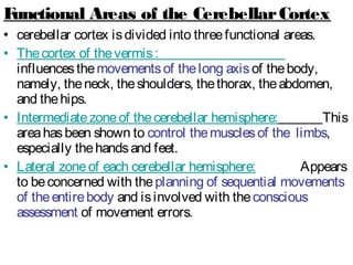
  • 42. Functional Areas of the CerebellarCortex • cerebellar cortex isdivided into threefunctional areas. • Thecortex of thevermis: influencesthemovementsof thelong axisof thebody, namely, theneck, theshoulders, thethorax, theabdomen, and thehips. • Intermediatezoneof thecerebellar hemisphere: This areahasbeen shown to control themusclesof the limbs, especially thehandsand feet. • Lateral zoneof each cerebellar hemisphere: Appears to beconcerned with theplanning of sequential movements of theentirebody and isinvolved with theconscious assessment of movement errors.
  • 43. Arterial supply of The cerebellum is by: 1. Superior cerebellar 2. Anterior inferior cerebellar, 3. Posterior inferior cerebellar Venous drainage by veinsthat empty into the • Great cerebral vein • Venoussinuses.
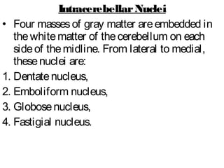
  • 44. IntracerebellarNuclei • Four massesof gray matter areembedded in thewhitematter of thecerebellum on each sideof themidline. From lateral to medial, thesenuclei are: 1. Dentatenucleus, 2. Emboliform nucleus, 3. Globosenucleus, 4. Fastigial nucleus.
  • 45. Fistugial nucleus Globosenucleus Emboliform nucleus Dentatenucleus 4th Ventricle Pons
  • 47. Information regarding theinitiation of movement in thecerebral cortex isprobably transmitted to thecerebellum so that the movement can bemonitored and appropriate adjustmentsin thevoluntary muscleactivity can bemade. 1. CerebellarAfferent Fibers From the Cerebral Cortex
  • 48. Pathway Function Origin Destination 1.1. CortiCo-CortiCo- pontoponto CerebellarCerebellar Conveyscontrol signalsfrom cerebral cortex Frontal, parietal, temporal, and occipital lobes Viapontine nuclei to cerebellar cortex 2.2. Cerebro-Cerebro- olivo-olivo- CerebellarCerebellar Conveyscontrol signalsfrom cerebral cortex Frontal, parietal, temporal, and occipital lobes Viainferior olivary nuclei to cerebellar cortex 3.3. Cerebro-Cerebro- retiCulo-retiCulo- CerebellarCerebellar Conveyscontrol signalsfrom cerebral cortex Sensorimotor areas Viareticular formation to cerebellar cortex
  • 49. Corticopontocerebellar pathway Corticoreticulocerebellar pathway Cortico-olivocerebellar pathway Reticular formation Pontine nuclie Inferior olivary nucleus
  • 50. • Thespinal cord sendsinformation to thecerebellum from somatosensory receptorsby threepathways: (1) theanterior spinocerebellar tract: isfound at all segmentsof thespinal cord, and itsfibers convey musclejoint information from theupper and lower limbs (2) theposterior spinocerebellar tract: receivesmusclejoint information from thetrunk and lower limbs. (3) thecuneocerebellar tract: receivesmusclejoint information from theupper limb and upper part of thethorax 2. CerebellarAfferent Fibers From Spinal Cord
  • 51. 3. CerebellarAfferent Fibers From the VestibularNerve • Thevestibular nervereceives information from theinner ear concerning: A. Motion from thesemicircular canals B. position relativeto gravity from: Utricle Saccule.
  • 52. 4. OtherAfferent Fibers • In addition, thecerebellum receivessmall bundlesof afferent fibersfrom: 1. thered nucleus 2. thetectum.
  • 53. Superior cerebellar peduncle Inferior cerebellar peduncle Cerebellum Inferior cerebellar peduncle Medulla Anterior spinocerebellar tract (minority of fibers) Vestibular nuclie Dentatenucleus Nucleuscunatus Vestibular nucleus Posterior spinocerebellar tract Anterior spinocerebellar tract (majority of fibers)
  • 55. Pathway Function Origin Destination Globose-Globose- emboliformemboliform -rubral-rubral Influences ipsilateral motor activity Globoseand emboliform nuclei contralateral red nucleus, then via crossed rubrospinal tract to ipsilateral motor neuronsin spinal cord
  • 56. Pathway Function Origin Destination Dento-Dento- thalamicthalamic Influences ipsilateral motor activity Dentate nucleus contralateral ventro- lateral nucleusof thalamus, contralateral motor cerebral cortex; corticospinal tract crossesmidlineand controlsipsilateral motor neuronsin spinal cord
  • 57. Pathway Function Origin Destination fastiGialfastiGial vestibularvestibular Influences ipsilateral extensor muscle tone Fastigial nucleus Mainly to ipsilateral and to contralateral lateral vestibular nuclei; vestibulo- spinal tract to ipsilateral motor neuronsin spinal cord
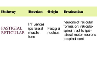
  • 58. Pathway Function Origin Destination fastiGialfastiGial reticularreticular Influences ipsilateral muscle tone Fastigial nucleus neuronsof reticular formation; reticulo- spinal tract to ipsi- lateral motor neurons to spinal cord
  • 60. General Considerations: • Each cerebellar hemisphereisconnected by nervous pathwaysprincipally with thesamesideof thebody; thus, a lesion in onecerebellar hemispheregivesriseto signsand symptomsthat arelimited to thesamesideof thebody. • Theessential function of thecerebellum isto coordinate, by synergistic action, all reflex and voluntary muscular activity. Thus, it graduatesand harmonizesmuscletoneand maintainsnormal body posture. It permitsvoluntary movements, such aswalking, to takeplacesmoothly with precision and economy of effort. • It must beunderstood that although thecerebellum playsan important rolein skeletal muscleactivity, it is not ableto initiatemusclemovement.
  • 61. Characteristic symptoms and signs of cerebellardysfunction: 1.hypotonia: Themusclesloseresilienceto palpation. Thereis diminished resistanceto passivemovementsof joints. Shaking thelimb producesexcessive movementsat theterminal joints. Thecondition is attributableto lossof cerebellar influenceon the simplestretch reflex.
  • 62. 2. Postural Changes and Alteration of Gait • Thehead isoften rotated and flexed, and the shoulder on thesideof thelesion islower than on thenormal side. • Thepatient assumesawidebasewhen heor she standsand isoften stiff legged to compensatefor lossof muscletone. • When theindividual walks, staggerstoward the affected side.
  • 63. 3. Disturbancesof Voluntary Movement (Ataxia) Themusclescontract irregularly and weakly. • Tremoroccurswhen finemovements, such as buttoning clothes, writing, and shaving, are attempted. Musclegroupsfail to work harmon- iously, and thereisdecomposition of movement. • Testsfor tremor : 1. Asking thepatient to touch thetip of thenosewith theindex finger, thefinger either passesthenose (past-pointing) or hitsthenose. 2. asking thepatient to placetheheel of onefoot on theshin of theoppositeleg, it will either hit the shin or not.
  • 64. 4. Dysdiadochokinesia: inability to perform alternating movementsregularly and rapidly. Ask thepatient to pronateand supinate theforearmsrapidly. On thesideof thecerebellar lesion, themovementsareslow, jerky, and incomplete.
  • 65. 5. Disturbances of Reflexes • Movement produced by tendon reflexestendsto continuefor alonger period of timethan normal, e.g. pendular kneejerk, for example, occurs following tapping of thepatellar tendon.
  • 66. 6. Disturbancesof Ocular Movement: • Nystagmus, which isan ataxia(incoordination) of theocular muscles, isarhythmical oscillation of the eyes. It ismoreeasily demonstrated when theeyes aredeviated in ahorizontal direction. 7. Disordersof Speech: Dysarthriaoccursin cerebellar diseasebecauseof ataxia(incoordination) of themusclesof thelarynx. Articulation isjerky, and thesyllablesoften are separated from oneanother. Speech tendsto be explosive, and thesyllablesoften areslurred.
  • 67. CerebellarSyndromes 1. Vermis Syndrome: • Themost common causeof vermissyndromeisa medulloblastomaof thevermisin children. • Involvement of theflocculonodular lobe resultsin signsand symptomsrelated to thevestibular system. • Sincethevermisisunpaired and influencesmidline structures, muscleincoordination involvesthehead and trunk and not thelimbs. • Thereisatendency to fall forward or backward. Thereisdifficulty in holding thehead steady and in an upright position. Therealso may be difficulty in holding thetrunk erect.
  • 68. 2. CerebellarHemisphere Syndrome: • Tumorsof onecerebellar hemispheremay bethecause of cerebellar hemispheresyndrome. • Thesymptomsand signsareusually unilateral and involvemuscleson thesideof thediseased cerebellar hemisphere. • Movementsof thelimbs, especially thearms, are disturbed. Swaying and falling to thesideof thelesion often occur. • Dysarthriaand nystagmusarealso common findings. • Disordersof thelateral part of thecerebellar hemispheresproducedelaysin initiating movements and inability to moveall limb segmentstogether in a coordinated manner but show atendency to moveone joint at atime.