SlideShare a Scribd company logo
Operative techniques: Nephrectomy
Presenter: Dr Sangamesh S K
Moderator: Col S Tripathy
Overview of presentation
• Surgical anatomy
• Indication of Nephrectomy
• Types of Nephrectomy
– Simple, Partial & Radical nephrectomy
– Donor Nephrectomy
• Different technique of Nephrectomy
• Complications
Surgical anatomy
• Bean shaped organ
• Location:
Retroperitoneal
• Extend :- T12-L3
• Two surface (anterior &
posterior)
• Two pole (superior &
inferior)
• Two border (medial &
lateral)
Position
• Lies psoas muscles :
Longitudinal axes
oblique
• Upper poles more
medial & posterior
• Medial aspect rotated
anteriorly : 30 degrees
• Right kidney : L1 top –
L3 bottom
• Left kidney : T12- L3
Anterior relation of kidney
Posterior relation of kidney
Axial section imaging
Renal vasculature
Segmental blood supply
Arterial supply
Indication of Nephrectomy
• Severe trauma
• Renal infection
– Xanthogranulomatous & Emphysematous Pyelonephritis
• Malignancy
• Non functioning kidney
– Stones & obstruction
• Renal fistula
• Renal vascular Hypertension
– All medical & surgical therapy have failed
• Transplantation
Types of Nephrectomy
• Simple Nephrectomy
– Removal of kidney within Gerota fascia
• Partial Nephrectomy
– Small-sized renal cancer
– Benign diseases- excised renal capsule (renorrhaphy)
• Radical Nephrectomy
– Complete removal of kidney outside Gerota fascia
– Ipsilateral adrenal gland
– Complete regional lymphadenectomy (crus of
diaphragm - aortic bifurcation)
Different surgical approach
1. Open approach of Nephrectomy
2. Laparoscopic
• Transperitoneal
• Retroperitoneal
• Hand assisted
3. Robotic assisted
Pre- op evaluation
• Global assessment of pt’s renal function is done:
– Urine analysis & culture
– Serum Creatinine
– GFR evaluation
– Cardiac & pulmonary status: Positioning & bleeding
– Cross sectional imaging (CT/MRI) :- Surgical planning
– Locally advanced metastatic lesion : Screen hepatic
status (stauffer syndrome)
– Renal Artery Embolisation (RAE) : Large tumor
Instruments
• General set
• Abdominal drainage tube
• Self-retaining retractors : omni tract, balfour
• Long genitourinary surgical instruments
• Bulldog &/or Satinsky vascular pedicle clamps
• Mixters right angle forceps
• Bulldog arterial clamp
• Retractor- doyen’s and deiver’s
• Kidney pedicle clamp
Positioning
• Induction & ET tube
placement
• Catheterization done
• Pt : Lateral decubitus
• Table flexed b/w iliac crest &
costal margin
• Head supported: Avoid
excessive flexion
• Pt back supported by blanket
• Dependent leg flexed & top
leg straight
• All pressure point : padded
surface
• Pt secured to table : tape
Surgical approach to kidney
Anterior approach of kidney
INCISION INDICATION BENEFIT LIMITATION
Midline
Transperitoneal
Trauma, IVC
Thrombus , B/L
Renal or Ureteral
disease , Horseshoe
Rapid ,Early
vascular control ,
Access both kidney
Limited exposure to
kidney & bowel
Subcostal Radical
nephrectomy ,
UPJO
Incision can extend
to chevron , early
vascular control
Bowel complication
Hilum access poor
Chevron B/L renal tumor ,
IVC thrombus
Excellent b/l
exposure ,
Injury to Liver ,
Spleen , Transection
of large muscle
Transverse
abdominal
Wilms tumor Easy access to
pedicle &
retroperitoneal
node
Modified
Thoracoabdominal
Radical
nephrectomy ,
lymphadenectomy
versatile Bowel
complication,
Transection of large
muscle
Flank incision
INCISION INDICATION BENEFIT LIMITATION
11th or 12th rib
supracostal
Partial
nephrectomy , &
Simple
nephrectomy
Good renal &
retroperitoneal
exposure
Pleural injury
11th rib Transcostal Partial and Simple
nephrectomy
Good renal &
retroperitoneal
exposure
Pleural injury &
Noticeable flank
defect
Thoracoabdominal Large renal mass ,
IVC thrombus ,
involvement of
surrounding
structure
Excellent exposure ,
Can approach
completely
extraperitoneally
Pleural injury ,
Transection of large
muscles,
Steps of flank approach
• After Gerota fascia incised & kidney is
dissected free from surrounding perinephric
fat.
• Renal artery identified & ligated before vein
• Renal vein ligated
Simple nephrectomy
Indications
• Poor functionaing kidney
– Obstruction, infection, trauma, stones,
nephrosclerosis, vesicoureteral reflux, polycystic
kidney, or congenital dysplasia
• Functional kidney
– Relieve intractable symptoms : bleeding, pain,
hypertension, or persistent infection
Simple nephrectomy
• Flank incision : retroperitoneal access
• Renal fascia incised , perirenal fat is separated
from kidney
• Aberrant vessels : near the poles
• Large hydronephrotic : puncture and aspirate
• Adrenal dissectd in upper pole
• Lower pole mobilised and ureter isolated
• Division of ureter : access to hilar structures
Right kidney approach
Left kidney approach
Radical nephrectomy : Right
• Additional mobilization of liver, avascular Rt triangular
ligament is incised
• White line of Toldt : pelvis to hepatic flexure
• 2nd part of duodenum : Kocher maneuver
• Dissection anterior to IVC : Identification of renal vein
& gonadal vein
• Ligature of ureter below : kidney lifted , artery more
exposed
• Difficult hilar dissections : Dissect in the interaortocaval
region
• Lumbar veins close approximation
Radical nephrectomy : left
• White line of Toldt : splenic flexure to descending
colon is reflected medially.
• Renocolic ligament is divided and extreme care is
taken to avoid injury to the tail of the pancreas
• Left renal vein is identified using the anterior
surface of the aorta as a guide
• Left renal artery is usually located cranial and
posterior to the left renal vein
• Further mobilization of the lower pole of the
kidney, the left ureter and the left gonadal vein
PARTIAL NEPHRECTOMY:- INDICATION
1) Absolute:- i) Single kidney
ii) Bilateral renal tumor
iii) Severe renal failure
2) Relative:- i) Abnormal contralateral
ii) Metabolic disease associated with
renal failure
iii) Genetic syndrome with tumor
multifocality (e.g VHL syndrome)
3) Elective:- i) Tumor < 4cm in young & healthy pt
ii) Peripheral tumor
Partial Nephrectomy : Relative
Contraindication
Technical issue
– Cold ischemia time >45min (consider extracorporeal
approach)
– Less than 20% of global nephron mass retained
Cancer related issue
– Diffuse encasement of renal pedicle by tumor
– Diffuse invasion of central collecting system
– Tumor thrombus involving major renal veins
– Adjacent organ invasion (stage cT4)
– Regional lymphadenopathy (stage cTxN1)
Partial nephrectomy
• Avascular plane : Segmental artery clamping
– 5ml Indigo carmine : clamped artey
– Cooled down to 20o
• Hyperfiltration injury :
– Over decades, FSGS : proteinuria and progressive
renal failure (nephron mass reduced by 80%)
• Vascular clamping: Ischemia & hypothermia
Partial Nephrectomy :- Wedge
Resection
Partial Nephrectomy: Segmental Polar
Resection
Complication of partial nephrectomy
• Urinary fistula
• Post operative bleeding
• Renal insufficiency
Wound closure
• Hemostasis and evaluate adjacent organs for any signs
of injury
• Pleural injury,
– retroperitoneum is filled to level of the flank incision with
saline.
– anesthesiologist then inflates lungs with high inspiratory
volumes
– Bubbling of saline irrigation in the retroperitoneum with
deep inspiration would suggest a pneumothorax
• Fascial layers approximated : two layers
– Transversus abdominis & internal oblique fasciae are
approximated together
– External oblique fascia is approximated as a separate layer
Donor Nephrectomy
• Kidney is mobilized, only remaining attachments are
the ureter, renal vein, and renal artery
• 12.5 g of mannitol and 20 mg of furosemide are rapidly
infused intravenously
• Immediately after dividing renal vessels, placed on
workbench : Pan of ice slush covered with a towel.
• Flushed intra-arterially by gravity flow with renal
preservation solution at 6°C
• Flushing should continue until it is cooled & renal
effluent is clear (~500 to 1000 mL)
• Kept in ice slush basin during procedure : Hypothermia
Donor nephrectomy
• Renal artery & vein are flushed independently
with preservation solution : Bleeding
• Retrograde flushing of ureter : Collecting system
leaks
• Transplanted into either lower quadrant,
transferred to iliac fossa
• Renal vein anastomosed : External iliac vein.
• Renal artery anastomosis
– End-to-end anastomosis to hypogastric artery
– End-to-side anastomosis with external iliac artery
Donor nephrectomy
• During anastomosis, vessels should be irrigated :
Heparin solution (10,000 U Heparin in 100 mL NS)
• Surgeon should consider injecting
– 10 mg Verapamil into renal artery following
anastomosis : Vasodilation.
• Ureter is implanted into dome of bladder with a
tension-free anastomosis
• Prior to completion ureteral anastomosis:
Ureteral stent is placed
Complication of Open Nephrectomy
• Hemorrhage
• Small bowel obstruction
• Pneumothorax
• Pneumonia
• DVT
• Superficial wound infection
• Bowel injury
LAPROSCOPIC NEPHRECTOMY
SURGICAL APPROACHES:-
1) Transperitoneal
2) Retroperitoneal
3) Hand-Assisted
TRANSPERITONEAL RETROPERITONEAL HAND-ASSISTED
Small incision & gives
advantage for trocar
placement
Limited working space
,result in difficulty in
orientation , trocar spacing
, & organ entrapment
Bridge b/w lap & open
surgery
Affords optimal working
space
Preferred in pt with h/o
multiple abdominal
procedure or pt with
peritonitis
Permits tactile feedback
Also preferred in pt with
abnormality of posterior
surface (exophytic cyst or
mass)
Allow the hand to assist
with dissection , retraction
, extraction & rapid control
of bleeding
PATIENT EVALUATION & PREPARATION
• Prior abdominal surgery
– Transperitoneal & retroperitoneal
– Pt positioning ,
– Placement of trocars
• Pre-op abdominal CT :- useful in surgical planning
• Rest of evaluation similar to open nephrectomy
Patient positioning & trocar sites
OPERATING ROOM
Procedure of transperitoneal approach
• Reflection of colon
• Dissection of ureter
• Identification of renal hilum
• Securing of renal blood vessel
• Isolation of upper pole
• Organ entrapment
Nephrectomy : Operative Technique
Retroperitoneal Approach
• Retroperitoneal approach used for posterior or
lower pole lesion
• Pt in full flank position ,10mm trocar is placed in
posterior axillary line , halfway b/w iliac crest &
12th rib
• Pneumo-peritonium is established & anterior wall
is identified with gentle dissection of
retroperitoneal fat
Retroperitoneal approach
Retroperitoneal Approach
• Second 12mm port is placed under direct
vision in anterior axillary line
• A third port 5mm is placed superior to 2nd port
below rib cage
• Using the perinephric fat to elevate kidney ,
surgeon readily sees the lower pole &
posterior surface
Complication of laparoscopic renal
surgery
1. Access related problems:-
i. Solid organ injury
ii. Bowel injury
iii. Abdominal wall hematoma
iv. Epigastric vessel injury
2. Hemorrhage
3. Pneumonia
4. Pulmonary embolus
5. Unrecognized bowel injury
6. Incisional hernia :- after intact specimen removal
Nephrectomy: Robotic Port Placement
Robotic Nephrectomy : Operating room
Advantage of robotic over laparoscopic
• Minimal invasive approach
• 3D visual & depth perception
Thank you

More Related Content

PPTX
PPTX
Discuss the operation of simple nephrectomy
PPTX
Deep vein thrombosis
PPT
Ureteric Injury at Gynaecological Surgery
PPTX
Percutaneous Nephrolithotomy PCNL by Dr. Majid Kakakhel IKD, Peshawar.
PDF
Pediatric urology pujo- pyeloplasty
PPTX
Prostate cancer
Discuss the operation of simple nephrectomy
Deep vein thrombosis
Ureteric Injury at Gynaecological Surgery
Percutaneous Nephrolithotomy PCNL by Dr. Majid Kakakhel IKD, Peshawar.
Pediatric urology pujo- pyeloplasty
Prostate cancer

What's hot (20)

PPTX
Hernia and herniorrhaphy
PPTX
Cholecystectomy class
DOC
Prostectomy
PPTX
Incisional hernia
PPTX
Laparoscopic cholecystectomy/ operative surgery
PPT
Appendicectomy
PPTX
Adrenalectomy new
PDF
Exploratory laparotomy
PPTX
Below knee amputation
PDF
Surgical incisions
PPTX
Colectomy
PPTX
Inguinal hernia repair
PPTX
Open Pyelolithotomy
PPTX
Testicular Torsion
PDF
Intestinal stomas
PDF
Colostomy & Ileostomy
PPT
Post cholecystectomy complications
PPTX
Thoracotomy
PPTX
Mastectomy
Hernia and herniorrhaphy
Cholecystectomy class
Prostectomy
Incisional hernia
Laparoscopic cholecystectomy/ operative surgery
Appendicectomy
Adrenalectomy new
Exploratory laparotomy
Below knee amputation
Surgical incisions
Colectomy
Inguinal hernia repair
Open Pyelolithotomy
Testicular Torsion
Intestinal stomas
Colostomy & Ileostomy
Post cholecystectomy complications
Thoracotomy
Mastectomy
Ad

Similar to Nephrectomy : Operative Technique (20)

PPTX
Upper gu trauma
PPT
Genito-urinary trauma
PDF
management of portal hypertension by Dr.Zarin
PPT
Ivu
PPTX
SURGICAL MANAGEMENT OF CARCINOMA ESOPHAGUS.pptx
PPTX
Recipient hepatectomy in liver transplantation
PPT
Liver trauma
PPTX
Liver trauma الدكتور طارق المنيزل
PPTX
Endoüroloji 2011 ankara
PPTX
Liver splenic and renal trauma presentation for postgraduates in radiology
PDF
Nutcracker syndrome
PPTX
Renal Trauma | Diagnosis, Management and Followup
PPTX
Portal Hypertension.pptx
PPTX
PORTAL HYPERTENSION by Dr.Abhijeet Majhi.pptx
PDF
Lecture 15- Renal Replacement Therapies.pdf
PPTX
Abdominal trauma ,an overview
PPTX
Surgical management of pancreatic pseudocyst..by dr chris alumona
PPTX
A practical guide to Renal Vascular Diseases.pptx
PPTX
Liver trauma
PPTX
Portal hypertension by Gowhar Ahmad
Upper gu trauma
Genito-urinary trauma
management of portal hypertension by Dr.Zarin
Ivu
SURGICAL MANAGEMENT OF CARCINOMA ESOPHAGUS.pptx
Recipient hepatectomy in liver transplantation
Liver trauma
Liver trauma الدكتور طارق المنيزل
Endoüroloji 2011 ankara
Liver splenic and renal trauma presentation for postgraduates in radiology
Nutcracker syndrome
Renal Trauma | Diagnosis, Management and Followup
Portal Hypertension.pptx
PORTAL HYPERTENSION by Dr.Abhijeet Majhi.pptx
Lecture 15- Renal Replacement Therapies.pdf
Abdominal trauma ,an overview
Surgical management of pancreatic pseudocyst..by dr chris alumona
A practical guide to Renal Vascular Diseases.pptx
Liver trauma
Portal hypertension by Gowhar Ahmad
Ad

Recently uploaded (20)

PDF
DAY-6. Summer class. Ppt. Cultural Nursing
PPTX
ABG advance Arterial Blood Gases Analysis
PPTX
Trichuris trichiura infection
PDF
Dermatology diseases Index August 2025.pdf
PPTX
First Aid and Basic Life Support Training.pptx
PPTX
different types of Gait in orthopaedic injuries
PPTX
1. Drug Distribution System.pptt b pharmacy
PDF
Structure Composition and Mechanical Properties of Australian O.pdf
PPTX
NUTRITIONAL PROBLEMS, CHANGES NEEDED TO PREVENT MALNUTRITION
PDF
Priorities Critical Care Nursing 7th Edition by Urden Stacy Lough Test Bank.pdf
PDF
MINERAL & VITAMIN CHARTS fggfdtujhfd.pdf
PDF
Megan Miller Colona Illinois - Passionate About CrossFit
PDF
2E-Learning-Together...PICS-PCISF con.pdf
PPTX
Current Treatment Of Heart Failure By Dr Masood Ahmed
PPTX
community services team project 2(4).pptx
DOCX
Copies if quanti.docxsegdfhfkhjhlkjlj,klkj
PPTX
PEDIATRIC OSCE, MBBS, by Dr. Sangit Chhantyal(IOM)..pptx
PPTX
First aid in common emergency conditions.pptx
PDF
Khaled Sary- Trailblazers of Transformation Middle East's 5 Most Inspiring Le...
PPTX
CBT FOR OCD TREATMENT WITHOUT MEDICATION
DAY-6. Summer class. Ppt. Cultural Nursing
ABG advance Arterial Blood Gases Analysis
Trichuris trichiura infection
Dermatology diseases Index August 2025.pdf
First Aid and Basic Life Support Training.pptx
different types of Gait in orthopaedic injuries
1. Drug Distribution System.pptt b pharmacy
Structure Composition and Mechanical Properties of Australian O.pdf
NUTRITIONAL PROBLEMS, CHANGES NEEDED TO PREVENT MALNUTRITION
Priorities Critical Care Nursing 7th Edition by Urden Stacy Lough Test Bank.pdf
MINERAL & VITAMIN CHARTS fggfdtujhfd.pdf
Megan Miller Colona Illinois - Passionate About CrossFit
2E-Learning-Together...PICS-PCISF con.pdf
Current Treatment Of Heart Failure By Dr Masood Ahmed
community services team project 2(4).pptx
Copies if quanti.docxsegdfhfkhjhlkjlj,klkj
PEDIATRIC OSCE, MBBS, by Dr. Sangit Chhantyal(IOM)..pptx
First aid in common emergency conditions.pptx
Khaled Sary- Trailblazers of Transformation Middle East's 5 Most Inspiring Le...
CBT FOR OCD TREATMENT WITHOUT MEDICATION

Nephrectomy : Operative Technique

  • 1. Operative techniques: Nephrectomy Presenter: Dr Sangamesh S K Moderator: Col S Tripathy
  • 2. Overview of presentation • Surgical anatomy • Indication of Nephrectomy • Types of Nephrectomy – Simple, Partial & Radical nephrectomy – Donor Nephrectomy • Different technique of Nephrectomy • Complications
  • 3. Surgical anatomy • Bean shaped organ • Location: Retroperitoneal • Extend :- T12-L3 • Two surface (anterior & posterior) • Two pole (superior & inferior) • Two border (medial & lateral)
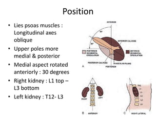
  • 4. Position • Lies psoas muscles : Longitudinal axes oblique • Upper poles more medial & posterior • Medial aspect rotated anteriorly : 30 degrees • Right kidney : L1 top – L3 bottom • Left kidney : T12- L3
  • 11. Indication of Nephrectomy • Severe trauma • Renal infection – Xanthogranulomatous & Emphysematous Pyelonephritis • Malignancy • Non functioning kidney – Stones & obstruction • Renal fistula • Renal vascular Hypertension – All medical & surgical therapy have failed • Transplantation
  • 12. Types of Nephrectomy • Simple Nephrectomy – Removal of kidney within Gerota fascia • Partial Nephrectomy – Small-sized renal cancer – Benign diseases- excised renal capsule (renorrhaphy) • Radical Nephrectomy – Complete removal of kidney outside Gerota fascia – Ipsilateral adrenal gland – Complete regional lymphadenectomy (crus of diaphragm - aortic bifurcation)
  • 13. Different surgical approach 1. Open approach of Nephrectomy 2. Laparoscopic • Transperitoneal • Retroperitoneal • Hand assisted 3. Robotic assisted
  • 14. Pre- op evaluation • Global assessment of pt’s renal function is done: – Urine analysis & culture – Serum Creatinine – GFR evaluation – Cardiac & pulmonary status: Positioning & bleeding – Cross sectional imaging (CT/MRI) :- Surgical planning – Locally advanced metastatic lesion : Screen hepatic status (stauffer syndrome) – Renal Artery Embolisation (RAE) : Large tumor
  • 15. Instruments • General set • Abdominal drainage tube • Self-retaining retractors : omni tract, balfour • Long genitourinary surgical instruments • Bulldog &/or Satinsky vascular pedicle clamps • Mixters right angle forceps • Bulldog arterial clamp • Retractor- doyen’s and deiver’s • Kidney pedicle clamp
  • 16. Positioning • Induction & ET tube placement • Catheterization done • Pt : Lateral decubitus • Table flexed b/w iliac crest & costal margin • Head supported: Avoid excessive flexion • Pt back supported by blanket • Dependent leg flexed & top leg straight • All pressure point : padded surface • Pt secured to table : tape
  • 18. Anterior approach of kidney INCISION INDICATION BENEFIT LIMITATION Midline Transperitoneal Trauma, IVC Thrombus , B/L Renal or Ureteral disease , Horseshoe Rapid ,Early vascular control , Access both kidney Limited exposure to kidney & bowel Subcostal Radical nephrectomy , UPJO Incision can extend to chevron , early vascular control Bowel complication Hilum access poor Chevron B/L renal tumor , IVC thrombus Excellent b/l exposure , Injury to Liver , Spleen , Transection of large muscle Transverse abdominal Wilms tumor Easy access to pedicle & retroperitoneal node Modified Thoracoabdominal Radical nephrectomy , lymphadenectomy versatile Bowel complication, Transection of large muscle
  • 19. Flank incision INCISION INDICATION BENEFIT LIMITATION 11th or 12th rib supracostal Partial nephrectomy , & Simple nephrectomy Good renal & retroperitoneal exposure Pleural injury 11th rib Transcostal Partial and Simple nephrectomy Good renal & retroperitoneal exposure Pleural injury & Noticeable flank defect Thoracoabdominal Large renal mass , IVC thrombus , involvement of surrounding structure Excellent exposure , Can approach completely extraperitoneally Pleural injury , Transection of large muscles,
  • 20. Steps of flank approach
  • 21. • After Gerota fascia incised & kidney is dissected free from surrounding perinephric fat. • Renal artery identified & ligated before vein • Renal vein ligated
  • 22. Simple nephrectomy Indications • Poor functionaing kidney – Obstruction, infection, trauma, stones, nephrosclerosis, vesicoureteral reflux, polycystic kidney, or congenital dysplasia • Functional kidney – Relieve intractable symptoms : bleeding, pain, hypertension, or persistent infection
  • 23. Simple nephrectomy • Flank incision : retroperitoneal access • Renal fascia incised , perirenal fat is separated from kidney • Aberrant vessels : near the poles • Large hydronephrotic : puncture and aspirate • Adrenal dissectd in upper pole • Lower pole mobilised and ureter isolated • Division of ureter : access to hilar structures
  • 26. Radical nephrectomy : Right • Additional mobilization of liver, avascular Rt triangular ligament is incised • White line of Toldt : pelvis to hepatic flexure • 2nd part of duodenum : Kocher maneuver • Dissection anterior to IVC : Identification of renal vein & gonadal vein • Ligature of ureter below : kidney lifted , artery more exposed • Difficult hilar dissections : Dissect in the interaortocaval region • Lumbar veins close approximation
  • 27. Radical nephrectomy : left • White line of Toldt : splenic flexure to descending colon is reflected medially. • Renocolic ligament is divided and extreme care is taken to avoid injury to the tail of the pancreas • Left renal vein is identified using the anterior surface of the aorta as a guide • Left renal artery is usually located cranial and posterior to the left renal vein • Further mobilization of the lower pole of the kidney, the left ureter and the left gonadal vein
  • 28. PARTIAL NEPHRECTOMY:- INDICATION 1) Absolute:- i) Single kidney ii) Bilateral renal tumor iii) Severe renal failure 2) Relative:- i) Abnormal contralateral ii) Metabolic disease associated with renal failure iii) Genetic syndrome with tumor multifocality (e.g VHL syndrome) 3) Elective:- i) Tumor < 4cm in young & healthy pt ii) Peripheral tumor
  • 29. Partial Nephrectomy : Relative Contraindication Technical issue – Cold ischemia time >45min (consider extracorporeal approach) – Less than 20% of global nephron mass retained Cancer related issue – Diffuse encasement of renal pedicle by tumor – Diffuse invasion of central collecting system – Tumor thrombus involving major renal veins – Adjacent organ invasion (stage cT4) – Regional lymphadenopathy (stage cTxN1)
  • 30. Partial nephrectomy • Avascular plane : Segmental artery clamping – 5ml Indigo carmine : clamped artey – Cooled down to 20o • Hyperfiltration injury : – Over decades, FSGS : proteinuria and progressive renal failure (nephron mass reduced by 80%) • Vascular clamping: Ischemia & hypothermia
  • 31. Partial Nephrectomy :- Wedge Resection
  • 33. Complication of partial nephrectomy • Urinary fistula • Post operative bleeding • Renal insufficiency
  • 34. Wound closure • Hemostasis and evaluate adjacent organs for any signs of injury • Pleural injury, – retroperitoneum is filled to level of the flank incision with saline. – anesthesiologist then inflates lungs with high inspiratory volumes – Bubbling of saline irrigation in the retroperitoneum with deep inspiration would suggest a pneumothorax • Fascial layers approximated : two layers – Transversus abdominis & internal oblique fasciae are approximated together – External oblique fascia is approximated as a separate layer
  • 35. Donor Nephrectomy • Kidney is mobilized, only remaining attachments are the ureter, renal vein, and renal artery • 12.5 g of mannitol and 20 mg of furosemide are rapidly infused intravenously • Immediately after dividing renal vessels, placed on workbench : Pan of ice slush covered with a towel. • Flushed intra-arterially by gravity flow with renal preservation solution at 6°C • Flushing should continue until it is cooled & renal effluent is clear (~500 to 1000 mL) • Kept in ice slush basin during procedure : Hypothermia
  • 36. Donor nephrectomy • Renal artery & vein are flushed independently with preservation solution : Bleeding • Retrograde flushing of ureter : Collecting system leaks • Transplanted into either lower quadrant, transferred to iliac fossa • Renal vein anastomosed : External iliac vein. • Renal artery anastomosis – End-to-end anastomosis to hypogastric artery – End-to-side anastomosis with external iliac artery
  • 37. Donor nephrectomy • During anastomosis, vessels should be irrigated : Heparin solution (10,000 U Heparin in 100 mL NS) • Surgeon should consider injecting – 10 mg Verapamil into renal artery following anastomosis : Vasodilation. • Ureter is implanted into dome of bladder with a tension-free anastomosis • Prior to completion ureteral anastomosis: Ureteral stent is placed
  • 38. Complication of Open Nephrectomy • Hemorrhage • Small bowel obstruction • Pneumothorax • Pneumonia • DVT • Superficial wound infection • Bowel injury
  • 39. LAPROSCOPIC NEPHRECTOMY SURGICAL APPROACHES:- 1) Transperitoneal 2) Retroperitoneal 3) Hand-Assisted
  • 40. TRANSPERITONEAL RETROPERITONEAL HAND-ASSISTED Small incision & gives advantage for trocar placement Limited working space ,result in difficulty in orientation , trocar spacing , & organ entrapment Bridge b/w lap & open surgery Affords optimal working space Preferred in pt with h/o multiple abdominal procedure or pt with peritonitis Permits tactile feedback Also preferred in pt with abnormality of posterior surface (exophytic cyst or mass) Allow the hand to assist with dissection , retraction , extraction & rapid control of bleeding
  • 41. PATIENT EVALUATION & PREPARATION • Prior abdominal surgery – Transperitoneal & retroperitoneal – Pt positioning , – Placement of trocars • Pre-op abdominal CT :- useful in surgical planning • Rest of evaluation similar to open nephrectomy
  • 42. Patient positioning & trocar sites
  • 44. Procedure of transperitoneal approach • Reflection of colon • Dissection of ureter • Identification of renal hilum • Securing of renal blood vessel • Isolation of upper pole • Organ entrapment
  • 46. Retroperitoneal Approach • Retroperitoneal approach used for posterior or lower pole lesion • Pt in full flank position ,10mm trocar is placed in posterior axillary line , halfway b/w iliac crest & 12th rib • Pneumo-peritonium is established & anterior wall is identified with gentle dissection of retroperitoneal fat
  • 48. Retroperitoneal Approach • Second 12mm port is placed under direct vision in anterior axillary line • A third port 5mm is placed superior to 2nd port below rib cage • Using the perinephric fat to elevate kidney , surgeon readily sees the lower pole & posterior surface
  • 49. Complication of laparoscopic renal surgery 1. Access related problems:- i. Solid organ injury ii. Bowel injury iii. Abdominal wall hematoma iv. Epigastric vessel injury 2. Hemorrhage 3. Pneumonia 4. Pulmonary embolus 5. Unrecognized bowel injury 6. Incisional hernia :- after intact specimen removal
  • 51. Robotic Nephrectomy : Operating room
  • 52. Advantage of robotic over laparoscopic • Minimal invasive approach • 3D visual & depth perception

Editor's Notes

  • #9: Level of IV disk between LV1 and LV2. Longer right renal artery passes posterior to IVC. Rt vein: 2-4cm, Left vein : 6-10cm
  • #10: A small Apical segmental branch : post br, but MC : Ant div. Posterior segmental artery : the posterior division passes posterior to the renal pelvis, while others pass anterior to the renal pelvis. Accessory renal arteries are seen in 25% to 28%
  • #34: renal artery is clamped with a vascular bulldo