NEURODEGENERATIVE
DISORDERS
PRESENTED BY:
Dr Dibyajyoti Prusty, PG, 2nd Yr
MODERATOR:
Dr Kalpalata Tripathy, Asso. Prof.
Dept. Of Pathology,
S.C.B. Medical College, Cuttack
DATE: 03.05.2018
INTRODUCTION
• characterized clinically by loss of neurological function
(dementia, loss of movement control, paralysis), and
pathologically by loss of neurons.
• Loss of neurons is accompanied by specific histopathological
findings.
• Gradual neuronal atrophy and loss, without specific
pathology.
• Involve specific anatomical systems or interconnected sets of
neurons.
• A biochemical basis.
• Many are inherited.
• The most common degenerative disease is Alzheimer's disease
and the second most frequent is Parkinson's disease.
Area wise classification Disease
Cortical 1. Alzheimer's Disease
2. Pick’s Disease
3. Nonspecific Lobar Atrophies
4. Cortical Or Diffuse Lewy Body Disease
Subcortical Nuclei 1. Huntington’s Disease
2. Parkinsonian Syndromes:
a. Idiopathic: Diffuse Lewy Body Disease
b. Progressive Supranuclear Palsy
c. Multisystem Atrophy
d. Acquired: Postencephalitic, Drugs, Trauma
Cerebellar, Brainstem And
Spinal Cord
1. Cerebello-olivary Degeneration
2. Olivopontocerebellar Atrophy
3. Spinocerebellar Syndromes: Fridreich’s Ataxia,
Heredetary Spastic Paraplegia, Machao-joseph Disease
Motor Neurons 1. Amyotrophic Lateral Sclerosis
- Variants
- Secondary: Toxic/Metabolic, Paraneoplastic,
Post Poliomyelitis
2. Spinal Muscular Atrophies
DEMENTIA
• a generic term, not a disease entity
• Any pathology that causes significant brain damage, at any age, can cause
dementia. The causes of dementia include:
Stroke and ischemic encephalopathy
Hippocampal sclerosis
Head trauma (subdural hematomas, diffuse axonal
injury, chronic traumatic encephalopathy)
Hydrocephalus
CNS infections (HIV encephalitis, Creutzfeldt-Jakob disease)
Metabolic CNS disorders (lysosomal storage and
peroxisomal diseases)
Demyelinative diseases (multiple sclerosis)
Neurodegenerative diseases
Neuropsychiatric disorders
Severe medical illness or organ failure
The effects of medications
DEGENERATIVE DISEASES AS
PROTEINOPATHIES
• the deposition of abnormal proteins in the brain
THE UBIQUITIN-PROTEASOME AND
LYSOSOMAL SYSTEM IN
NEURODEGENERATIVE DISEASES
Protein recycling in cells
• Proteasome(UPS) : Short lived proteins
• Lysosomes : long lived proteins, cytoplasm, oganelles.
ALZHEIMER'S DISEASE
• the most common cause of dementia in old people.
• 13% of persons over 65 years and 45% of those over 85
have AD.
• characterized by loss of memory, inability to learn new
things, loss of language function, deranged perception of
space, inability to do calculations, indifference,
depression, delusions, and other manifestations.
• Mild cognitive impairment (MCI)
memory and other cognitive impairments that do not
affect social functioning or daily living.
PATHOGENESIS OF AD
• two processes: extracellular deposition of beta amyloid-Aβ and
intracellular accumulation of tau protein.
• BETA AMYLOID- Aβ is toxic to neurons
• TAU- Over-phosphorylated microtubule associated protein tau
• Aβ deposition is specific for AD and is thought to be primary.
• Tau accumulation is also seen in other degenerative diseases and is
thought to be secondary.
Neurodegenerative disorders
PATHOLOGY OF AD
• senile plaques (SPs)- Diffuse Aβ plaques (AβPs) and neuritic
plaques (NPs).
• neurofibrillary tangles(NFTs)-deposits of tau filaments in the
neuronal body
Large numbers of
plaques and tangles
cause massive loss of
neurons and
synapses.
Cortical atrophy in advanced AD
Cortical atrophy and
hydrocephalus in AD
The
neurofibrillary
tangles are
composed of
paired helical
filaments
Diffuse plaque.
Beta amyloid immunostain Neurofibrillary tangles. Bielschowsky
silver stain.
Gliosis in and around plaques.
GFAP immunostain
Beta amyloid immunostain. Amyloid
in plaques and vessels in the cerebral
cortex
ETIOLOGY OF AD-GENETICS
• The gene for APP is on chromosome 21
• the presenillin 1 and 2 genes (PSEN1 and PSEN2) on
chromosomes 14 and 1 respectively
• the vast majority (95%) of AD cases appear in old age
and do not follow Mendelian inheritance.
• an interaction of genetic and other intrinsic and
environmental factors.
• the most important genetic risk factor is the
Apolipoprotein E (ApoE) genotype
• three isoforms: ApoE2, ApoE3, and ApoE4-
(chromosome 19)
ETIOLOGY OF AD-OTHER
CONTRIBUTING FACTORS
• environment, diet, and general state of health
• Neuroinflammation
• Free radicals
• Diabetes
• Traumatic brain injury
• Homocysteine
• OLD AGE AND AD
• VASCULAR DISEASE IN AD
• NEUROTRANSMITTERS IN AD
• decreased acetylcholine (Ach), choline acetyltransferase (CHAT)
DIAGNOSIS OF AD
• There are no specific clinical findings in AD- progressive
dementia
• Elevation of tau protein and decrease of Aβ in the CSF
• pathological examination of brain tissue
• Autopsy studies show that the brains of most people over 65,
even without clinical dementia, contain a few SPs and NFTs in
the hippocampus and entorrhinal cortex
NEUROIMAGING MARKERS IN AD
• PiB PET-Fibrillar Aβ deposits are imaged
• FDG-PET-measures cerebral glucose metabolism and blood
flow, which are indicators of synaptic function.
• fMRI-by measuring blood-oxygen-level-dependent (BOLD)
signal.
• DTI(Diffusion tensor imaging)-detects axon density
• Should cases with a few SPs and NFTs be diagnosed as
AD, even if there is no clinical dementia?
• The NIA-Reagan and CERAD diagnostic criteria for
AD
• not only the numbers of SPs and NFTs but also age and
intellectual function
FRONTOTEMPORAL LOBAR
DEGENERATIONS
• refers to the neuropathology of the Frontotemporal
dementia (FTD)
• that affect the frontal and temporal lobes causing
personality change (apathy, disinhibition, loss of insight
and emotional control), loss of the ability to recognize
the meaning of words and objects, language
dysfunction, and global cognitive decline.
Three clinical FTD syndromes:
• behavioral variant of FTD, semantic dementia, and
progressive nonfluent aphasia
• Pathologically, the FTLDs are characterized by atrophy
of the frontal and temporal lobes (lobar atrophy)
• neuronal loss and gliosis, vacuolization of the superficial
cortex (spongiosis), and ballooned neurons.
Three groups of FTLDs are recognized:
• 1.FTLD-TAU with tau-positive inclusions (tauopathies)
• 2. FTLD-TDP with tau-negative, alpha-synuclein-
negative inclusions; RNA binding
• 3. FTLD-FUS, containing fused in sarcoma protein; RNA
binding
• The pathological diagnosis of FTLDs requires extensive
tissue sampling and immunohistochemistry targeting
tau, TDP-43, and ubiquitin.
FTLD-TAU (TAUOPATHIES)
• Tau (the Greek letter) is a microtubule associated protein
• the gene MAPT gene on 17q21: Normally, tau is
phosphorylated and is present mainly in axons
• In FTLDs, abnormal deposits of hyperphosphorylated
tau
• The FTLD-TAU group includes:
– Pick's Disease
FTD with Parkinsonism Linked to Chromosome 17
(FTDP-17), a MAPT mutation
Corticobasal Degeneration
Progressive supranuclear palsy
PICK DISEASE
• the prototype of FTLDs
• It presents between 45-65 years with confusion and FTLD symptoms
and has a progressive course lasting 2-5 years
• Pick bodies (tau-positive spherical cytoplasmic neuronal inclusions,
composed of straight filaments)
• Pick cells (ballooned neurons with dissolution of chromatin)
Frontotemporal
atrophy
Pick inclusion bodies
-tau immunostain
Ballooned neuron
(Pick cell)
FTLD-TDP
(TDP-43 PROTEINOPATHIES)
• TDP-43, encoded by the TARDBP gene on chromosome 1
• Genetic forms(3 types) and sporadic forms
• Deposited in neurons in the form of intranuclear(NII),
cytoplasmic(NCI), and neuritic inclusions.
• Overlap between TDP-43 proteinopathies and motor
neuron diseases.
-ALS patients present with FTD symptoms:
hexaneucleotide repeat(C9orf72) and TDP-43 mutation )
• Progranulin mutation
• TDP-43 deposits are also seen in some cases of
Alzheimer's disease, Pick's disease, Parkinson's disease,
and diffuse Lewy body disease.
PARKINSON'S DISEASE
• second most common neurodegenerative disease after AD, and the
most frequent subcortical degenerative disease. Loss of
dopaminergic neurons in substantia niagra
• motor symptoms (rigidity, tremor at rest, slowness of voluntary
movement, stooped posture, a shuffling, small-step gait, difficulty
with balance)
• nonmotor symptoms (expressionless face, soft voice, olfactory loss,
mood disturbances, dementia, sleep disorders, and autonomic
dysfunction, including constipation, cardiac arrhythmias, and
hypotension)
• Sporadic
• 5-10 % of patients have a monogenic, autosomal dominant or recessive form
of PD
• The first autosomal dominant form of PD : α-synuclein, a synaptic protein
encoded by SNCA on 4q22; other: LRRK2 Mutation
• Autosomal recessive PD is Parkin. Others: DJ-1, PINK1-Roll in clearance of
dysfunctional mitochondria
• association with Gaucher disease (GD)
Pathology in PD
• The key pathology in PD is α-synuclein accumulation
(lipid binding protein normally associated with
synapses)
• Lewy bodies (LBs): aggregates form round lamellated
eosinophilic cytoplasmic inclusions in the neuronal body
-By Fredrich Henry Lewey
• Lewy neurites: fibrils made of insoluble polymers of α-
synuclein
-Deposited in neuronal processes and in astrocytes and
oligodendroglial cells
• Its accumulation impairs the functions of mitochondria,
lysosomes, and endoplasmic reticulum, and interferes
with microtubular transport.
Left:PD; Right: normal SN
Normal sustantia nigra-zona compacta Loss of sustantia nigra neurons in PD
Lewy body in a SN
neuron
• antibodies to α-synuclein made it easier to detect LBs
and synuclein deposits outside the SN.
• PD should be viewed as a systemic disorder that affects
the entire CNS and PNS, not just the dopaminergic
system.
• explains the diverse motor and nonmotor manifestations
of PD
• AD and PD (and in a broader sense tauopathies and
synucleinopathies) overlap clinically and pathologically.
• Trials of neural implantation
DIFFUSE LEWY BODY
DISEASE
• Lewy body dementia
• sporadic neurodegenerative disease
• second most common cause of dementia after AD
• It combines the neurological manifestations of dementia and parkinsonism.
• shows small, inconspicuous Lewy bodies in the neocotex, limbic system,
and brainstem
Cortical Lewy bodies.
Alpha-synuclein
immunostain.
MPTP PARKINSONISM
• The pyridine analogue MPTP (1-methyl-1-4-phenyl-
1,2,3,6-tetrahydropyridine)
• taken up selectively by dopaminergic neurons.
• active compound, MPP (1-methyl-4-
phenylpyridinium) inhibits mitochondrial function and
induces cell death
• Self-made synthetic heroin, contaminated by MPTP
• rotenone
• Caffeine and nicotine : ?protective
GUAM PARKINSON-DEMENTIA (GPD)
• Chamorro people of Guam
• combination of tauopathy and synucleinopathy
• this disease in successive generations implicates genetic factors.
•
GUAM
In recent years, the incidence of GPD has declined
dramatically
Hypothesis :
• GPD is caused by a toxic amino acid in the seed of a
cycad plant which is used to make flour and is a staple
in the local diet.
• deficiencies of calcium and magnesium, and high
concentrations of aluminum and other minerals in
drinking water, wth aluminium deposition in neurons
• GPD underlines the important role of genetics and
environmental neurotoxins in the pathogenesis of
neurodegenerative diseases.
• Other conditions that damage the SN:
striatonigral degeneration, postencephalitic
parkinsonism, manganese poisoning, carbon monoxide
poisoning, hypoxic-ischemic encephalopathy, traumatic
brain injury, and stroke.
ATYPICAL PARKINSONISM
SYNDROMES
- Progressive supranuclear palsy
- corticobasal degenerations
- multisystem atrophy
HUNTINGTON'S DISEASE
• Fatal autosomal dominant condition;
• Average course-15yrs
• behavioral changes, chorea, and dementia.
• Atrophy of the caudate nucleus and putamen and
dilatation of the anterior horns of the lateral ventricles
• Degeneration begins in the caudal portions of the
caudate and putamen and advances rostrally.
• Cortical atrophy.
• Microscopically:
– there is loss of medium size spiny neurons those use
GABA, enkephalin, dynorphin, substance P as
neurotransmitters-in the caudate nucleus and putamen
– loss of cortical neurons and gliosis
– Proliferation of reactive astrocytes
– neuronal intranuclear inclusions
Normal caudate nuclei, small
lateral ventricles.
HD- Atrophy of the caudate
nuclei and dilatation of the
lateral ventricles.
from
Huntington's
disease; right
GFAP IHC:
Caudate
nucleus from
normal brain;
left.
Molecular abnormality of HD
• Huntingtin- protein involved in cellular transport
• CAG trinucleotide expansion of the HTT(HUNTINGTIN) gene on
chromosome 4p: adds a polyglutamine segment
• the CAG-expanded huntingtin has a toxic function
• CAG triplet expansion is probably due to malfunction of cellular processes
that repair strand breaks and remove mispaired bases from DNA.
• expanded huntingtin, conjugated with ubiquitin, forms aggregates
(inclusions) in the nuclei cytoplasm and dendrites of affected neurons.
-Detected by IHC using antibodies to huntingtin
• Adult-onset patients have 40-50 repeats
• A high number of repeats is associated with early onset and more rapidly
progressing disease
• expanded CAG repeat is unstable and may increase in size in successive
generations.
• the disease to appear at a younger age and more severe form, a
phenomenon known as anticipation
• CAG repeats on other genes:
spinobulbar muscular atrophy (Kennedy's disease),
machado-Joseph disease
cerebellar degenerations
*Biochemical analysis of the striatum in HD shows loss of
neurotransmitters, including GABA, acetylcholine and
glutamate
*Decrease of GABA and unbalanced dopamine activity
result in chorea.
-Chorea with acanthocytosis- With RBC abnormality
-Sydenham Chorea- antibodies act against neuronal
antigens in Basal ganglia
ATAXIA AND SPINOCEREBELLAR
DEGENERATION
• spinal or sensory ataxia
• cerebellar ataxia
• spinocerebellar ataxia
• Grouped according to genetics
Autosomal dominant spinocerebellar
ataxias (ADSCAs)
1.CAG triplet expansion
• If the expansion lies in a coding sequence, it is translated into
a polyglutamine (polyQ) stretch of the affected protein
• phenomenon of anticipation
• The expansion occurs more often with paternal transmission.
2. Expansion of non-coding region repeats
3. Point mutations
Characteristics:
• cause parkinsonism and other extrapyramidal manifestations
• neuropathology is cerebellar degeneration
• additional degenerations involving the basal ganglia, substantia
nigra, motor neurons of the brainstem and spinal cord, the
spinocerebellar and olivocerebellar tracts, cerebral cortex, and other
systems
• M/S: shows loss of Purkinje cells and neurons in other affected
nuclei
Friedreich's ataxia (FRDA)
• The most frequent inherited ataxia
• an autosomal recessive disease which begins usually
before age 20 with gait ataxia, proprioceptive and
superficial sensory loss, weakness and atrophy of the
extremities, spasticity with extensor plantar responses
and cardiomyoathy.
• GAA trinucleotide repeat expansion of
the frataxin (FXN) gene on chromosome 9q21:
Mitochondrial matrix protein which is involved in iron
homeostasis in iron sulfur enzyme clusters in complex I
& II.
• Loss of sensory ganglion cells and degeneration of their
axons in peripheral nerves, dorsal roots, and posterior
columns deprives the cerebellum of sensory input that is
necessary to coordinate movement.
Ataxia-telangiectasia
• a childhood disease
-ataxia, extrapyramidal dysfunction, peripheral
neuropathy and other neurologic deficits, vascular
dilatation in CNS, skin and conjuctiva, and
immunodeficiency
• mutations of a gene(ATM) that regulates the cell cycle
result in defective DNA repair
• Cerebellar deegeneration (loss of Purkinje and granule
cells). Loss of anterior horns, degeneration of brainstem
nuclei, substantia nigra, and other neuronal groups, and
loss of dorsal root ganglionic neurons with dorsal
column degeneration
• Amphicytes
• A-T patients frequently develop opportunistic infections
and T-cell leukemias. (? B-cell lymphomas).
Cerebellar ataxias
• Cerebellar cortical (cerebello-olivary)
degeneration consists of loss of Purkinje cells and
inferior olivary neurons.
• Olivopontocerebellar atrophy (OPCA) there is, in
addition, loss of neurons in the pontine nuclei, and
atrophy of the transverse fibers of the pons and middle
cerebellar peduncles.
• Multiple system atrophy-MSA: combination of
parkinsonism, ataxia, and autonomic dysfunction
– loss of normal pigmentation in the substantia nigra
– atrophy of the cerebellum, basis pontis, and inferior
olives and atrophy of the putamen
– m/s: neuronal loss, axon and myelin degeneration,
and gliosis
– glial cytoplasmic inclusions-GCI: filamentous α-
synuclein
• Pontocerebellar hypoplasia- In children
– degeneration of motor neurons and other
systems, congenital glycosylation disorders
MOTOR NEURON DISEASES
• A fatal degenerative disorder of upper and lower motor neurons
• Primary lateral sclerosis : ALS variant that affects upper motor neurons
only
• Progressive bulbar palsy :ALS begins with brainstem symptoms
(dysarthria, difficulty swallowing) followed by extremity weakness.
• Progressive muscular atrophy : have only lower motor neuron involvement
• Frontotemporal dementia
• The majority of patients die, usually from respiratory paralysis, within 2-3
years
• Sporadic
• Familial ALS (FALS) :
• 1. hexanucleotide (GGGGCC) repeat expansion in a noncoding region of
C9ORF72 gene on 9p21- FTD and TDP-43 inclusions.
2. chromosome 21q that encodes Cu/Zn superoxide dismutase (SOD1).
3. Glutamate toxicity
Amyotrophic Lateral Sclerosis (ALS)
• The pathology
of ALS
Loss of motor
neurons in the
hypoglossal
nuclei
Denervation atrophy
Degeneration of the corticospinal
tracts. Myelin stain.
NORMAL MILD
MODERATE SEVERE
Myelin stain: k- pal
Spinal Muscular atrophy (SMA)
• Genetic disorder that causes degeneration and loss of spinal
and brain stem motor neurons (lower motor neurons)
resulting in progressive muscle weakness
• most common fatal recessive disorder in children after cystic
fibrosis
• deficiency of a protein encoded by the Survival Motor Neuron
gene on chromosome 5q11-q13
• SMN protein is important in RNA processing in all cells and
appears to have a critical but unknown function in survival of
motor neurons
• Two forms of the SMN gene, the telomeric (SMN1) and
centromeric (SMN2)
• Most SMA cases are caused by homozygous deletion of SMN1
clinical spectrum SMA
• denervation atrophy of muscle, manifested by severe
hypotonia, weakness, and inability to breathe.
• Degenerating brain stem and spinal motor neurons undergo
chromatoysis and die.
• Neuronophagia, gliosis
SUMMARY
• Although each neurodegenerative disease is presented as a distinct
entity there is often overlap.
• Studying disease heterogeneity at autopsy is key to understanding
discrepancies between clinical and pathological diagnoses.
• The increasing complexity of neuropathological findings
necessitates that all aspects of the antemortem clinical picture be
considered to study their molecular and structural correlates.
• Neuropathologists need: Age of onset, symptom duration, clinical
diagnosis, clinical indices of disease severity, and the density and
distribution of the various pathologic processes.
• Many efforts to develop biomarkers
• Neuropathology will continue to be the gold standard for the
foreseeable future.
Neurodegenerative disorders

More Related Content

PPTX
PPTX
Ch 4 hemorragic disorders,thromboembolic diseases, shock
PPT
Supranuclear eye movement control (1)
PPT
Chronic obstructive pulmonary disease
PPTX
Thalamus and hypothalamus
PPTX
THE DIALYSIS TEAM
PPTX
Heart failure
PPTX
Seminar prenatal genetic screening
Ch 4 hemorragic disorders,thromboembolic diseases, shock
Supranuclear eye movement control (1)
Chronic obstructive pulmonary disease
Thalamus and hypothalamus
THE DIALYSIS TEAM
Heart failure
Seminar prenatal genetic screening

What's hot (20)

PPTX
Inborn errors of metabolism
PPTX
Pathology of Neurodegenerative disorders
PPTX
Neurodegeneration ppt
PPTX
Alzheimer powerpoint
PPT
PPTX
Fabry disease
PPTX
Homocystinuria
PPT
Maple syrup urine disease MSUD and Homocystinuria
PDF
Inborn error of metabolism
PPTX
Interpretation of nerve biopsy
PPTX
Enzyme replacement therapy in neurological disorders
PPTX
Alzheimer disease
PPTX
Lysosomal storage disorders
PPT
Lesson 7.1 inborn errors of metabolism
PPTX
Creutzfeldt jakob disease
PPTX
Pathophysiology: Alzheimer's Disease
PPT
Lymphocyte production and maturation
PPTX
DOC
Chediak higashi syndrome
Inborn errors of metabolism
Pathology of Neurodegenerative disorders
Neurodegeneration ppt
Alzheimer powerpoint
Fabry disease
Homocystinuria
Maple syrup urine disease MSUD and Homocystinuria
Inborn error of metabolism
Interpretation of nerve biopsy
Enzyme replacement therapy in neurological disorders
Alzheimer disease
Lysosomal storage disorders
Lesson 7.1 inborn errors of metabolism
Creutzfeldt jakob disease
Pathophysiology: Alzheimer's Disease
Lymphocyte production and maturation
Chediak higashi syndrome
Ad

Similar to Neurodegenerative disorders (20)

PPTX
Taupathy 08
PPTX
Parkinsonism Disease
PPTX
Parkinson Plus Seminar PPT.pptx
PPTX
Approach to evaluation of a child with upper motor neuron disorder
PPTX
Polyglutaminedisorders
PPTX
approach to polyneuropathy
PPTX
ALS_Detailed_Academic_Presentation .pptx
PPTX
mnd-151204195837-lva1-app6892.pptx
PPTX
Motor Neuron Disease
PDF
Drugs used in the management of Dementia.pdf
PPTX
Mitochondria related diseases
PPTX
Adulthood leukodystrophies .pptx
PPTX
Degenerative diseases of cns
PPTX
ALZHEIMER'S DEMENTIA anu_11034nhjjjjjjjjkk5.pptx
PPT
Cp gdementiafor kelantan2012
PPT
Neuro degenerative disease, pediatric neurologist, dr amit vatkar
PPT
Prion disease
PPTX
Parkinsonism
PPTX
Progressive myoclonic epilepsy
PPTX
Progressive myoclonic epilepsy
Taupathy 08
Parkinsonism Disease
Parkinson Plus Seminar PPT.pptx
Approach to evaluation of a child with upper motor neuron disorder
Polyglutaminedisorders
approach to polyneuropathy
ALS_Detailed_Academic_Presentation .pptx
mnd-151204195837-lva1-app6892.pptx
Motor Neuron Disease
Drugs used in the management of Dementia.pdf
Mitochondria related diseases
Adulthood leukodystrophies .pptx
Degenerative diseases of cns
ALZHEIMER'S DEMENTIA anu_11034nhjjjjjjjjkk5.pptx
Cp gdementiafor kelantan2012
Neuro degenerative disease, pediatric neurologist, dr amit vatkar
Prion disease
Parkinsonism
Progressive myoclonic epilepsy
Ad

More from Dibyajyoti Prusty (8)

PPTX
Sickle cell disease; Dr Dibyajyoti Prusty.pptx
PPTX
CBC interpretation in routine clinical practice.pptx
PPTX
BLOOD TRANSFUSION-I (Basics) .pptx
PPTX
Blood Basics TLS.pptx
PPTX
Combination therapy of hydroxyurea and thalidomide in β-thalassemia
PPTX
Lymphoma WHO 2016
PPTX
Pineoblastoma
PPTX
Angiogenesis in health and disease
Sickle cell disease; Dr Dibyajyoti Prusty.pptx
CBC interpretation in routine clinical practice.pptx
BLOOD TRANSFUSION-I (Basics) .pptx
Blood Basics TLS.pptx
Combination therapy of hydroxyurea and thalidomide in β-thalassemia
Lymphoma WHO 2016
Pineoblastoma
Angiogenesis in health and disease

Recently uploaded (20)

PPTX
etomidate and ketamine action mechanism.pptx
PPTX
المحاضرة الثالثة Urosurgery (Inflammation).pptx
PPTX
Local Anesthesia Local Anesthesia Local Anesthesia
PPTX
Nutrition needs in a Surgical Patient.pptx
PPTX
gut microbiomes AND Type 2 diabetes.pptx
PDF
neonatology-for-nurses.pdfggghjjkkkkkkjhhg
PDF
periodontaldiseasesandtreatments-200626195738.pdf
PPTX
Approach to Abdominal trauma Gemme(COMMENT).pptx
PPTX
PLANNING in nursing administration study
PPTX
ANTI BIOTICS. SULPHONAMIDES,QUINOLONES.pptx
PDF
FMCG-October-2021........................
PPTX
CASE PRESENTATION CLUB FOOT management.pptx
PPTX
AWMI case presentation ppt AWMI case presentation ppt
PPTX
GAIT IN HUMAN AMD PATHOLOGICAL GAIT ...............
PPTX
Peripheral Arterial Diseases PAD-WPS Office.pptx
PPTX
presentation on dengue and its management
PPTX
Genetics and health: study of genes and their roles in inheritance
PPTX
Indications for Surgical Delivery...pptx
PDF
NCCN CANCER TESTICULAR 2024 ...............................
PPTX
1.-THEORETICAL-FOUNDATIONS-IN-NURSING_084023.pptx
etomidate and ketamine action mechanism.pptx
المحاضرة الثالثة Urosurgery (Inflammation).pptx
Local Anesthesia Local Anesthesia Local Anesthesia
Nutrition needs in a Surgical Patient.pptx
gut microbiomes AND Type 2 diabetes.pptx
neonatology-for-nurses.pdfggghjjkkkkkkjhhg
periodontaldiseasesandtreatments-200626195738.pdf
Approach to Abdominal trauma Gemme(COMMENT).pptx
PLANNING in nursing administration study
ANTI BIOTICS. SULPHONAMIDES,QUINOLONES.pptx
FMCG-October-2021........................
CASE PRESENTATION CLUB FOOT management.pptx
AWMI case presentation ppt AWMI case presentation ppt
GAIT IN HUMAN AMD PATHOLOGICAL GAIT ...............
Peripheral Arterial Diseases PAD-WPS Office.pptx
presentation on dengue and its management
Genetics and health: study of genes and their roles in inheritance
Indications for Surgical Delivery...pptx
NCCN CANCER TESTICULAR 2024 ...............................
1.-THEORETICAL-FOUNDATIONS-IN-NURSING_084023.pptx

Neurodegenerative disorders

  • 1. NEURODEGENERATIVE DISORDERS PRESENTED BY: Dr Dibyajyoti Prusty, PG, 2nd Yr MODERATOR: Dr Kalpalata Tripathy, Asso. Prof. Dept. Of Pathology, S.C.B. Medical College, Cuttack DATE: 03.05.2018
  • 2. INTRODUCTION • characterized clinically by loss of neurological function (dementia, loss of movement control, paralysis), and pathologically by loss of neurons. • Loss of neurons is accompanied by specific histopathological findings. • Gradual neuronal atrophy and loss, without specific pathology. • Involve specific anatomical systems or interconnected sets of neurons. • A biochemical basis. • Many are inherited. • The most common degenerative disease is Alzheimer's disease and the second most frequent is Parkinson's disease.
  • 3. Area wise classification Disease Cortical 1. Alzheimer's Disease 2. Pick’s Disease 3. Nonspecific Lobar Atrophies 4. Cortical Or Diffuse Lewy Body Disease Subcortical Nuclei 1. Huntington’s Disease 2. Parkinsonian Syndromes: a. Idiopathic: Diffuse Lewy Body Disease b. Progressive Supranuclear Palsy c. Multisystem Atrophy d. Acquired: Postencephalitic, Drugs, Trauma Cerebellar, Brainstem And Spinal Cord 1. Cerebello-olivary Degeneration 2. Olivopontocerebellar Atrophy 3. Spinocerebellar Syndromes: Fridreich’s Ataxia, Heredetary Spastic Paraplegia, Machao-joseph Disease Motor Neurons 1. Amyotrophic Lateral Sclerosis - Variants - Secondary: Toxic/Metabolic, Paraneoplastic, Post Poliomyelitis 2. Spinal Muscular Atrophies
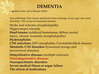
  • 4. DEMENTIA • a generic term, not a disease entity • Any pathology that causes significant brain damage, at any age, can cause dementia. The causes of dementia include: Stroke and ischemic encephalopathy Hippocampal sclerosis Head trauma (subdural hematomas, diffuse axonal injury, chronic traumatic encephalopathy) Hydrocephalus CNS infections (HIV encephalitis, Creutzfeldt-Jakob disease) Metabolic CNS disorders (lysosomal storage and peroxisomal diseases) Demyelinative diseases (multiple sclerosis) Neurodegenerative diseases Neuropsychiatric disorders Severe medical illness or organ failure The effects of medications
  • 5. DEGENERATIVE DISEASES AS PROTEINOPATHIES • the deposition of abnormal proteins in the brain
  • 6. THE UBIQUITIN-PROTEASOME AND LYSOSOMAL SYSTEM IN NEURODEGENERATIVE DISEASES Protein recycling in cells • Proteasome(UPS) : Short lived proteins • Lysosomes : long lived proteins, cytoplasm, oganelles.
  • 7. ALZHEIMER'S DISEASE • the most common cause of dementia in old people. • 13% of persons over 65 years and 45% of those over 85 have AD. • characterized by loss of memory, inability to learn new things, loss of language function, deranged perception of space, inability to do calculations, indifference, depression, delusions, and other manifestations. • Mild cognitive impairment (MCI) memory and other cognitive impairments that do not affect social functioning or daily living.
  • 8. PATHOGENESIS OF AD • two processes: extracellular deposition of beta amyloid-Aβ and intracellular accumulation of tau protein. • BETA AMYLOID- Aβ is toxic to neurons • TAU- Over-phosphorylated microtubule associated protein tau • Aβ deposition is specific for AD and is thought to be primary. • Tau accumulation is also seen in other degenerative diseases and is thought to be secondary.
  • 10. PATHOLOGY OF AD • senile plaques (SPs)- Diffuse Aβ plaques (AβPs) and neuritic plaques (NPs). • neurofibrillary tangles(NFTs)-deposits of tau filaments in the neuronal body
  • 11. Large numbers of plaques and tangles cause massive loss of neurons and synapses. Cortical atrophy in advanced AD Cortical atrophy and hydrocephalus in AD The neurofibrillary tangles are composed of paired helical filaments
  • 12. Diffuse plaque. Beta amyloid immunostain Neurofibrillary tangles. Bielschowsky silver stain. Gliosis in and around plaques. GFAP immunostain Beta amyloid immunostain. Amyloid in plaques and vessels in the cerebral cortex
  • 13. ETIOLOGY OF AD-GENETICS • The gene for APP is on chromosome 21 • the presenillin 1 and 2 genes (PSEN1 and PSEN2) on chromosomes 14 and 1 respectively • the vast majority (95%) of AD cases appear in old age and do not follow Mendelian inheritance. • an interaction of genetic and other intrinsic and environmental factors. • the most important genetic risk factor is the Apolipoprotein E (ApoE) genotype • three isoforms: ApoE2, ApoE3, and ApoE4- (chromosome 19)
  • 14. ETIOLOGY OF AD-OTHER CONTRIBUTING FACTORS • environment, diet, and general state of health • Neuroinflammation • Free radicals • Diabetes • Traumatic brain injury • Homocysteine
  • 15. • OLD AGE AND AD • VASCULAR DISEASE IN AD • NEUROTRANSMITTERS IN AD • decreased acetylcholine (Ach), choline acetyltransferase (CHAT)
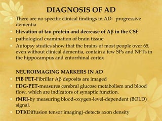
  • 16. DIAGNOSIS OF AD • There are no specific clinical findings in AD- progressive dementia • Elevation of tau protein and decrease of Aβ in the CSF • pathological examination of brain tissue • Autopsy studies show that the brains of most people over 65, even without clinical dementia, contain a few SPs and NFTs in the hippocampus and entorrhinal cortex NEUROIMAGING MARKERS IN AD • PiB PET-Fibrillar Aβ deposits are imaged • FDG-PET-measures cerebral glucose metabolism and blood flow, which are indicators of synaptic function. • fMRI-by measuring blood-oxygen-level-dependent (BOLD) signal. • DTI(Diffusion tensor imaging)-detects axon density
  • 17. • Should cases with a few SPs and NFTs be diagnosed as AD, even if there is no clinical dementia? • The NIA-Reagan and CERAD diagnostic criteria for AD • not only the numbers of SPs and NFTs but also age and intellectual function
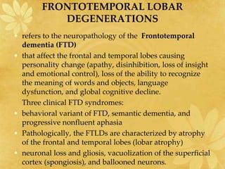
  • 18. FRONTOTEMPORAL LOBAR DEGENERATIONS • refers to the neuropathology of the Frontotemporal dementia (FTD) • that affect the frontal and temporal lobes causing personality change (apathy, disinhibition, loss of insight and emotional control), loss of the ability to recognize the meaning of words and objects, language dysfunction, and global cognitive decline. Three clinical FTD syndromes: • behavioral variant of FTD, semantic dementia, and progressive nonfluent aphasia • Pathologically, the FTLDs are characterized by atrophy of the frontal and temporal lobes (lobar atrophy) • neuronal loss and gliosis, vacuolization of the superficial cortex (spongiosis), and ballooned neurons.
  • 19. Three groups of FTLDs are recognized: • 1.FTLD-TAU with tau-positive inclusions (tauopathies) • 2. FTLD-TDP with tau-negative, alpha-synuclein- negative inclusions; RNA binding • 3. FTLD-FUS, containing fused in sarcoma protein; RNA binding • The pathological diagnosis of FTLDs requires extensive tissue sampling and immunohistochemistry targeting tau, TDP-43, and ubiquitin.
  • 20. FTLD-TAU (TAUOPATHIES) • Tau (the Greek letter) is a microtubule associated protein • the gene MAPT gene on 17q21: Normally, tau is phosphorylated and is present mainly in axons • In FTLDs, abnormal deposits of hyperphosphorylated tau • The FTLD-TAU group includes: – Pick's Disease FTD with Parkinsonism Linked to Chromosome 17 (FTDP-17), a MAPT mutation Corticobasal Degeneration Progressive supranuclear palsy
  • 21. PICK DISEASE • the prototype of FTLDs • It presents between 45-65 years with confusion and FTLD symptoms and has a progressive course lasting 2-5 years • Pick bodies (tau-positive spherical cytoplasmic neuronal inclusions, composed of straight filaments) • Pick cells (ballooned neurons with dissolution of chromatin) Frontotemporal atrophy Pick inclusion bodies -tau immunostain Ballooned neuron (Pick cell)
  • 22. FTLD-TDP (TDP-43 PROTEINOPATHIES) • TDP-43, encoded by the TARDBP gene on chromosome 1 • Genetic forms(3 types) and sporadic forms • Deposited in neurons in the form of intranuclear(NII), cytoplasmic(NCI), and neuritic inclusions. • Overlap between TDP-43 proteinopathies and motor neuron diseases. -ALS patients present with FTD symptoms: hexaneucleotide repeat(C9orf72) and TDP-43 mutation ) • Progranulin mutation • TDP-43 deposits are also seen in some cases of Alzheimer's disease, Pick's disease, Parkinson's disease, and diffuse Lewy body disease.
  • 23. PARKINSON'S DISEASE • second most common neurodegenerative disease after AD, and the most frequent subcortical degenerative disease. Loss of dopaminergic neurons in substantia niagra • motor symptoms (rigidity, tremor at rest, slowness of voluntary movement, stooped posture, a shuffling, small-step gait, difficulty with balance) • nonmotor symptoms (expressionless face, soft voice, olfactory loss, mood disturbances, dementia, sleep disorders, and autonomic dysfunction, including constipation, cardiac arrhythmias, and hypotension) • Sporadic • 5-10 % of patients have a monogenic, autosomal dominant or recessive form of PD • The first autosomal dominant form of PD : α-synuclein, a synaptic protein encoded by SNCA on 4q22; other: LRRK2 Mutation • Autosomal recessive PD is Parkin. Others: DJ-1, PINK1-Roll in clearance of dysfunctional mitochondria • association with Gaucher disease (GD)
  • 24. Pathology in PD • The key pathology in PD is α-synuclein accumulation (lipid binding protein normally associated with synapses) • Lewy bodies (LBs): aggregates form round lamellated eosinophilic cytoplasmic inclusions in the neuronal body -By Fredrich Henry Lewey • Lewy neurites: fibrils made of insoluble polymers of α- synuclein -Deposited in neuronal processes and in astrocytes and oligodendroglial cells • Its accumulation impairs the functions of mitochondria, lysosomes, and endoplasmic reticulum, and interferes with microtubular transport.
  • 25. Left:PD; Right: normal SN Normal sustantia nigra-zona compacta Loss of sustantia nigra neurons in PD Lewy body in a SN neuron
  • 26. • antibodies to α-synuclein made it easier to detect LBs and synuclein deposits outside the SN. • PD should be viewed as a systemic disorder that affects the entire CNS and PNS, not just the dopaminergic system. • explains the diverse motor and nonmotor manifestations of PD • AD and PD (and in a broader sense tauopathies and synucleinopathies) overlap clinically and pathologically. • Trials of neural implantation
  • 27. DIFFUSE LEWY BODY DISEASE • Lewy body dementia • sporadic neurodegenerative disease • second most common cause of dementia after AD • It combines the neurological manifestations of dementia and parkinsonism. • shows small, inconspicuous Lewy bodies in the neocotex, limbic system, and brainstem Cortical Lewy bodies. Alpha-synuclein immunostain.
  • 28. MPTP PARKINSONISM • The pyridine analogue MPTP (1-methyl-1-4-phenyl- 1,2,3,6-tetrahydropyridine) • taken up selectively by dopaminergic neurons. • active compound, MPP (1-methyl-4- phenylpyridinium) inhibits mitochondrial function and induces cell death • Self-made synthetic heroin, contaminated by MPTP • rotenone • Caffeine and nicotine : ?protective
  • 29. GUAM PARKINSON-DEMENTIA (GPD) • Chamorro people of Guam • combination of tauopathy and synucleinopathy • this disease in successive generations implicates genetic factors. • GUAM
  • 30. In recent years, the incidence of GPD has declined dramatically Hypothesis : • GPD is caused by a toxic amino acid in the seed of a cycad plant which is used to make flour and is a staple in the local diet. • deficiencies of calcium and magnesium, and high concentrations of aluminum and other minerals in drinking water, wth aluminium deposition in neurons • GPD underlines the important role of genetics and environmental neurotoxins in the pathogenesis of neurodegenerative diseases. • Other conditions that damage the SN: striatonigral degeneration, postencephalitic parkinsonism, manganese poisoning, carbon monoxide poisoning, hypoxic-ischemic encephalopathy, traumatic brain injury, and stroke.
  • 31. ATYPICAL PARKINSONISM SYNDROMES - Progressive supranuclear palsy - corticobasal degenerations - multisystem atrophy
  • 32. HUNTINGTON'S DISEASE • Fatal autosomal dominant condition; • Average course-15yrs • behavioral changes, chorea, and dementia. • Atrophy of the caudate nucleus and putamen and dilatation of the anterior horns of the lateral ventricles • Degeneration begins in the caudal portions of the caudate and putamen and advances rostrally. • Cortical atrophy. • Microscopically: – there is loss of medium size spiny neurons those use GABA, enkephalin, dynorphin, substance P as neurotransmitters-in the caudate nucleus and putamen – loss of cortical neurons and gliosis – Proliferation of reactive astrocytes – neuronal intranuclear inclusions
  • 33. Normal caudate nuclei, small lateral ventricles. HD- Atrophy of the caudate nuclei and dilatation of the lateral ventricles. from Huntington's disease; right GFAP IHC: Caudate nucleus from normal brain; left.
  • 34. Molecular abnormality of HD • Huntingtin- protein involved in cellular transport • CAG trinucleotide expansion of the HTT(HUNTINGTIN) gene on chromosome 4p: adds a polyglutamine segment • the CAG-expanded huntingtin has a toxic function • CAG triplet expansion is probably due to malfunction of cellular processes that repair strand breaks and remove mispaired bases from DNA. • expanded huntingtin, conjugated with ubiquitin, forms aggregates (inclusions) in the nuclei cytoplasm and dendrites of affected neurons. -Detected by IHC using antibodies to huntingtin • Adult-onset patients have 40-50 repeats • A high number of repeats is associated with early onset and more rapidly progressing disease • expanded CAG repeat is unstable and may increase in size in successive generations. • the disease to appear at a younger age and more severe form, a phenomenon known as anticipation
  • 35. • CAG repeats on other genes: spinobulbar muscular atrophy (Kennedy's disease), machado-Joseph disease cerebellar degenerations *Biochemical analysis of the striatum in HD shows loss of neurotransmitters, including GABA, acetylcholine and glutamate *Decrease of GABA and unbalanced dopamine activity result in chorea. -Chorea with acanthocytosis- With RBC abnormality -Sydenham Chorea- antibodies act against neuronal antigens in Basal ganglia
  • 36. ATAXIA AND SPINOCEREBELLAR DEGENERATION • spinal or sensory ataxia • cerebellar ataxia • spinocerebellar ataxia • Grouped according to genetics
  • 37. Autosomal dominant spinocerebellar ataxias (ADSCAs) 1.CAG triplet expansion • If the expansion lies in a coding sequence, it is translated into a polyglutamine (polyQ) stretch of the affected protein • phenomenon of anticipation • The expansion occurs more often with paternal transmission. 2. Expansion of non-coding region repeats 3. Point mutations Characteristics: • cause parkinsonism and other extrapyramidal manifestations • neuropathology is cerebellar degeneration • additional degenerations involving the basal ganglia, substantia nigra, motor neurons of the brainstem and spinal cord, the spinocerebellar and olivocerebellar tracts, cerebral cortex, and other systems • M/S: shows loss of Purkinje cells and neurons in other affected nuclei
  • 38. Friedreich's ataxia (FRDA) • The most frequent inherited ataxia • an autosomal recessive disease which begins usually before age 20 with gait ataxia, proprioceptive and superficial sensory loss, weakness and atrophy of the extremities, spasticity with extensor plantar responses and cardiomyoathy. • GAA trinucleotide repeat expansion of the frataxin (FXN) gene on chromosome 9q21: Mitochondrial matrix protein which is involved in iron homeostasis in iron sulfur enzyme clusters in complex I & II. • Loss of sensory ganglion cells and degeneration of their axons in peripheral nerves, dorsal roots, and posterior columns deprives the cerebellum of sensory input that is necessary to coordinate movement.
  • 39. Ataxia-telangiectasia • a childhood disease -ataxia, extrapyramidal dysfunction, peripheral neuropathy and other neurologic deficits, vascular dilatation in CNS, skin and conjuctiva, and immunodeficiency • mutations of a gene(ATM) that regulates the cell cycle result in defective DNA repair • Cerebellar deegeneration (loss of Purkinje and granule cells). Loss of anterior horns, degeneration of brainstem nuclei, substantia nigra, and other neuronal groups, and loss of dorsal root ganglionic neurons with dorsal column degeneration • Amphicytes • A-T patients frequently develop opportunistic infections and T-cell leukemias. (? B-cell lymphomas).
  • 40. Cerebellar ataxias • Cerebellar cortical (cerebello-olivary) degeneration consists of loss of Purkinje cells and inferior olivary neurons. • Olivopontocerebellar atrophy (OPCA) there is, in addition, loss of neurons in the pontine nuclei, and atrophy of the transverse fibers of the pons and middle cerebellar peduncles.
  • 41. • Multiple system atrophy-MSA: combination of parkinsonism, ataxia, and autonomic dysfunction – loss of normal pigmentation in the substantia nigra – atrophy of the cerebellum, basis pontis, and inferior olives and atrophy of the putamen – m/s: neuronal loss, axon and myelin degeneration, and gliosis – glial cytoplasmic inclusions-GCI: filamentous α- synuclein • Pontocerebellar hypoplasia- In children – degeneration of motor neurons and other systems, congenital glycosylation disorders
  • 42. MOTOR NEURON DISEASES • A fatal degenerative disorder of upper and lower motor neurons • Primary lateral sclerosis : ALS variant that affects upper motor neurons only • Progressive bulbar palsy :ALS begins with brainstem symptoms (dysarthria, difficulty swallowing) followed by extremity weakness. • Progressive muscular atrophy : have only lower motor neuron involvement • Frontotemporal dementia • The majority of patients die, usually from respiratory paralysis, within 2-3 years • Sporadic • Familial ALS (FALS) : • 1. hexanucleotide (GGGGCC) repeat expansion in a noncoding region of C9ORF72 gene on 9p21- FTD and TDP-43 inclusions. 2. chromosome 21q that encodes Cu/Zn superoxide dismutase (SOD1). 3. Glutamate toxicity Amyotrophic Lateral Sclerosis (ALS)
  • 43. • The pathology of ALS Loss of motor neurons in the hypoglossal nuclei Denervation atrophy Degeneration of the corticospinal tracts. Myelin stain.
  • 45. Spinal Muscular atrophy (SMA) • Genetic disorder that causes degeneration and loss of spinal and brain stem motor neurons (lower motor neurons) resulting in progressive muscle weakness • most common fatal recessive disorder in children after cystic fibrosis • deficiency of a protein encoded by the Survival Motor Neuron gene on chromosome 5q11-q13 • SMN protein is important in RNA processing in all cells and appears to have a critical but unknown function in survival of motor neurons • Two forms of the SMN gene, the telomeric (SMN1) and centromeric (SMN2) • Most SMA cases are caused by homozygous deletion of SMN1
  • 46. clinical spectrum SMA • denervation atrophy of muscle, manifested by severe hypotonia, weakness, and inability to breathe. • Degenerating brain stem and spinal motor neurons undergo chromatoysis and die. • Neuronophagia, gliosis
  • 47. SUMMARY • Although each neurodegenerative disease is presented as a distinct entity there is often overlap. • Studying disease heterogeneity at autopsy is key to understanding discrepancies between clinical and pathological diagnoses. • The increasing complexity of neuropathological findings necessitates that all aspects of the antemortem clinical picture be considered to study their molecular and structural correlates. • Neuropathologists need: Age of onset, symptom duration, clinical diagnosis, clinical indices of disease severity, and the density and distribution of the various pathologic processes. • Many efforts to develop biomarkers • Neuropathology will continue to be the gold standard for the foreseeable future.