ADVANCED  PATHOPHYSIOLOGY
FUNDAMENTALS OF   PATHOPHYSIOLOGY PHYSICAL RESPONSE TO STRESS According to Hans Selye’s General Adaptation Model, the body reacts to stress in the stages depicted below .
PHYSICAL OR PSYCHOLOGICAL STRESSOR ALARM REACTION (FIGHT-OR-FLIGHT RESPONSE) * Arousal of the central nervous system begins. * Epinephrine and norepinephrine, along with other hormones, are released, causing an increase in heart  rate, force of heart  contractions, oxygen intake, and mental activity. RESISTANCE * The body responds to the stressor and attempts to return to homeostasis. * Coping mechanisms come into play. RECOVERY * If stress ceases, the body should return to a normal state,  leading to recovery. EXHAUSTION * The body can no longer produce hormones as it did in the alarm stage. * Organ damage begins.
HEMATOLOGIC SYSTEM KEY FACTS ABOUT THE HEMATOLOGIC SYSTEM Blood is a major body tissue  Plasma factors and platelets control clotting Erythrocytes carry oxygen; remove carbon dioxide Leukocytes act in inflammatory and immune responses Plasma carries antibodies and nutrients to tissues and carries away waste Hematopoiesis occurs primarily in marrow Average person has 5 to 6 L of circulating blood
PATHOPHYSIOLOGIC CHANGES Bone marrow cells particularly vulnerable to physiologic changes Disease can affect structure or concentration of any hematologic cell
CHARACTERISTICS   OF DIC Life-threatening disorder Occurs as a complication of diseases and conditions that accelerate clotting Clotting in the microcirculation affects kidneys, extremities, brain, lungs, pituitary and adrenal glands, GI mucosa Also called consumption coagulopathy or defibrination syndrome Generally acute but may be chronic in cancer patients Prognosis depends on early detection and treatment, severity of hemorrhage, treatment of underlying disease
Understanding Disseminated Intravascular Coagulation and its Treatment Precipitating mechanism Tissue damage Treat the underlying problem Endothelial damage Increased tissue  thromboplastin Intrinsic pathway of coagulation Extrinsic pathway of coagulation Heparin to prevent microclotting (controversial) Intravascular coagulation (production of microthrombi) Production of thrombi Activation of fibrinolytic system Digestion of fibrin clots Inhibition of platelet function Consumption of clotting factors Decreased clotting factors Cryoprecipitate factor VIII Fresh frozen plasma Platelets Occlusion of small blood vessels Tissue necrosis Thrombocytopenia Bleeding Blood Key:  = treatment
Causes * Infection * Obstetric complications * Neoplastic disease How it Happens Foreign protein enters circulation or vascular endothelial injury occurs Accelerated clotting, activation of prothrombin, excess of thrombin Thrombin converts fibrinogen to fibrin Fibrin clots in microcirculation Hypofibrinogenemia, hypopro-thrombinemia, thrombocytopenia, deficiencies in factors V and VIII Fibrinolytic system activates, dissolves fibrin clots Hemorrhage is result of fibrin degradation products, depletion of plasma coagulation factors
Key signs and symptoms Abnormal bleeding Cutaneous oozing of serum Petechiae Bleeding Complications Acute tubular necrosis Shock Multiple organ failure Diagnosis Based on decreased platelet count and low fibrinogen level Prothrombin time >15 seconds Partial thromboplastin time >60 seconds Increased fibrin degration products Treatment Treatment of underlying disorder  Administration of blood Fresh frozen plasma, platelet, packed RBCs Heparin therapy (controversial)
Key Nursing Actions Patient care must focus on early recognition of abnormal bleeding, prompt treatment o the underlying disorders, and prevention of further bleeding. Don’t scrub bleeding areas. Use pressure, cold compresses, and topical hemostatic agents to control bleeding. Enforce complete bed rest during episodes. Check all I.V. and venipuncture sites frequently for bleeding. Monitor intake and output hourly in acute DIC. Weigh dressings and linen to measure blood loss. Watch for transfusion reactions and signs of fluid overload. Weigh the patient daily. Watch for bleeding from the GI and genitourinary tracts.
CARDIOVASCULAR SYSTEM Begins when the fetus is barely 4 weeks old; last system to cease activity Helps define presence of life Comprises the heart, arteries, veins, and lymphatics Serves as the body’s transport system Brings oxygen and nutrients to cells, removes metabolic waste products, and carries hormones Commonly called the circulatory system Pulmonary circulation: blood picks up oxygen and eliminates carbon dioxide Systemic circulation: blood carries oxygen and nutrients to active cells and transports waste for excretion Blood circulates through arteries, veins, and capillaries
Characteristics of Acute Coronary Syndromes (ACS) Involve the rupture or erosion of plaque as initiating event Rupture results in platelet adhesions, fibrin clot formation, and activation of thrombin Acute MI and unstable angina now part of this group Death commonly results
CAUSES Family history Obesity Smoking High-fat / high-carbohydrate diet Sedentary lifestyle
HOW IT HAPPENS Thrombus progresses and occludes blood flow Degree of blockage and time affected vessel determines type of infarct that occurs Imbalance in myocardial oxygen supply and demand Distal microthrombi cause necrosis in some myocytes Smaller vessels infarct, placing patient at risk for non-Q-wave MI When infarct occurs myocardial cells have died Infracted muscle becomes edematous and cyanotic; leukocytes infiltrate necrotic area and begin to remove necrotic cells, thinning ventricular wall; scar formation begins
CORONARY VESSELS CONCERNS Area of the heart supplied by the affected vessel Demand for oxygen in the affected area of the heart Collateral circulation in the affected area
KEY SIGNS AND SYMPTOMS Angina as burning, squeezing, and a crushing tightness in chest Usually follows physical exertion, emotional excitement, exposure to cold, or a large meal Four major forms: stable, unstable, Prinzmetal’s (or variant), and microvascular Patient experiences severe, persistent chest pain not relieved by rest in MI Patient may have feeling of impending doom
THROMBUS FORMATION Area of plaque ruptures or erodes Platelets adhere to the damaged area and are exposed to activating factors Activation causes expression of glycoprotein llb/lla receptors Platelet aggregation and adhesion occurs Thrombus is enlarged
COMPLICATIONS   Arrhythmias  Cardiogenic shock Heart failure Pericarditis Rupture of atrial or ventricular septum, ventricular wall, or valves Death
DIAGNOSIS ECG during an anginal episode shows ischemia Coronary angiography  Myocardial perfusion imaging with thallium-201 during treadmill exercise With MI, serial serum cardiac marker measurements show elevated CK, especially CK-MB isoenzyme, troponins T and I, and myoglobin With a Q-wave MI, echocardiography shows decreased ventricular wall movement
TREATMENT Angina   Nitrates to reduce myocardial oxygen consumption Beta-adrenergic blockers to reduce workload and oxygen demands Antiplatelet drugs Coronary artery bypass grafting or PTCA Laser angioplasty Stent placement  EECP
TYPES OF MYOCARDIAL INFARCTION (MI) Inferior Anterior Septal Lateral Anterolateral Posterior Right ventricular
TREATMENT MI Thrombolytic therapy should be started within 3 hours of onset of symptoms PTCA Oxygen and nitroglycerin, unless contraindicated  Transcutaneous pacing patches (or a transvenous pacemaker) Defibrillation Epinephrine I.V. beta-adrenergic blocker followed by oral therapy ACE inhibitors Stent placement Transmyocardial revascularization Lipid-lowering drugs
KEY NURSING ACTIONS Assess and record the pain’s severity, location, type, and duration. Obtain a 12-lead ECG and assess heart rate and blood pressure when the patient is experiencing acute chest pain. During chest pain, monitor the ECG, blood pressure and pulmonary catheter measurements. Assess urine output hourly for at least the first 4 hours. Monitor the patient’s oxygen saturation levels and continue oxygen administration as ordered. Notify the physician is oxygen saturation falls below 90%. Check the patient’s blood pressure after giving nitroglycerin. Advise the patient to report typical or atypical chest pain.
TREATING MYOCARDIAL INFARCTION *Aspirin *Antiplatelet Aggregates *Glycoprotein IIB/IIIA Receptor Blocking Agents *Thrombolytic Therapy  *Percussions Transluminal CoronaryAngio-plasty *Nitrates *Beta- Adrenergic Blockers *Oxygen  *Bed Rest *Vasodilators *Morphine *Beta –Adrenergic Blockers *Nitrates *Vasodilators Change in the condition of plaque in the coronary artery Altered depolarization of the myocardium Pathologic Q wave appears Elevated ST segment T wave changes Elevated CK-MB  Elevated lactate dehydrogenase Elevated troponin and myoglobin Activation of platelets Altered repolarization of the myocardium Formation of thrombus Release of lysosomal enzymes Coronary blood supply less than demand Anaerobic glycolysis Lactic acid production Myocardial irritability Angina  Ischemia of tissue in the region supplied by the artery Myocardial cell death Arrhythmias  Decreased contractility Stimulation of the sympathetic nervous system Decreased left ventricular function Increased heart rate Increased afterload Increased 0 2  needs Vasoconstriction Increased preload Decreased cardiac output *Angiotensin- Converting Enzyme Inhibitors Decreased left ventricular ejection fraction *Fluid Restriction *Diuretics Increased central venous pressure Increased pulmonary artery wedge pressure Key:  = treatment
CHARACTERISTICS OF HYPERTENSION Elevation in diastolic or systolic blood pressure Two types: essential or secondary Major cause of stroke, cardiac disease, and renal failure Risk increases with age; higher for blacks than whites
ALERT! Elderly people may have isolated systolic hypertension, in which just the systolic blood pressure is elevated. Blacks are at an increased risk for primary hypertension when predisposition to low plasma renin levels diminishes the ability to excrete excess sodium.
CAUSES Primary hypertension Sleep apnea Diabetes mellitus Race Tobacco use Excessive alcohol consumption Obesity Secondary hypertension Coarctation of aorta Renal artery stenosis Renal parenchymal disease Head injury Pregnancy Hormonal contraceptives
Intrinsic blood pressure regulators Renin- angiotensin system Autoregulation Sympathetic nervous system Antidiuretic hormone
HOW IT HAPPENS Primary hypertension Peripheral resistance is increased blood viscosity or reduce lumen size of vessels Changes in arteriolar bed cause increased peripheral vascular resistance Increased blood volume results from renal or hormonal dysfunction Increase in arteriolar thickening caused by genetic factors Abnormal renin release, resulting in formation of angiostensin II Increases heart’s workload as resistance to left ventricular ejection increases Left ventricle hypertrophies, raising heart’s oxygen demands and workload Causes vascular damage, leading to accelerated atherosclerosis and target organ damage
HOW IT HAPPENS Secondary hypertension Insult to kidney from chronic renal disease interferes with systems causing blood pressure to increase Primary aldosteronism Pheochromocytoma increasing blood pressure
ALERT! Because many older adults have a wide auscultatory gap, failure to pump the blood pressure cuff up high enough can lead to missing the first beat and underestimating systolic blood pressure. Palpate the radial artery and inflate the cuff to a point approximately 20mm beyond which the pulse beat disappear.
KEY SIGNS AND SYMPTOMS Frequently asymptomatic  Occipital headache Associated nausea and vomiting Epistaxis Dizziness Bruits Cushing’s syndrome Pheochromocytoma
COMPLICATIONS Hypertensive crisis Peripheral artery disease  Dissecting aortic aneurysm MI Heart failure HYPERTENSIVE CRISIS Disturbance occurs Prolonged hypertension Inflammation and necrosis of arterioles Narrowing of blood vessels Restriction of blood flow to major organs Organ damage Renal, cardiac, or cerebral complications occur
CLOSER LOOK What happens in hypertensive crisis Abnormal renal function   * Myocardial ischemia  Hypertensive encephalopathy   * Eclampsia  * Intracerebral hemorrhage    * Pheochromocytoma  Withdrawal of antihypertensive  * Monoamine oxidase inhibitor interactions  drugs (abrupt)  Prolonged hypertension Inflammation and necrosis of arterioles Narrowing of blood vessels Restriction of blood flow to major organs Organ damage RENAL *Decreased renal perfusion  *Progressive deterioration of nephrons  * Decreased ability to concentrate urine  * Increased serum creatinine and blood urea nitrogen  * Increased renal tubule permeability with protein leakage into tubules  * Renal insufficiency  * Uremia  * Renal failure CARDIAC Decreased cardiac perfusion  * Coronary artery disease  * Angina or myocardial infarction  * Increased cardiac workload  * Left ventricular hypertrophy  * Heart failure CEREBRAL Decreased cerebral perfusion  * Increased stress on vessel wall  *   Arterial spasm  * Ischemia  * Transient ischemic attacks  * Weakening of vessel intima  * Aneurysm  formation  * Intracranial hemorrhage
DIAGNOSIS   Serial blood pressure measurements Urinalysis laboratory testing  Elevated blood urea nitrogen Elevated creatinine levels Hypokalemia Polycythemia Anemia
TREATMENT   PRIMARY HYPERTENSION Weight loss Dietary sodium restrictions  Physical activity Moderation of alcohol consumption DASH eating plan Thiazide diuretics  Antihypertensives Beta-adrenergic blockers Calcium channel blockers SECONDARY HYPERTENSION Parenteral administration of a vasodilator or an adrenergic inhibitor
RENAL SYSTEM Key facts about the renal system Kidneys produce and excrete urine to maintain homeostasis Ureters transport urine to bladder from kidneys Bladder: reservoir for urine until it leaves body through urethra PATHOPHYSICAL CHANGES Filtration and reabsorption changes affect total filtration effort Capillary pressure and interstitial fluid colloid osmotic pressure affect filtration Interstitial fluid pressure and plasma colloid osmotic pressure affect filtration Altered renal perfusion; disease affecting vessels, glomeruli, tubules; obstruction to urine slow the GFR
CHARACTERISTICS OF GLOMERULONEPHRITIS Bilateral inflammation of glomeruli after streptococcal infection Acute glomerulonephritis Most common in boys ages 3 to 7, but can occur at any age  Prognosis good except in elderly  RPGN Commonly occurs between ages 50 and 60 May be idiopathic or associated with proliferative glomerular Chronic glomerulonephritis   Slowly progressive disease Characterized by inflammation, sclerosis, scarring, renal failure
Key types of glomerular lesions Diffuse Focal Segmental-local ALERT! Goodpasture’s syndrome is rare but occurs most commonly in men 20 to 30 CAUSES   Acute glomerulonephritis and RPGN Streptoccocal infection of respiratory tract Impetigo Chronic glomerulonephritis  Membranoproliferative glomerulonephritis  Membranous glomerulopathy
HOW IT HAPPENS Epithelial layer of glomerular membrane is disturbed  Loss of negative charge Acute poststreptoccal glomerulonephritis   Entrapment and collection of antigen-antibody complexes in glomerular capillary membranes  Occurs after group A beta- hemolytic streptococcus infection Antigens stimulate formation of antibodies Circulating antigen- antibody complexes become lodged in glomerular capillaries Complexes initiate complement activation, release of immunologic substances that lyse cells, increase membrane permeability Severity is related to size, number, location, duration of exposure, type of antigen- antibody complexes
HOW IT HAPPENS   ( continued ) Membrane damage leads to platelet aggregation, and platelet degranulation releases substances that increase glomerular permeability Proteinuria and hematuria result  Fibrin deposits in Browman’s space, leading to diminished renal blood flow and GFR Fluid retention and decreased urine output, extracellular fluid volume expansion, and hypertension result
Goodpasture’s syndrome An RPGN in which antibodies are produced against the pulmonary capillaries and glomerular basement membrane IgA nephropathy  Berger’s disease is usually idiopathic; plasma IgA level is elevated, and IgA and inflammatory cells are deposited into Bowman’s space The result is sclerosis and fibrosis of the glomerulus and a reduced GFR Lipid nephrosis Lipid nephrosis causes disruption of the capillary filtration membrane and loss of its negative charge
KEY SIGNS AND SYMPTOMS Decreased urination or oliguria  Smoky, coffee-colored urine Dyspnea Orthopnea Hypertension ALERT ! The presenting features of glomerulonephritis in children may be encephalopathy with seizures and local neurologic deficits. An elderly patient with glomerulonephritis may report vague, nonspecific symptoms, such as nausea, malaise, and arthralgia.
COMPLICATIONS Pulmonary edema Heart failure Sepsis  Renal failure  DIAGNOSIS   Elevated electrolyte, blood urea nitrogen, creatinine levels Decreased serum protein level  Decreased hemoglobin levels in chronic glomerulonephritis Elevated antistreptolysin-O titers Elevated streptozyme ALERT Significant proteinuria isn’t a common finding in an elderly patient.
AVERTING RENAL FAILURE IN GLOMERULONEPHRITIS   Antigen –antibody reaction Antibiotics, corticosteroids, plasmapheresis Glomerular proliferation and damage  Tension builds within the rigid medullary cavity Decreased glomerular filtration rate Increased vasopressor activity Capillary damage Increased aldosterone Vasoconstriction Release of protein molecules and RBCs Sodium retention Hypertention Proteinuria or hematuria Water retention Edema Diuretics Dialysis, renal transplant Renal failure Vasolidators Key:  = treatment
TREATMENT Antibiotics  Anticoagulants Bed rest Loop diuretics Vasodilators Dialysis or kidney transplantation KEY NURSING ACTIONS Check vital signs and electrolyte values. Consult the dietitian to provide a diet high in calories and low in protein, sodium, potassium, and fluids. Administer medications as ordered. Protect the debilitated patient against secondary infection by providing good nutrition, using good hygienic technique, and preventing contact with infected people. Bed rest is necessary during the acute phase. Advise the patient with a history of chronic upper respiratory tract infections to immediately report signs of infection. Encourage pregnant women with a history of glomerulonephritis to have frequent medical evaluation.
RESPIRATORY SYSTEM KEY FACTS ABOUT THE RESPIRATORY SYSTEM Major function is gas exchange Upper airway allows airflow into lungs; warm, humidify, filter air Lower airway consists of trachea, mainstream bronchi, secondary bronchi, bronchioles, terminal bronchioles Structures are anatomic dead spaces, function only as passageways for moving air into and out of the lungs Distal to terminal bronchioles are acinus- respiratory bronchioles, alveolar ducts, alveolar sacs. Bronchioles and ducts function as conduits Alveoli: chief units of gas exchange Clearance mechanisms: cough reflex and mucociliary system.
KEY FACTS ABOUT THE RESPIRATORY SYSTEM ( continued ) Lower airway protects lungs with defense mechanisms Respiration delivers inspired air to lower respiratory tract and alveoli Contraction and relaxation of respiratory muscles moves air into and out of lungs Normal expiration is passive Adult lung contains 300 million alveoli; each supplied by many capillaries To reach capillary lumen, oxygenmust cross alveolar capillary membrane The pulmonary alveoli promote gas exchange by diffusion  Circulating blood delivers oxygen to cells for metabolism and transport metabolic wastes and CO 2  back to lungs CO 2  reaches alveolar capillaries, diffuses into the alveoli; removed from the alveoli during exhalation
CHARACTERISTICS OF ASTHMA   Chronic inflammatory disorder Airflow obstruction and hyper- responsiveness to various stimuli Obstruction caused by bronchospasm, edema of airway mucosa, and increased mucus production with plugging and airway remodeling COPD characterized by increased airflow resistance Caused by sensitivity to allergens Extrinsic asthma begins in childhood Typically, patients sensitive to specific external allergens ALERT ! 50% of asthmatics are younger than age 10; twice as many boys as girls are affected in this group.
CHARACTERISTICS OF ASTHMA ( continued ) Intrinsic asthma reacts to internal, nonallergenic factors  External substances not implicated ALERT ! Extrinsic asthma is commonly accompanied by other hereditary allergies. CAUSES Extrinsic allergens Pollen Animal dander House dust or mold Kapok or feather pillows Intrinsic allergens Irritants Emotional stress Fatigue
HOW IT HAPPENS Locus of chromosome 11 contains an abnormal gene Environmental factors interact with inherited factors Bronchial linings overreact to stimuli; when exposed to antigen IgE antibody joins with antigen Mast cells degranulate, release mediators, and release histamine and leukotrienes Histamine causes swelling in smooth muscles Mucous membranes become inflamed, irritated, and swollen Leukotrienes cause swelling in bronchi; enhance histamine effect Wheeze during coughing occurs Histamine stimulates excessive mucus secretion Goblet cells secrete viscous mucus – difficult to cough up Mucosal edema and thickened secretions further block airways On inhalation, narrowed bronchial lumen can still expand slightly On exhalation, increased intrathotacic pressure closes bronchial lumen completely Air enters but can’t escape
HOW IT HAPPENS (continued) Patient develops a barrel chest; mucus fills lung bases Blood shunted to alveoli in other lung parts Intrapleural and alveolar gas pressures rise Uneven ventillation-perfusion ratios and mismatching within different lung segments result Hypoxia triggers hyperventilation by respiratory center stimulation Respiratory alkalosis results Ventilation and perfusion remain inadequate; CO 2  retention develops Respiratory acidosis results  Respiratory failure occurs Becomes life-threatening as no air becomes audible upon auscultation and Paco 2  rises to over 70mm Hg
KEY SIGNS AND SYMPTOMS Sudden dyspnea Wheezing Tightness in chest Coughing that produces thick, clear, or yellow sputum Tachypnea Rapid pulse Profuse perspiration Hyperresonant lung fields Diminished breath sounds MEDICATIONS USED TO TREAT ASTHMA Mast cell stabilizers Antihistamines Bronchodilators
AVERTING AN ASTHMA ATTACK Exposure to allergens and causative factors  Avoidance of allergens  Allergy injections  Reduction of causative factors (stress-reduction classes)  Corticosteroids Mast cell degranulation  Immunoglobunin E stimulation   Mast cell stabilizers Histamine  Leukotrienes  Prostaglandins  Bradykinins Antihistamines Mucus secretion Inflammation  Bronchospasm Wheezing and narrowing of airways Airway obstruction Bronchodilators Key:  = treatment
4 LEVELS OF ASTHMA SEVERITY Mild intermittent: symptoms occur fewer than two times per week Mild persistent: symptoms occur more than 2 times per week Moderate persistent: symptoms occur daily Severe persistent: symptoms occur continuosly COMPLICATIONS Status asthmaticus Respiratory failure
DIAGNOSIS Signs of airway obstructive disease Serum IgE levels may increase from allergic reaction Sputum analysis may indicate presence of Curschmann’s spirals, Charcot-Leyden crystals, and eosinophils Blood count reveals increased eosinophil count Chest X-rays can be used to diagnose or monitor asthma’s progress ABG analysis detects hypoxemia Skin test may identify allergens Bronchial challenge testing evaluates allergens ECG shows sinus tachycardia during attack
TREATMENT   Drug therapy Desensitization to antigens Bronchodilators Corticosteroids  Mast cell stabilizers LTRAs Anticholinergic bronchodilators Low-flow humidified oxygen KEY NURSING ACTIONS During an acute attack Assess the severity of asthma. Administer prescribed treatments; assess the patient’s response Place the  聰 atient in high Fowler’s position. Encourage pursed-lip and diaphragmatic breathing. Monitor the patient’s vital signs. Keep in mind that developing or increasing tachypnea may indicate worsening asthma or drug toxicity.  Administer prescribed humidified oxygen by nasal cannula at 2 L/minute to ease breathing and to increase Sa02. Observe the frequency and severity of the patient’s cough, and note whether it’s productive.
KEY NURSING ACTIONS During long-term care Monitor the patient’s respiratory status to detect baseline changes, to assess response to treatment, and to prevent or detect complications. Auscultate the lungs frequently, noting the degree of wheezing and quality of air movement. Review ABG levels, pulmonary function test results, and Sa0 2  readings. Control exercise- induced asthma by instructing the patient to use a bronchodilator or cromolyn 30 minutes before exercise.  During patient education Teach the patient and his family to avoid known allergens and irritants. Teach the patient how to use a metered-dose inhaler.  Explain how to use a peak flow meter to measure the degree of airway obstruction. Tell him to keep a record of peak flow readings.
CHARACTERISTICS OF CHRONIC BRONCHITIS   Inflammation of the bronchi caused by irritants or infection  Acute or chronic In chronic bronchitis, hyper-secretion of mucus and chronic productive cough last 3 months of the year and occur for at least 2 consecutive years ALERT ! COPD is more prevalent in urban than rural environments and is also related to occupational factors.  Children of parents who smoke are at higher risk for respiratory tract infection that can lead to chronic bronchitis.
CAUSES Exposure to irritants Cigarette smoking Genetic predisposition Organic or inorganic dusts Noxious gases Respiratory tract infection HOW IT HAPPENS Irritants inhaled for prolonged time Inflamed tracheobronchial tree leads to increased mucus production and narrowed or blocked airway Respiratory changes result in V/Q imbalance Results in hypertrophy and hyperplasia of mucous glands, increased goblet cells, ciliary damage, squamous metaplasia of columnar epithelium, and chronic leukocytic or lymphocytic infiltration of bronchial walls Hypersecretion of goblet cells blocks movement of the cilia Individual is prone to respiratory tract infections
KEY SIGNS AND SYMPTOMS Gray, white, or yellow sputum  Dyspnea Cyanosis Use of accessory muscles for breathing  Tachypnea Pedal edema Jugular vein distention Weight gain Wheezing Prolonged expiratory time Rhonchi Pulmonary hypertension COMPLICATIONS Recurrent respiratory tract infections  Cor pulmonale Pulmonary hypertension Heart failure Acute respiratory failure
DIAGNOSIS   Chest X-rays show hyperinflation and increased bronchovascular markings Pulmonary function test reveals increased residual volume, decreased vital capacity, forced expiratory flow, normal static compliance and diffusing capacity ABGs show decreased Pao 2  and normal or increased Paco 2 TREATMENT   Avoidance of air pollutants Bronchodilators Chest physiotherapy Ultrasonic or mechanical nebulizers Corticosteroids
KEY NURSING ACTIONS Encourage smoking- cessation Perform chest physiotherapy. Warn the patient that exposures to blasts of cold air may precipitate bronchospasm. Provide adequate fluid intake.
CHARACTERISTICS OF EMPHYSEMA Form of COPD Abnormal, permanent enlargement of the acini with destruction of alveolar walls Results from tissue changes Marked by airflow limitation, lack of elastic recoil in the lungs More prevalent in males ALERT ! Aging is a risk factor for emphysema.
CAUSES   AAT deficiency  Smoking ABNORMAL ALVEOLI Can’t recoil normally after expanding Cause bronchioles to collapse on expiration As walls are destroyed, lungs become enlarged Total lung capacity and residual volume increase
HOW IT HAPPENS Recurrent pulmonary inflammation damages and destroys alveolar walls Large air spaces created Amount of air expired passively is diminished, trapping air Hyperinflation of alveoli produces bullae adjacent to pleura Septal destruction decreases airway calibration Damage occurs to respiratory bronchioles, alveolar ducts Can involve entire acinus with damage more random and involving lower lobes Pulmonary capillary destruction usually allows ventilation to perfusion match Total lung capacity and residual volume increase
ALERT! Panacinar emphysema tends to occur in elderly people with an AAT deficiency. Centriacinar emphysema occurs in smokers with chronic bronchitis KEY SIGNS AND SYMPTOMS Shortness of breath; chronic cough Anorexia and feeling of malaise  Barrel-chest Breathing through pursed lips Decreased tactile fremitus and chest expansion Hyperresonance Decreased breath sounds
COMPLICATIONS Cor pulmonale Respiratory failure Recurrent respiratory tract infections DIAGNOSIS Chest X-ray shows flattened diaphragm, reduced vascular markings at lung periphery, overaeration of the lungs, vertical heart, enlarged anteroposterior chest diameter, and large retrosternal air space Pulmonary function studies reveal reduced diffusing capacity and increase inspiratory flow ABG analysis reveals reduced Pa0 2 .
TREATMENT Avoid smoking and air pollution  Bronchodilators Antibiotics Polyvalent pneumococcal vaccine Adequate hydration  Mucolytics Aerosolized or systemic corticosteroids
KEY NURSING ACTIONS Assess for changes in baseline respiratory function. As needed, perform chest physiotherapy several times daily. Weigh the patient three times weekly and assess of edema. Make sure the patient receives adequate fluids to loosen secretions. Schedule respiratory therapy at least 1 hour before or after meals. Warn the patient that exposure to blasts of cold air may precipitate bronchospasm.
ENDOCRINE SYSTEM KEY FACTS ABOUT THE ENDOCRINE SYSTEM Consists of glands, specialized cell clusters, hormones, tissues Glands and cell clusters secrete hormones, chemical transmitters in response to stimulation With nervous system, regulates metabolic activities, maintains internal homeostatis Hormones connect with receptors in target tissues Resulting hormone-receptor complex triggers target cell’s response
HORMONAL REGULATION Hypothalamus helps control some endocrine glands Neural stimulation of posterior pituitary causes secretion of ADH, oxytocin Hypothalamic hormones stimulate pituitary gland to synthesize and release trophic hormones. Trophic hormones stimulate adrenal cortex, thyroid gland, gonads. Hypothalamic hormones stimulate pituitary to release or inhibit release of effector hormones Negative feedback system regulates endocrine system Simple feedback occurs when level of one substance regulates secretion of a hormone
RHYTHMS   Circadian rhythm increases and decreases hormone levels by time of day Infradian rhythm” biorhythm that repeats in patterns > 24 hours HORMONAL EFFECTS Oxytocin: stimulates contractionof uterus, milk- letdown reflex ADH: controls concentration of body fluids Proclactin: controls milk secretion , GH GH: triggers growth Iodinated hormones: affect growth, development PTH: regulates calcium, phosphate metabolism.
CHARACTERISTICS OF DIABETES MELLITUS Metabolic disorder characterized by hyperglycemia resulting from lack of insulin or insulin effect Three general classifications: type 1, type 2, gestational diabetes Type 1 occurs before age 30 Type 2 occurs in obese adults after age 40 CAUSES Etiology of type 1 and type 2 unknown Genetic factors may play role Autoimmune disease, viral infections may be risk factors in type 1 Obesity Physiologic or emotional stress  Pregnancy causes weight gain, increases levels of hormones, which antagonize insulin Medications that antagonize effects of insulin
HOW IT HAPPENS Type 1 Triggering event in susceptible person causes production of autoantibodies against pancreas beta cells Leads to decline in and lack of insulin secretion Leads to hyperglycemia, enhanced lipolysis, protein catabolism Characteristics occur when more than 90% of beta cells destroyed Type 2 Impaired insulin secretion, inappropriate hepatic glucose production, peripheral insulin receptor insensitivity Genetic factors significant Onset accelerated by obesity, sedentary lifestyle Gestational   Woman without diabetes shows glucose intolerance during pregnancy May occur if placental hormones counteract insulin Significant risk factor for future occurrence of type 2
ALERT! Type 1 diabetes usually presents rapidly. Type 2 diabetes is typically slow and insidious in onset. KEY SIGNS AND SYMPTOMS Polyuria and polydipsia Anorexia or polyphagia Headaches, fatigue, lethargy, reduced energy levels Muscle cramps, irritability, emotional lability Vision changes Numbness and tingling
COMPLICATIONS   Ketoacidosis and hyperosmolar coma Cardiovascular disease Peripheral vascular disease Retinopathy Nephropathy DIAGNOSIS   In adult men and nonpregnant women Fasting plasma glucose level of 126 mg/dl or more on at least two occasions Documentation showing typical symptoms of uncontrolled diabetes Random blood glucose level of 200 mg/dl or more 2 hours after ingesting 75g of oral dextrose Urinalysis for acetone and glycosylated hemoglobin
TREATMENT Type 1 Insulin replacement Meal planning Exercise Type 2 Oral antidiabetic drugs Meal planning  Maintain appropriate body weight Gestational   Medical nutrition therapy Injectable insulin if needed Postpartum counseling, regular exercise, prevention of weight gain
Treatment of type 1 diabetes mellitus Genetic predisposition  Environmental or viral stressor  Destruction of alpha and beta cells of the pancreas Pancreatic transplantation Failure to produce insulin Production of excess glucagon Production of glucose from protein and fat stores Wasting of lean body mass Chronic elevations in blood glucose levels Increased ketones Acidosis Fatigue Weight loss Acetone breath odor Increased osmolarity due to glucose Elevated blood glucose level Polydipsia Polyuria Polyphagia Weight loss Small-vessel disease Accelerated atherosclerosis Impaired immune function Diabetic retinopathy Diabetic nephropathy Diabetic retinopathy Infection Delayed wound healing Dialysis, transplantation End- stage renal failure Laser therapy Hypertension Coronary artery disease Increased low-density lipoprotein levels Loss of vision, blindness Symmetrical loss of sensation Numbness and tingling in the extremities Wasting of intrinsic muscles Charcot’s joint (neopathic joint disease) Autonomic neuropathy Diabetic and foot ulceration Insulin, meal planning, exercise Key:  = treatment
KEY NURSING ACTIONS Stress the importance of complying with the prescribed treatment program Be alert for signs of ketoacidosis and hyperosmolar coma Watch for complications of diabetic therapy, especially hypoglycemia Obtain blood glucose, glycosylated hemoglobin, lipid levels, and blood pressure measurements regularly. Watch for diabetic effects on the cardiovascular system. Treat all injuries, cuts, and blisters meticously. Urge regular ophthalmologic examiantions to detect diabetic retinopathy. Assess for signs of diabetic neuropathy. Advise genetic counseling for young adults with diabetes who are planning families.
REPRODUCTIVE SYSTEM KEY FACTS ABOUT THE REPRODUCTIVE SYSTEM Must function properly to ensure survival of species Male reproductive system produces sperm, delivers them to female reproductive tract Female reproductive system produces ova, nurtures and protects embryo and fetus; delivers it at birth Functioning is determined by anatomic structure, hormonal, neurologic, vascular, psychogenic factors
CHARACTERISTICS OF AMENORRHEA Abnormal absence or suppression of menstruation  Primary in adolescent; secondary if at least 3 months after normal onset of menarche Prognosis variable, depending on specific cause Surgical correction of outflow tract obstruction usually curative CAUSES  Anovulation due to hormonal abnormalities Absence of uterus Endometrial damage Ovarian, adrenal, pituitary tumors Emotional disorders
HOW IT HAPPENS Mechanism varies depending on cause Adequate estrogen but deficient progesterone; infertility results Hypothalamic- pituitary- ovarian axis often dysfunctional in primary disease Ovary doesn’t receive hormonal signals from CNS Secondary amenorrhea results from central factors, uterine factors, cervical stenosis, premature ovarian failure, others KEY SIGNS AND SYMPTOMS Depend on specific cause  Absence of menstruation Vasomotor flushes
COMPLICATIONS Infertility Endometrial adenocarcinoma DIAGNOSIS Confirmed by history of failure to menstruate in females ages 16 and older Secondary disease diagnosed after absence of menses for 3 months in previously established pattern Rule out pregnancy and anatomic abnormalities Diagnosis confirmed with onset of menstruation within 1 week after giving pure progestational agents Blood and urine show hormonal imbalances
TREATMENT Appropriate hormone replacemnet Inducing ovulation  FSH and human menopausal gonadotropins Improving nutritional status Modifying daily exercise routine KEY NURSING ACTIONS Explain all diagnostic procedures Provide reassurance and emotional support Psychiatric counseling may be necessary if amenorrhea results from emotional disturbances  After treatment, teach the patient how to keep an accurate record of her mentrual cycles to aid early detection of recurrent amenorrhea.
CHARACTERISTICS OF BPH Also known as benign  prostatic hypertrophy Prostate gland enlarges enough to compress urethra, causes urinary obstruction Treatment depends on size of prostate, age and health of patient CAUSES Age- associated changes in hormone activity Arteriosclerosis Inflammation
HOW IT HAPPENS Begins with nonmalignant changes in periurethral glandular tissue Growth of fibroadenomatous nodules compress normal gland Prostate enlarges, may extend into bladder and obstruct urinary outflow Progressive bladder distention may cause pouch to form in bladder that retains urine  Retained urine may lead to calculi, cystitis.
COMPLICATIONS Complete urinary obstruction  Infection DIAGNOSIS   Visible midline mass above symphysis pubis  Rectal palpation reveals enlarged prostate  Elevated blood urea nitrogen and serum creatinine levels Elevated PSA TREATMENT   Prostate massages  Sitz baths Fluid restrictions Antimicrobials Regular ejaculation Alpha – adrenergic blockers
KEY NURSING ACTIONS Monitor and record vital signs, intake and output, and daily weight. Watch closely for signs of post-obstructive diuretics.  Insert an indwelling urinary catheter  for urine retention. If the catheter can’t be passed transurethrally, assist with suprapubic cystostomy under local anesthetic. After prostatic surgery Observe for immediate dangers of prostatic bleeding Keep the catheter open at a rate sufficient to maintain clear, light pink returns. Irrigate a catheter with stopped drainage due to clots with 80 to 100 ml normal saline solution, as ordered, maintaining strict sterile technique. Administer belladonna and opium suppositories or other anti-cholinergics as ordered. Suppositories and rectal temperatures are sometimes contraindicated after open prostatic procedures; confirm orders with the physician. Administer stool softeners and laxatives as ordered to prevent straining. Reassure the patient that temporary frequency, dribbling, and occasional hematuria will likely occur after the catheter is removed. Encourage annual digital rectal exams and screening for PSA to identify a possible malignancy.
FLUIDS AND ELECTROLYTES Key facts about fluid balance and exchange Kidneys regulate fluid components Fluid inside each cell is ICF; blood plasma and other fluid in the spaces outside cells is ECF ECF transport nutrients and oxygen to cells and carries away waste products for elimination Fluid is moved out of vessels by hydrostatic pressure of blood and osmotic pressure and hydrostatic pressure of interstitial fluid When the capillary wall is normal, fluid escapes at the arteriolar end of the capillary and is returned at the venular end.
KEY FACTS ABOUT PATHOPHYSIOLOGIC CHANGES IN ACID-BASE IMBALANCE Acidemia   Arterial pH<7.35 – excess acid in the blood Hydrogen ion content increases  Potassium leaves cells to neutralize, causing hypokalemia Acidosis  Systemic increase in hydrogen ion concentration Occurs when lung can’t eliminate C02, or if diarrhea causes loss of bicarbonate anions or if the kidneys fail to reabsorb bicarbonate or secrete hydrogen ions Alkalemia Arterial blood pH> 7.45 – excess base in the blood Potassium moves into cells to neutralize, causing hypokalemia
Alkalosis Bodywide decrease in hydrogen ion concentration Caused by hyperventilation and loss of nonvital acids during vomiting or from excessive ingestion of base Compensation Lungs, kidneys, other chemical buffer systems work together to maintain normal plasma pH range Buffer systems consists of a weak acid and corresponding base Four major buffers or buffer systems work to restore normal pH The kidneys normalize pH by altering handling of hydrogen and bicarbonate ions Responds in hours or days to respiratory alteration of pH Compensation by the lungs regulates respiratory rate to adjust pH; respiration increases or decreases to raise or lower Paco 2  levels
CHARACTERISTIC OF RESPIRATORY ACIDOSIS Characterized by alveolar ventilation Patient can’t clear enough C0 2  from the body  Paco 2  buildup causes hypercapnia (Paco 2  > 45 mm Hg) and acidosis May be acute or chronic CAUSES   Opioids  General anesthetics Hypnotics Injury to the medulla Reduced cardiac output Neuromuscular or respiratory disease Sleep apnea
HOW IT HAPPENS Pulmonary ventilation decreases, Paco 2  increases, and C0 2  levels rise in all tissues Respiration increases; pH falls  Respiratory mechanisms fail; kidney buffer mechanisms take over, then fail Electrolyte imbalances critically depress neurologic and cardiac functions  KEY SIGNS AND SYMPTOMS Restlessness Confusion Apprehension Somnolence Asterixis  Coma Headache Dyspnea and tachypnea Papilledema
COMPLICATIONS Profound CNS and cardiovascular deterioration Myocardial depression Elevated Paco 2  levels. DIAGNOSIS   ABG confirms when Paco 2  > 45 mm Hg  pH typically < 7.35 Chest X-ray may reveal causes Potassium > 5 mEq/L Low serum chloride Acidic urine pH
TREATMENT   Remove airway obstructions Create artificial airway if necessary Mechanical ventilation Increasing arterial oxygen to 60 mm Hg Bronchodilators Antibiotics Chest tubes PEEP Thrombolytic or anticoagulant therapy Bronchoscopy  Drug therapy KEY NURSING ACTIONS Be alert for and immediately report critical changes in respiratory, CNS, and cardiovascular functions. Maintain the patient’s airway and perform adequate humidification if on ventilation. Perform tracheal suctioning regularly and chest physiotherapy.
CHARACTERISTICS OF RESPIRATORY ALKALOSIS Paco 2  < 35 mm Hg and blood pH > 7.45 Poor prognosis when severe CAUSES Pulmonary – pneumonia, interstitial lung disease, pulmonary vascular disease, and acute asthma Non-pulmonary – anxiety, fever, aspirin toxicity, metabolic acidosis, CNS disease, sepsis, hepatic failure, and pregnancy.
HOW IT HAPPENS Pulmonary ventilation increases to maintain normal C02 levels, leading to higher pH Increased ECF hydrogen combines with bicarbonate to form acid, raising pH level Heart rate increases, blood to brain decreases, and autonomic nervous system overexcites Continued low Paco2 and vaso-constriction increase hypoxia Alkalosis oerwhelmes CNS and heart KEY SIGNS AND SYMPTOMS Deep, rapid breathing Light-headedness or dizziness Agitation Paresthesia
COMPLICATIONS Cardiac arrythmias not responsive to conventional treatment  Hypocalcemic tetany Seizures Periods of apnea DIAGNOSIS ABG analysis Paco 2  < 35 mm Hg; blood pH increases ECG indicates arrhythmias  Toxicology screening may reveal salicylate poisoning Basic urine pH detected TREATMENT Removal of ingested toxins Treatment of fever, sepsis, CNS  Oxygen administration  Breathing into a paper bag to treat hyperventilation Adjustment of mechanical ventilation
KEY NURSING ACTIONS Watch for and report changes in neurologic, neuromuscular, or cardiovascular functions Be alert for twitching and cardiac arrythmias, which may be associated with alkalemia and electrolyte imbalances.
CHARACTERISTICS OF METABOLIC ACIDOSIS Characterized by excess acid and deficient bicarbonate Can occur with increased production or decreased clearance of nonvolatile acid, or loss of bicarbonate; can be fatal ALERT ! More prevalent in children, who are more vulnerable to acid-base imbalances. CAUSES Excessive fat metabolism in absence of usable carbohydrates  Cardiac pump failure Pulmonary or hepatic disease Anemia Renal insufficiency or failure Aspirin overdose Addison’s disease Hypoaldosteronism
HOW IT HAPPENS Excess hydrogen ions decrease blood pH and respiration increases Paco 2  fall freeshydrogen ions to bind with bicarbonate  Respiration can’t compensate  Kidneys try to compensate by secreting excess hydrogen into renal tubules Excess hydrogen ions in ECF diffuse into cells; cells release potassium ions Imbalance impairs neural excitability KEY SIGNS AND SYMPTOMS Headache Lethargy  Drowsiness CNS depression Kussmaul’s respirations Hypotension Stupor
COMPLICATIONS Weakness  Flaccid paralysis Coma Ventricular arrhythmias  Cardiac arrest Growth retardation in children Bone disorders DIAGNOSIS Arterial pH < 7.35 Paco 2  < 34 mm Hg Bicarbonate levels < 22 mEq/L Urine pH <4.5 in absence of renal disease Serum potassium > 5.5 mEq/L Glucose levels > 150 mg/dl Anion gap:  ≤ 12 mEq/L indicates normal anion gap metabolic acidosis; ≥ 14 mEq/L indicates high- anion gap metabolic acidosis
TREATMENT Address underlying cause  Sodium bicarbonate I.V. I.V. lactated Ringer’s solution Mechanical ventilation Antibiotic therapy Dialysis Antidiarrheal administration  KEY NURSING ACTIONS Keep sodium bicarbonate ampules handy for emergencies. Position the patient to prevent aspiration in case of vomiting. Teach diabetic patients how to test urine; encourage adherence to hypoglycemic therapy.
CHARACTERISTICS OF METABOLIC ALKALOSIS Low acid or high bicarbonate levels cause metabolic, respiratory, and renal responses, producing characteristic symptoms Always secondary to underlying cause Prognosis good with prompt diagnosis and treatment Can lead to coma and death CAUSES Loss of acid, retention of base, or renal mechanisms associated  with low serum levels of potassium and chloride Chronic vomiting NG tube drainage or lavage without adequate electrolyte replacement Fistulas Use of steroids Massive blood transfusions Cushing’s disease
HOW IT HAPPENS Excess unbound bicarbonate raises blood pH, inhibiting respiration and raising Paco 2 Blood bicarbonate rises to > 28 mEq/L, is excreted into urine; hydrogen is retained Hydrogen in ECF drops and moves out of ICF to maintain balance; potassium moves into ICF Calcium ionization decreases; sodium moves into cells Neural impulses triggered in peripheral nervous system and CNS KEY SIGNS AND SYMPTOMS Irritability  Confusion Nausea Vomiting Diarrhea
DIAGNOSIS Blood pH > 7.45 and bicarbonate level > 26 mEq/L Paco 2  > 45 mm Hg Potassium < 3.5 mEq/L Calcium < 8.9 mg/dl Chloride < 98 mEq/L TREATMENT Cautious use of ammonium chloride I.V. or hydrochloric acid I.V. potassium chloride and normal saline Discontinue diuretics and administer supplementary potassium chloride  Oral or I.V. acetazolamide KEY NURSING ACTIONS Monitor patient’s status. Dilute potassium for I.V. fluids containing potassium salts Don’t give ammonium chloride  to patients with renal or hepatic disease. Watch for muscle weakness, tetany, or decreased activity. Irrigate NG tubes with isotonic saline solution.
GASTROINTESTINAL SYSTEM KEY FACTS ABOUT THE GI SYSTEM Supplies essential nutrients  Serves to digest food and eliminate waste Profoundly affects quality of life Two major components: GI tract and accessory organs Malfunction in system can produce life – threatening metabolic effects GI TRACT Hollow muscular tube from mouth to anus Oral cavity, pharynx, esophagus, stomach, small and large intestine Peristalsis propels ingested, material; sphincters prevent reflux PATHOPHYSIOLOGIC CHANGES Disorders typically nonspecific, reflect disruption in functions
CHARACTERISTICS OF INTESTINAL OBSTRUCTION Partial or complete blockage of lumen in small or large bowel Small- bowel obstruction more common, more serious Complete obstruction can cause death within hours Most likely to occur after abdominal surgery or in persons with congenital bowel deformities CAUSES Adhesions and strangulated hernias Carcinomas Foreign bodies Compression of the bowel wall physiologic disturbances
HOW IT HAPPEN Simple intestinal obstruction: blockage prevents intestinal contents from passing Strangulated intestinal obstruction: blood supply to obstructed section cut off, in addition to blockage of lumen Closed-loop intestinal obstruction: both ends of bowel section are occluded When obstruction occurs, fluid, air, gas collect near site Peristalsis increases temporarily, injuring intestinal mucosa, causing distention Distention blocks flow of venous blood, halts normal absorptive processes Bowel begins to secrete water, sodium, potassium into fluid pooled in lumen Small intestine obstruction results in metabolic alkalosis Lower bowel obstruction results in metabolic acidosis Obstruction may lead to ischemia, necrosis, death
PARALYTIC ILEUS Causes Trauma, toxemia, or peritonitis Electrolyte deficiencies Vascular causes Treatment Intubation  Intestinal tube Cholinergic agents ALERT ! Watch for air-fluid lock syndrome in older adults who remain recumbent for extended periods.
KEY SIGNS AND SYMPTOMS Colicky pain Nausea and vomiting Constipation Abdominal distention Drowsiness Intense thirst Malaise Aching Dry oral mucous membranes, tongue  Bowel sound, borborygmi rushes Abdominal tenderness, moderate distention
SYMPTOM PROGRESSION ON INTESTINAL OBSTRUCTION Obstruction Fluid, gas, and air collect behind obstruction Peristalsis temporarily increases in attempt to force contents past obstruction. Distention increases at and above obstruction site. Distention impedes blood supply to bowel, halting absorption. Bowel wall swells as water, sodium, and potassium are secreted into intestine and not absorbed from it. Gas-forming bacteria collect above obstruction, increasing distention. Dehydration results because fluids aren’t  absorbed into bloodstream. With no treatment, severe hypovolemia occurs. Shock Sepsis  Death
COMPLICATIONS Perforation Peritonitis Septicemia Secondary infection Metabolic alkalosis or acidosis DIAGNOSIS Progressive, colicky, abdominal pain, distention, with or without nausea and vomiting X-rays show presence and location of intestinal gas or fluid “ Stepladder” pattern emerges in small bowel on X-ray Barium enema reveals distended, air-filled colon or closed loop of sigmoid with extreme distention
TREATMENT Correction of fluid and electrolyte imbalances Decompression of bowel  Treatment of shock, peritonitis  Blood replacement I.V. fluid administration Passage of NG tube Miller- Abbott or Cantor tube  Surgical resection with anastomosis, colostomy, ileostomy KEY NURSING ACTIONS Monitor vital signs and observe for signs of shock. Stay alert for signs and symptoms of metabolic alkalosis or acidosis. Watch for signs and symptoms of secondary infections. Monitor urine output carefully to access renal function, circulating blood volume, and possible urine retention caused by bladder compression by the distended intestine. Look for signs of dehydration Record the amount and color of drainage from the decompression tube. Keep the patient in Fowler’s position as much as possible.
CHARACTERISTICS OF PEPTIC ULCERS Circumscribed lesions in mucosal membrane extending below epithelium Can develop in lower esophagus, stomach, pylorus, duodenum, jejunum Erosions, commonly called ulcers but don’t extend below epithelium  May be acute or chronic Chronic ulcers identified by scar tissue at  base 80% of peptic ulcers: duodenal  Most common in men between ages 20 and 50 Usually follow chronic course with remissions, exacerbations  5% to 10% of patients need surgery Gastric ulcers most common in middle- age and elderly men, users of NSAIDs, tobacco CAUSES H. pylori infection Use of NSAIDs  Pathologic hypersecretory disorders
HOW IT HAPPENS May result from destruction of mucosal barrier Duodenal ulcers appear to result from excessive acid production H. pylori  releases toxin that destroys gastric and duodenal mucosa Epithelium’s resistance to acid digestion reduced Results in gastritis, ulcer disease Salicylates and NSAIDs inhibit secretion of prostaglandins KEY SIGNS AND SYMPTOMS Vary by type of ulcer Gastric ulcer: pain, worsens with eating; nausea and anorexia Duodenal ulcer: gnawing, dull, aching, “hungerlike” epigastric pain; relived by food or antacids; usually recurs 2 to 4 hours later
COMPLICATIONS Hemorrhage  Shock Gastric perforation Gastric outlet obstruction DIAGNOSIS Barium swallow Upper GI and small bowel series Urea breath test results reflect activity of  H. pylori
TREATMENT Antimicrobial agents Misoprostol Antacids Anticholinergic drugs Sucralfate Proton gastric acid pump inhibitor Dietary measures NG tube Gastroscopy surgery
KEY NURSING ACTIONS Watch for adverse reactions to H 2 -receptor antagonists and omeprazole Advise any patient who uses antacids, has a history of heart disease, or follows a sodium-restricted diet to take only those antacids that contain low amounts of sodium Warn the patient to avoid steroids and NSAIDs because they irritate the gastric mucosa. AFTER GASTRIC SURGERY Keep the NG tube patent Monitor intake and output, including NG tube drainage. Replace fluids and electrolytes Monitor for possible complications.
TREATING PEPTIC ULCER DISEASE Bile salts, aspirin, nonsteroidal anti-inflammation, alcohol, ischema Damaged mucosal barrier Increased number of parietal and chief cells Inade q uate mucosal blood supply Decreased function of mucosal cells Decreased  q uality of mucus Loss of tight junctions between cells Increased sensitivity to food and other stimuli Decreased inhibition of gastric secretions Bile or pancreatic enzyme reflux from duodenum Conversion of pepsinogen to pepsin Antimicrobials Further mucosal erosion Destruction of blood vessels Bleeding Colonization by  Helicobacter pylori Ulceration Mucosal injury Dietary measures Misoprostol Sucralfate Histamine-2 blockers Antacids Formation and liberation of histamine Increased acid secretion Local Vasodilatation Anticholinergic drugs Excessive vagal stimulation Stimulation of cholinergic intramural plexus, causing muscle spasm Increased capillary permeability Loss of plasma proteins Mucosal edema Loss of plasma in gastric  lumen Key  = treatment
NERVOUS SYSTEM KEY FACTS ABOUT THE NERVOUS SYSTEM Coordinates and organizes functions of all body systems Has three main divisions: CNS, peripheral nervous system, ANS Neurons transmit nerve impulses through body Most neurons have several dendrites, only one axon Three types: sensory, motor, and interneurons Nervous system disorders can cause signs and symptoms in any body system; hard to diagnose
REVIEWING THE CNS Cerebrum – frontal lobe, temporal lobe, parietal lobe, occipital lobe Cerebellum – maintains muscle tone, coordination Brain stem – pons, midbrain, medulla oblongata Primitive structures – thalamus, hypothalamus, limbic system, RAS Spinal cord – relays sensory, motor impulses REVIEWING THE PERIPHERAL NERVOUS SYSTEM Cranial nerves –  1 2 pairs: olfactory, optic, oculomotor, trochlear, trigeminal, abducens, facial, acoustic, glossopharyngeal, vagus, spinal accessory, hypoglossal Spinal nerves - 3 1  pairs with sensory and motor neurons ANS – innervates internal organs; sympathetic nervous system
PATHOPHYSIOLOGIC CHANGES Typically involve alteration in arousal, cognition, movement, muscle tone, homeostatic mechanisms or pain Most cause more than one alteration One alteration may lead to another KEY FACTS ABOUT AROUSAL Level of consciousness or state of awareness SEVERAL MECHANISMS CAN ALTER AROUSAL Direct destruction of RAS and pathways Destruction of brain stem Compression of RAS by disease process Mechanisms may result from structural, metabolic, or psychogenic disturbances
6 STAGES OF ALTERED AROUSAL Usually begins with interruption or disruption in diencephalon Patient shows evidence of dullness, confusion, lethargy, and stupor Continued decreases result from midbrain dysfunction Coma results if medulla and pons are effected Patient will progress through rostal-caudal progression Levels: confusion, disorientation, lethargy, obtundation, stupor, coma 5 NEUROLOGIC FUNCTION AREAS USED TO IDENTIFY CAUSE Level of consciousness Pattern of breathing Pupillary changes Eye movement and reflex responses Motor responses
KEY FACTS ABOUT COGNITION Ability to be aware, perceive, reason, judge, remember, use intuition Reflects higher functioning of cerebral cortex Alteration results from direct or indirect destruction May manifest as agnosia, aphasia, or dysphasia ALTERED AROUSAL STAGES Confusion Disorientation Lethargy Obtundation Stupor coma
CHARACTERISTICS OF ALZHEIMER’S DISEASE Degenerative disorder of cerebral cortex, especially frontal lobe Accounts for more than 50% of dementia cases Found in elderly population Primary progressive dementia has poor prognosis CAUSES Exact cause unknown NEUROCHEMICAL FACTORS Deficiencies in acetylcholine, somatostatin, substance P, and norephineprine ENVIRONMENTAL FACTORS Repeated head trauma Exposure to aluminum or manganese GENETIC FACTORS Family history Down syndrome
HOW IT HAPPENS Brain tissue exhibits neurofibrillatory tangles, neuritic pla q ues, granulovascular changes Structural changes: cortical atrophy, ventricular dilation, amyloid deposits around cortical blood vessels, and reduced brain volume KEY SIGNS AND SYMPTOMS Reflect neurologic abnormalities associates with the disease Loss of memory Flattening of affect and personality Deterioration of personal hygiene Progressive impaired communication COMPLICATIONS Injury from violent behavior Pneumonia and other infections Malnutrition
DIAGNOSIS Diagnosed by exclusion PET can shows changes in metabolism of cerebral cortex CT scan shows evidence of early brain atrophy MRI shows no lesion as cause of dementia TREATMENT No cure or definitive treatment Cerebral vasodilators Hyperbaric oxygen Psychostimulants Antidepressants KEY NURSING ACTIONS Focus on supporting the patient’s remaining abilities Establish a communication system with the patient and his family Provide the patient with a safe environment

More Related Content

PPTX
Atrial Myxoma
PPTX
Bradyarrhythmia Management
PPTX
Cardiomyopathy
PPT
Valvular heart disease
ODP
Myxoma powerpoint
PPTX
Post resuscitation care
PPTX
Rheumatic heart disease
PPTX
Myocardial Infarction
Atrial Myxoma
Bradyarrhythmia Management
Cardiomyopathy
Valvular heart disease
Myxoma powerpoint
Post resuscitation care
Rheumatic heart disease
Myocardial Infarction

What's hot (20)

PPTX
Acute coronary syndrome
PPTX
Infectious diseases of the heart
PPTX
Sinus tachycardia
PPTX
Management of cardiac arrhythmias
PPT
Patent ductus arteriosus
PDF
Nursing Care of Clients with Valvular Disorders
PPTX
Valve transplant and its nursing management
PPT
Myocardial infarction
PPTX
Icu psychosis
PPTX
Acute viral myocarditis
PPTX
Pulmonary embolism
PPTX
ETHICAL AND LEGAL ISSUES IN CARDIOVASCULAR AND THORACIC NURSING.pptx
PPT
Heart failure 2013 Pathophysiology
PPT
Tricuspid atresia
PPTX
Cardiac Emergencies
PDF
Ventricular assist devices
PPTX
Cardiac tamponade
PPSX
Acute Coronary Syndrome (NSTEMI)
PPSX
Tricuspid Valvular Heart Disease for post graduates
PPT
Management of Cardiogenic shock
Acute coronary syndrome
Infectious diseases of the heart
Sinus tachycardia
Management of cardiac arrhythmias
Patent ductus arteriosus
Nursing Care of Clients with Valvular Disorders
Valve transplant and its nursing management
Myocardial infarction
Icu psychosis
Acute viral myocarditis
Pulmonary embolism
ETHICAL AND LEGAL ISSUES IN CARDIOVASCULAR AND THORACIC NURSING.pptx
Heart failure 2013 Pathophysiology
Tricuspid atresia
Cardiac Emergencies
Ventricular assist devices
Cardiac tamponade
Acute Coronary Syndrome (NSTEMI)
Tricuspid Valvular Heart Disease for post graduates
Management of Cardiogenic shock
Ad

Similar to Pathophysiology (20)

PPT
Advanced Pathophysiology
PPTX
MUCLecture_2022_12319533. Medical surgical nursing pptx
PPTX
Extrac disease part 1.pptx
PPT
Coronary Heart Disease, Myocardial Infarction, and Heart Failure, A Review of...
PPT
Atherosclerosis
PPT
disorders 224466.cardiac66335566.function.ppt
PPTX
Pathology cardiovascular diseases .pptx
PPT
1 Cardiovascular Disorders
PPTX
Acute myocardial infarction
PDF
power point myocardial infaction
PPTX
myocardial infarction
PPT
PPTX
Coronary artery diseases
PPTX
Acute coronary syndrome (2).pptx
PPTX
Cardiovascular emergencies - solo.pptx
PPTX
CAD/CAHD/CHD/IHD coronary artery disease introduction
PPT
Heart pathology
PPTX
Cardiovsacular Disorders
PPTX
Myocardial infraction sushila
Advanced Pathophysiology
MUCLecture_2022_12319533. Medical surgical nursing pptx
Extrac disease part 1.pptx
Coronary Heart Disease, Myocardial Infarction, and Heart Failure, A Review of...
Atherosclerosis
disorders 224466.cardiac66335566.function.ppt
Pathology cardiovascular diseases .pptx
1 Cardiovascular Disorders
Acute myocardial infarction
power point myocardial infaction
myocardial infarction
Coronary artery diseases
Acute coronary syndrome (2).pptx
Cardiovascular emergencies - solo.pptx
CAD/CAHD/CHD/IHD coronary artery disease introduction
Heart pathology
Cardiovsacular Disorders
Myocardial infraction sushila
Ad

Recently uploaded (20)

PDF
Calcified coronary lesions management tips and tricks
PDF
OSCE SERIES - Set 7 ( Questions & Answers ).pdf
PDF
Copy of OB - Exam #2 Study Guide. pdf
PPTX
HYPERSENSITIVITY REACTIONS - Pathophysiology Notes for Second Year Pharm D St...
PDF
MNEMONICS MNEMONICS MNEMONICS MNEMONICS s
PDF
SEMEN PREPARATION TECHNIGUES FOR INTRAUTERINE INSEMINATION.pdf
PPTX
NUCLEAR-MEDICINE-Copy.pptxbabaabahahahaahha
PDF
Transcultural that can help you someday.
PPTX
Hearthhhhhhhhhhhhhhhhhhhhhhhhhhhhhhhhhhhhhhhhhhhhhhhhhh
PPTX
CARDIOVASCULAR AND RENAL DRUGS.pptx for health study
PDF
Plant-Based Antimicrobials: A New Hope for Treating Diarrhea in HIV Patients...
PPT
Dermatology for member of royalcollege.ppt
PPT
Rheumatology Member of Royal College of Physicians.ppt
PPTX
Vaccines and immunization including cold chain , Open vial policy.pptx
PPTX
Neonate anatomy and physiology presentation
PDF
focused on the development and application of glycoHILIC, pepHILIC, and comm...
PDF
The Digestive System Science Educational Presentation in Dark Orange, Blue, a...
PPTX
NRP and care of Newborn.pptx- APPT presentation about neonatal resuscitation ...
PPTX
09. Diabetes in Pregnancy/ gestational.pptx
PDF
Comparison of Swim-Up and Microfluidic Sperm Sorting.pdf
Calcified coronary lesions management tips and tricks
OSCE SERIES - Set 7 ( Questions & Answers ).pdf
Copy of OB - Exam #2 Study Guide. pdf
HYPERSENSITIVITY REACTIONS - Pathophysiology Notes for Second Year Pharm D St...
MNEMONICS MNEMONICS MNEMONICS MNEMONICS s
SEMEN PREPARATION TECHNIGUES FOR INTRAUTERINE INSEMINATION.pdf
NUCLEAR-MEDICINE-Copy.pptxbabaabahahahaahha
Transcultural that can help you someday.
Hearthhhhhhhhhhhhhhhhhhhhhhhhhhhhhhhhhhhhhhhhhhhhhhhhhh
CARDIOVASCULAR AND RENAL DRUGS.pptx for health study
Plant-Based Antimicrobials: A New Hope for Treating Diarrhea in HIV Patients...
Dermatology for member of royalcollege.ppt
Rheumatology Member of Royal College of Physicians.ppt
Vaccines and immunization including cold chain , Open vial policy.pptx
Neonate anatomy and physiology presentation
focused on the development and application of glycoHILIC, pepHILIC, and comm...
The Digestive System Science Educational Presentation in Dark Orange, Blue, a...
NRP and care of Newborn.pptx- APPT presentation about neonatal resuscitation ...
09. Diabetes in Pregnancy/ gestational.pptx
Comparison of Swim-Up and Microfluidic Sperm Sorting.pdf

Pathophysiology

  • 2. FUNDAMENTALS OF PATHOPHYSIOLOGY PHYSICAL RESPONSE TO STRESS According to Hans Selye’s General Adaptation Model, the body reacts to stress in the stages depicted below .
  • 3. PHYSICAL OR PSYCHOLOGICAL STRESSOR ALARM REACTION (FIGHT-OR-FLIGHT RESPONSE) * Arousal of the central nervous system begins. * Epinephrine and norepinephrine, along with other hormones, are released, causing an increase in heart rate, force of heart contractions, oxygen intake, and mental activity. RESISTANCE * The body responds to the stressor and attempts to return to homeostasis. * Coping mechanisms come into play. RECOVERY * If stress ceases, the body should return to a normal state, leading to recovery. EXHAUSTION * The body can no longer produce hormones as it did in the alarm stage. * Organ damage begins.
  • 4. HEMATOLOGIC SYSTEM KEY FACTS ABOUT THE HEMATOLOGIC SYSTEM Blood is a major body tissue Plasma factors and platelets control clotting Erythrocytes carry oxygen; remove carbon dioxide Leukocytes act in inflammatory and immune responses Plasma carries antibodies and nutrients to tissues and carries away waste Hematopoiesis occurs primarily in marrow Average person has 5 to 6 L of circulating blood
  • 5. PATHOPHYSIOLOGIC CHANGES Bone marrow cells particularly vulnerable to physiologic changes Disease can affect structure or concentration of any hematologic cell
  • 6. CHARACTERISTICS OF DIC Life-threatening disorder Occurs as a complication of diseases and conditions that accelerate clotting Clotting in the microcirculation affects kidneys, extremities, brain, lungs, pituitary and adrenal glands, GI mucosa Also called consumption coagulopathy or defibrination syndrome Generally acute but may be chronic in cancer patients Prognosis depends on early detection and treatment, severity of hemorrhage, treatment of underlying disease
  • 7. Understanding Disseminated Intravascular Coagulation and its Treatment Precipitating mechanism Tissue damage Treat the underlying problem Endothelial damage Increased tissue thromboplastin Intrinsic pathway of coagulation Extrinsic pathway of coagulation Heparin to prevent microclotting (controversial) Intravascular coagulation (production of microthrombi) Production of thrombi Activation of fibrinolytic system Digestion of fibrin clots Inhibition of platelet function Consumption of clotting factors Decreased clotting factors Cryoprecipitate factor VIII Fresh frozen plasma Platelets Occlusion of small blood vessels Tissue necrosis Thrombocytopenia Bleeding Blood Key: = treatment
  • 8. Causes * Infection * Obstetric complications * Neoplastic disease How it Happens Foreign protein enters circulation or vascular endothelial injury occurs Accelerated clotting, activation of prothrombin, excess of thrombin Thrombin converts fibrinogen to fibrin Fibrin clots in microcirculation Hypofibrinogenemia, hypopro-thrombinemia, thrombocytopenia, deficiencies in factors V and VIII Fibrinolytic system activates, dissolves fibrin clots Hemorrhage is result of fibrin degradation products, depletion of plasma coagulation factors
  • 9. Key signs and symptoms Abnormal bleeding Cutaneous oozing of serum Petechiae Bleeding Complications Acute tubular necrosis Shock Multiple organ failure Diagnosis Based on decreased platelet count and low fibrinogen level Prothrombin time >15 seconds Partial thromboplastin time >60 seconds Increased fibrin degration products Treatment Treatment of underlying disorder Administration of blood Fresh frozen plasma, platelet, packed RBCs Heparin therapy (controversial)
  • 10. Key Nursing Actions Patient care must focus on early recognition of abnormal bleeding, prompt treatment o the underlying disorders, and prevention of further bleeding. Don’t scrub bleeding areas. Use pressure, cold compresses, and topical hemostatic agents to control bleeding. Enforce complete bed rest during episodes. Check all I.V. and venipuncture sites frequently for bleeding. Monitor intake and output hourly in acute DIC. Weigh dressings and linen to measure blood loss. Watch for transfusion reactions and signs of fluid overload. Weigh the patient daily. Watch for bleeding from the GI and genitourinary tracts.
  • 11. CARDIOVASCULAR SYSTEM Begins when the fetus is barely 4 weeks old; last system to cease activity Helps define presence of life Comprises the heart, arteries, veins, and lymphatics Serves as the body’s transport system Brings oxygen and nutrients to cells, removes metabolic waste products, and carries hormones Commonly called the circulatory system Pulmonary circulation: blood picks up oxygen and eliminates carbon dioxide Systemic circulation: blood carries oxygen and nutrients to active cells and transports waste for excretion Blood circulates through arteries, veins, and capillaries
  • 12. Characteristics of Acute Coronary Syndromes (ACS) Involve the rupture or erosion of plaque as initiating event Rupture results in platelet adhesions, fibrin clot formation, and activation of thrombin Acute MI and unstable angina now part of this group Death commonly results
  • 13. CAUSES Family history Obesity Smoking High-fat / high-carbohydrate diet Sedentary lifestyle
  • 14. HOW IT HAPPENS Thrombus progresses and occludes blood flow Degree of blockage and time affected vessel determines type of infarct that occurs Imbalance in myocardial oxygen supply and demand Distal microthrombi cause necrosis in some myocytes Smaller vessels infarct, placing patient at risk for non-Q-wave MI When infarct occurs myocardial cells have died Infracted muscle becomes edematous and cyanotic; leukocytes infiltrate necrotic area and begin to remove necrotic cells, thinning ventricular wall; scar formation begins
  • 15. CORONARY VESSELS CONCERNS Area of the heart supplied by the affected vessel Demand for oxygen in the affected area of the heart Collateral circulation in the affected area
  • 16. KEY SIGNS AND SYMPTOMS Angina as burning, squeezing, and a crushing tightness in chest Usually follows physical exertion, emotional excitement, exposure to cold, or a large meal Four major forms: stable, unstable, Prinzmetal’s (or variant), and microvascular Patient experiences severe, persistent chest pain not relieved by rest in MI Patient may have feeling of impending doom
  • 17. THROMBUS FORMATION Area of plaque ruptures or erodes Platelets adhere to the damaged area and are exposed to activating factors Activation causes expression of glycoprotein llb/lla receptors Platelet aggregation and adhesion occurs Thrombus is enlarged
  • 18. COMPLICATIONS Arrhythmias Cardiogenic shock Heart failure Pericarditis Rupture of atrial or ventricular septum, ventricular wall, or valves Death
  • 19. DIAGNOSIS ECG during an anginal episode shows ischemia Coronary angiography Myocardial perfusion imaging with thallium-201 during treadmill exercise With MI, serial serum cardiac marker measurements show elevated CK, especially CK-MB isoenzyme, troponins T and I, and myoglobin With a Q-wave MI, echocardiography shows decreased ventricular wall movement
  • 20. TREATMENT Angina Nitrates to reduce myocardial oxygen consumption Beta-adrenergic blockers to reduce workload and oxygen demands Antiplatelet drugs Coronary artery bypass grafting or PTCA Laser angioplasty Stent placement EECP
  • 21. TYPES OF MYOCARDIAL INFARCTION (MI) Inferior Anterior Septal Lateral Anterolateral Posterior Right ventricular
  • 22. TREATMENT MI Thrombolytic therapy should be started within 3 hours of onset of symptoms PTCA Oxygen and nitroglycerin, unless contraindicated Transcutaneous pacing patches (or a transvenous pacemaker) Defibrillation Epinephrine I.V. beta-adrenergic blocker followed by oral therapy ACE inhibitors Stent placement Transmyocardial revascularization Lipid-lowering drugs
  • 23. KEY NURSING ACTIONS Assess and record the pain’s severity, location, type, and duration. Obtain a 12-lead ECG and assess heart rate and blood pressure when the patient is experiencing acute chest pain. During chest pain, monitor the ECG, blood pressure and pulmonary catheter measurements. Assess urine output hourly for at least the first 4 hours. Monitor the patient’s oxygen saturation levels and continue oxygen administration as ordered. Notify the physician is oxygen saturation falls below 90%. Check the patient’s blood pressure after giving nitroglycerin. Advise the patient to report typical or atypical chest pain.
  • 24. TREATING MYOCARDIAL INFARCTION *Aspirin *Antiplatelet Aggregates *Glycoprotein IIB/IIIA Receptor Blocking Agents *Thrombolytic Therapy *Percussions Transluminal CoronaryAngio-plasty *Nitrates *Beta- Adrenergic Blockers *Oxygen *Bed Rest *Vasodilators *Morphine *Beta –Adrenergic Blockers *Nitrates *Vasodilators Change in the condition of plaque in the coronary artery Altered depolarization of the myocardium Pathologic Q wave appears Elevated ST segment T wave changes Elevated CK-MB Elevated lactate dehydrogenase Elevated troponin and myoglobin Activation of platelets Altered repolarization of the myocardium Formation of thrombus Release of lysosomal enzymes Coronary blood supply less than demand Anaerobic glycolysis Lactic acid production Myocardial irritability Angina Ischemia of tissue in the region supplied by the artery Myocardial cell death Arrhythmias Decreased contractility Stimulation of the sympathetic nervous system Decreased left ventricular function Increased heart rate Increased afterload Increased 0 2 needs Vasoconstriction Increased preload Decreased cardiac output *Angiotensin- Converting Enzyme Inhibitors Decreased left ventricular ejection fraction *Fluid Restriction *Diuretics Increased central venous pressure Increased pulmonary artery wedge pressure Key: = treatment
  • 25. CHARACTERISTICS OF HYPERTENSION Elevation in diastolic or systolic blood pressure Two types: essential or secondary Major cause of stroke, cardiac disease, and renal failure Risk increases with age; higher for blacks than whites
  • 26. ALERT! Elderly people may have isolated systolic hypertension, in which just the systolic blood pressure is elevated. Blacks are at an increased risk for primary hypertension when predisposition to low plasma renin levels diminishes the ability to excrete excess sodium.
  • 27. CAUSES Primary hypertension Sleep apnea Diabetes mellitus Race Tobacco use Excessive alcohol consumption Obesity Secondary hypertension Coarctation of aorta Renal artery stenosis Renal parenchymal disease Head injury Pregnancy Hormonal contraceptives
  • 28. Intrinsic blood pressure regulators Renin- angiotensin system Autoregulation Sympathetic nervous system Antidiuretic hormone
  • 29. HOW IT HAPPENS Primary hypertension Peripheral resistance is increased blood viscosity or reduce lumen size of vessels Changes in arteriolar bed cause increased peripheral vascular resistance Increased blood volume results from renal or hormonal dysfunction Increase in arteriolar thickening caused by genetic factors Abnormal renin release, resulting in formation of angiostensin II Increases heart’s workload as resistance to left ventricular ejection increases Left ventricle hypertrophies, raising heart’s oxygen demands and workload Causes vascular damage, leading to accelerated atherosclerosis and target organ damage
  • 30. HOW IT HAPPENS Secondary hypertension Insult to kidney from chronic renal disease interferes with systems causing blood pressure to increase Primary aldosteronism Pheochromocytoma increasing blood pressure
  • 31. ALERT! Because many older adults have a wide auscultatory gap, failure to pump the blood pressure cuff up high enough can lead to missing the first beat and underestimating systolic blood pressure. Palpate the radial artery and inflate the cuff to a point approximately 20mm beyond which the pulse beat disappear.
  • 32. KEY SIGNS AND SYMPTOMS Frequently asymptomatic Occipital headache Associated nausea and vomiting Epistaxis Dizziness Bruits Cushing’s syndrome Pheochromocytoma
  • 33. COMPLICATIONS Hypertensive crisis Peripheral artery disease Dissecting aortic aneurysm MI Heart failure HYPERTENSIVE CRISIS Disturbance occurs Prolonged hypertension Inflammation and necrosis of arterioles Narrowing of blood vessels Restriction of blood flow to major organs Organ damage Renal, cardiac, or cerebral complications occur
  • 34. CLOSER LOOK What happens in hypertensive crisis Abnormal renal function * Myocardial ischemia Hypertensive encephalopathy * Eclampsia * Intracerebral hemorrhage * Pheochromocytoma Withdrawal of antihypertensive * Monoamine oxidase inhibitor interactions drugs (abrupt) Prolonged hypertension Inflammation and necrosis of arterioles Narrowing of blood vessels Restriction of blood flow to major organs Organ damage RENAL *Decreased renal perfusion *Progressive deterioration of nephrons * Decreased ability to concentrate urine * Increased serum creatinine and blood urea nitrogen * Increased renal tubule permeability with protein leakage into tubules * Renal insufficiency * Uremia * Renal failure CARDIAC Decreased cardiac perfusion * Coronary artery disease * Angina or myocardial infarction * Increased cardiac workload * Left ventricular hypertrophy * Heart failure CEREBRAL Decreased cerebral perfusion * Increased stress on vessel wall * Arterial spasm * Ischemia * Transient ischemic attacks * Weakening of vessel intima * Aneurysm formation * Intracranial hemorrhage
  • 35. DIAGNOSIS Serial blood pressure measurements Urinalysis laboratory testing Elevated blood urea nitrogen Elevated creatinine levels Hypokalemia Polycythemia Anemia
  • 36. TREATMENT PRIMARY HYPERTENSION Weight loss Dietary sodium restrictions Physical activity Moderation of alcohol consumption DASH eating plan Thiazide diuretics Antihypertensives Beta-adrenergic blockers Calcium channel blockers SECONDARY HYPERTENSION Parenteral administration of a vasodilator or an adrenergic inhibitor
  • 37. RENAL SYSTEM Key facts about the renal system Kidneys produce and excrete urine to maintain homeostasis Ureters transport urine to bladder from kidneys Bladder: reservoir for urine until it leaves body through urethra PATHOPHYSICAL CHANGES Filtration and reabsorption changes affect total filtration effort Capillary pressure and interstitial fluid colloid osmotic pressure affect filtration Interstitial fluid pressure and plasma colloid osmotic pressure affect filtration Altered renal perfusion; disease affecting vessels, glomeruli, tubules; obstruction to urine slow the GFR
  • 38. CHARACTERISTICS OF GLOMERULONEPHRITIS Bilateral inflammation of glomeruli after streptococcal infection Acute glomerulonephritis Most common in boys ages 3 to 7, but can occur at any age Prognosis good except in elderly RPGN Commonly occurs between ages 50 and 60 May be idiopathic or associated with proliferative glomerular Chronic glomerulonephritis Slowly progressive disease Characterized by inflammation, sclerosis, scarring, renal failure
  • 39. Key types of glomerular lesions Diffuse Focal Segmental-local ALERT! Goodpasture’s syndrome is rare but occurs most commonly in men 20 to 30 CAUSES Acute glomerulonephritis and RPGN Streptoccocal infection of respiratory tract Impetigo Chronic glomerulonephritis Membranoproliferative glomerulonephritis Membranous glomerulopathy
  • 40. HOW IT HAPPENS Epithelial layer of glomerular membrane is disturbed Loss of negative charge Acute poststreptoccal glomerulonephritis Entrapment and collection of antigen-antibody complexes in glomerular capillary membranes Occurs after group A beta- hemolytic streptococcus infection Antigens stimulate formation of antibodies Circulating antigen- antibody complexes become lodged in glomerular capillaries Complexes initiate complement activation, release of immunologic substances that lyse cells, increase membrane permeability Severity is related to size, number, location, duration of exposure, type of antigen- antibody complexes
  • 41. HOW IT HAPPENS ( continued ) Membrane damage leads to platelet aggregation, and platelet degranulation releases substances that increase glomerular permeability Proteinuria and hematuria result Fibrin deposits in Browman’s space, leading to diminished renal blood flow and GFR Fluid retention and decreased urine output, extracellular fluid volume expansion, and hypertension result
  • 42. Goodpasture’s syndrome An RPGN in which antibodies are produced against the pulmonary capillaries and glomerular basement membrane IgA nephropathy Berger’s disease is usually idiopathic; plasma IgA level is elevated, and IgA and inflammatory cells are deposited into Bowman’s space The result is sclerosis and fibrosis of the glomerulus and a reduced GFR Lipid nephrosis Lipid nephrosis causes disruption of the capillary filtration membrane and loss of its negative charge
  • 43. KEY SIGNS AND SYMPTOMS Decreased urination or oliguria Smoky, coffee-colored urine Dyspnea Orthopnea Hypertension ALERT ! The presenting features of glomerulonephritis in children may be encephalopathy with seizures and local neurologic deficits. An elderly patient with glomerulonephritis may report vague, nonspecific symptoms, such as nausea, malaise, and arthralgia.
  • 44. COMPLICATIONS Pulmonary edema Heart failure Sepsis Renal failure DIAGNOSIS Elevated electrolyte, blood urea nitrogen, creatinine levels Decreased serum protein level Decreased hemoglobin levels in chronic glomerulonephritis Elevated antistreptolysin-O titers Elevated streptozyme ALERT Significant proteinuria isn’t a common finding in an elderly patient.
  • 45. AVERTING RENAL FAILURE IN GLOMERULONEPHRITIS Antigen –antibody reaction Antibiotics, corticosteroids, plasmapheresis Glomerular proliferation and damage Tension builds within the rigid medullary cavity Decreased glomerular filtration rate Increased vasopressor activity Capillary damage Increased aldosterone Vasoconstriction Release of protein molecules and RBCs Sodium retention Hypertention Proteinuria or hematuria Water retention Edema Diuretics Dialysis, renal transplant Renal failure Vasolidators Key: = treatment
  • 46. TREATMENT Antibiotics Anticoagulants Bed rest Loop diuretics Vasodilators Dialysis or kidney transplantation KEY NURSING ACTIONS Check vital signs and electrolyte values. Consult the dietitian to provide a diet high in calories and low in protein, sodium, potassium, and fluids. Administer medications as ordered. Protect the debilitated patient against secondary infection by providing good nutrition, using good hygienic technique, and preventing contact with infected people. Bed rest is necessary during the acute phase. Advise the patient with a history of chronic upper respiratory tract infections to immediately report signs of infection. Encourage pregnant women with a history of glomerulonephritis to have frequent medical evaluation.
  • 47. RESPIRATORY SYSTEM KEY FACTS ABOUT THE RESPIRATORY SYSTEM Major function is gas exchange Upper airway allows airflow into lungs; warm, humidify, filter air Lower airway consists of trachea, mainstream bronchi, secondary bronchi, bronchioles, terminal bronchioles Structures are anatomic dead spaces, function only as passageways for moving air into and out of the lungs Distal to terminal bronchioles are acinus- respiratory bronchioles, alveolar ducts, alveolar sacs. Bronchioles and ducts function as conduits Alveoli: chief units of gas exchange Clearance mechanisms: cough reflex and mucociliary system.
  • 48. KEY FACTS ABOUT THE RESPIRATORY SYSTEM ( continued ) Lower airway protects lungs with defense mechanisms Respiration delivers inspired air to lower respiratory tract and alveoli Contraction and relaxation of respiratory muscles moves air into and out of lungs Normal expiration is passive Adult lung contains 300 million alveoli; each supplied by many capillaries To reach capillary lumen, oxygenmust cross alveolar capillary membrane The pulmonary alveoli promote gas exchange by diffusion Circulating blood delivers oxygen to cells for metabolism and transport metabolic wastes and CO 2 back to lungs CO 2 reaches alveolar capillaries, diffuses into the alveoli; removed from the alveoli during exhalation
  • 49. CHARACTERISTICS OF ASTHMA Chronic inflammatory disorder Airflow obstruction and hyper- responsiveness to various stimuli Obstruction caused by bronchospasm, edema of airway mucosa, and increased mucus production with plugging and airway remodeling COPD characterized by increased airflow resistance Caused by sensitivity to allergens Extrinsic asthma begins in childhood Typically, patients sensitive to specific external allergens ALERT ! 50% of asthmatics are younger than age 10; twice as many boys as girls are affected in this group.
  • 50. CHARACTERISTICS OF ASTHMA ( continued ) Intrinsic asthma reacts to internal, nonallergenic factors External substances not implicated ALERT ! Extrinsic asthma is commonly accompanied by other hereditary allergies. CAUSES Extrinsic allergens Pollen Animal dander House dust or mold Kapok or feather pillows Intrinsic allergens Irritants Emotional stress Fatigue
  • 51. HOW IT HAPPENS Locus of chromosome 11 contains an abnormal gene Environmental factors interact with inherited factors Bronchial linings overreact to stimuli; when exposed to antigen IgE antibody joins with antigen Mast cells degranulate, release mediators, and release histamine and leukotrienes Histamine causes swelling in smooth muscles Mucous membranes become inflamed, irritated, and swollen Leukotrienes cause swelling in bronchi; enhance histamine effect Wheeze during coughing occurs Histamine stimulates excessive mucus secretion Goblet cells secrete viscous mucus – difficult to cough up Mucosal edema and thickened secretions further block airways On inhalation, narrowed bronchial lumen can still expand slightly On exhalation, increased intrathotacic pressure closes bronchial lumen completely Air enters but can’t escape
  • 52. HOW IT HAPPENS (continued) Patient develops a barrel chest; mucus fills lung bases Blood shunted to alveoli in other lung parts Intrapleural and alveolar gas pressures rise Uneven ventillation-perfusion ratios and mismatching within different lung segments result Hypoxia triggers hyperventilation by respiratory center stimulation Respiratory alkalosis results Ventilation and perfusion remain inadequate; CO 2 retention develops Respiratory acidosis results Respiratory failure occurs Becomes life-threatening as no air becomes audible upon auscultation and Paco 2 rises to over 70mm Hg
  • 53. KEY SIGNS AND SYMPTOMS Sudden dyspnea Wheezing Tightness in chest Coughing that produces thick, clear, or yellow sputum Tachypnea Rapid pulse Profuse perspiration Hyperresonant lung fields Diminished breath sounds MEDICATIONS USED TO TREAT ASTHMA Mast cell stabilizers Antihistamines Bronchodilators
  • 54. AVERTING AN ASTHMA ATTACK Exposure to allergens and causative factors Avoidance of allergens Allergy injections Reduction of causative factors (stress-reduction classes) Corticosteroids Mast cell degranulation Immunoglobunin E stimulation Mast cell stabilizers Histamine Leukotrienes Prostaglandins Bradykinins Antihistamines Mucus secretion Inflammation Bronchospasm Wheezing and narrowing of airways Airway obstruction Bronchodilators Key: = treatment
  • 55. 4 LEVELS OF ASTHMA SEVERITY Mild intermittent: symptoms occur fewer than two times per week Mild persistent: symptoms occur more than 2 times per week Moderate persistent: symptoms occur daily Severe persistent: symptoms occur continuosly COMPLICATIONS Status asthmaticus Respiratory failure
  • 56. DIAGNOSIS Signs of airway obstructive disease Serum IgE levels may increase from allergic reaction Sputum analysis may indicate presence of Curschmann’s spirals, Charcot-Leyden crystals, and eosinophils Blood count reveals increased eosinophil count Chest X-rays can be used to diagnose or monitor asthma’s progress ABG analysis detects hypoxemia Skin test may identify allergens Bronchial challenge testing evaluates allergens ECG shows sinus tachycardia during attack
  • 57. TREATMENT Drug therapy Desensitization to antigens Bronchodilators Corticosteroids Mast cell stabilizers LTRAs Anticholinergic bronchodilators Low-flow humidified oxygen KEY NURSING ACTIONS During an acute attack Assess the severity of asthma. Administer prescribed treatments; assess the patient’s response Place the 聰 atient in high Fowler’s position. Encourage pursed-lip and diaphragmatic breathing. Monitor the patient’s vital signs. Keep in mind that developing or increasing tachypnea may indicate worsening asthma or drug toxicity. Administer prescribed humidified oxygen by nasal cannula at 2 L/minute to ease breathing and to increase Sa02. Observe the frequency and severity of the patient’s cough, and note whether it’s productive.
  • 58. KEY NURSING ACTIONS During long-term care Monitor the patient’s respiratory status to detect baseline changes, to assess response to treatment, and to prevent or detect complications. Auscultate the lungs frequently, noting the degree of wheezing and quality of air movement. Review ABG levels, pulmonary function test results, and Sa0 2 readings. Control exercise- induced asthma by instructing the patient to use a bronchodilator or cromolyn 30 minutes before exercise. During patient education Teach the patient and his family to avoid known allergens and irritants. Teach the patient how to use a metered-dose inhaler. Explain how to use a peak flow meter to measure the degree of airway obstruction. Tell him to keep a record of peak flow readings.
  • 59. CHARACTERISTICS OF CHRONIC BRONCHITIS Inflammation of the bronchi caused by irritants or infection Acute or chronic In chronic bronchitis, hyper-secretion of mucus and chronic productive cough last 3 months of the year and occur for at least 2 consecutive years ALERT ! COPD is more prevalent in urban than rural environments and is also related to occupational factors. Children of parents who smoke are at higher risk for respiratory tract infection that can lead to chronic bronchitis.
  • 60. CAUSES Exposure to irritants Cigarette smoking Genetic predisposition Organic or inorganic dusts Noxious gases Respiratory tract infection HOW IT HAPPENS Irritants inhaled for prolonged time Inflamed tracheobronchial tree leads to increased mucus production and narrowed or blocked airway Respiratory changes result in V/Q imbalance Results in hypertrophy and hyperplasia of mucous glands, increased goblet cells, ciliary damage, squamous metaplasia of columnar epithelium, and chronic leukocytic or lymphocytic infiltration of bronchial walls Hypersecretion of goblet cells blocks movement of the cilia Individual is prone to respiratory tract infections
  • 61. KEY SIGNS AND SYMPTOMS Gray, white, or yellow sputum Dyspnea Cyanosis Use of accessory muscles for breathing Tachypnea Pedal edema Jugular vein distention Weight gain Wheezing Prolonged expiratory time Rhonchi Pulmonary hypertension COMPLICATIONS Recurrent respiratory tract infections Cor pulmonale Pulmonary hypertension Heart failure Acute respiratory failure
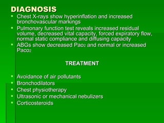
  • 62. DIAGNOSIS Chest X-rays show hyperinflation and increased bronchovascular markings Pulmonary function test reveals increased residual volume, decreased vital capacity, forced expiratory flow, normal static compliance and diffusing capacity ABGs show decreased Pao 2 and normal or increased Paco 2 TREATMENT Avoidance of air pollutants Bronchodilators Chest physiotherapy Ultrasonic or mechanical nebulizers Corticosteroids
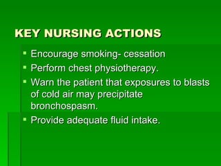
  • 63. KEY NURSING ACTIONS Encourage smoking- cessation Perform chest physiotherapy. Warn the patient that exposures to blasts of cold air may precipitate bronchospasm. Provide adequate fluid intake.
  • 64. CHARACTERISTICS OF EMPHYSEMA Form of COPD Abnormal, permanent enlargement of the acini with destruction of alveolar walls Results from tissue changes Marked by airflow limitation, lack of elastic recoil in the lungs More prevalent in males ALERT ! Aging is a risk factor for emphysema.
  • 65. CAUSES AAT deficiency Smoking ABNORMAL ALVEOLI Can’t recoil normally after expanding Cause bronchioles to collapse on expiration As walls are destroyed, lungs become enlarged Total lung capacity and residual volume increase
  • 66. HOW IT HAPPENS Recurrent pulmonary inflammation damages and destroys alveolar walls Large air spaces created Amount of air expired passively is diminished, trapping air Hyperinflation of alveoli produces bullae adjacent to pleura Septal destruction decreases airway calibration Damage occurs to respiratory bronchioles, alveolar ducts Can involve entire acinus with damage more random and involving lower lobes Pulmonary capillary destruction usually allows ventilation to perfusion match Total lung capacity and residual volume increase
  • 67. ALERT! Panacinar emphysema tends to occur in elderly people with an AAT deficiency. Centriacinar emphysema occurs in smokers with chronic bronchitis KEY SIGNS AND SYMPTOMS Shortness of breath; chronic cough Anorexia and feeling of malaise Barrel-chest Breathing through pursed lips Decreased tactile fremitus and chest expansion Hyperresonance Decreased breath sounds
  • 68. COMPLICATIONS Cor pulmonale Respiratory failure Recurrent respiratory tract infections DIAGNOSIS Chest X-ray shows flattened diaphragm, reduced vascular markings at lung periphery, overaeration of the lungs, vertical heart, enlarged anteroposterior chest diameter, and large retrosternal air space Pulmonary function studies reveal reduced diffusing capacity and increase inspiratory flow ABG analysis reveals reduced Pa0 2 .
  • 69. TREATMENT Avoid smoking and air pollution Bronchodilators Antibiotics Polyvalent pneumococcal vaccine Adequate hydration Mucolytics Aerosolized or systemic corticosteroids
  • 70. KEY NURSING ACTIONS Assess for changes in baseline respiratory function. As needed, perform chest physiotherapy several times daily. Weigh the patient three times weekly and assess of edema. Make sure the patient receives adequate fluids to loosen secretions. Schedule respiratory therapy at least 1 hour before or after meals. Warn the patient that exposure to blasts of cold air may precipitate bronchospasm.
  • 71. ENDOCRINE SYSTEM KEY FACTS ABOUT THE ENDOCRINE SYSTEM Consists of glands, specialized cell clusters, hormones, tissues Glands and cell clusters secrete hormones, chemical transmitters in response to stimulation With nervous system, regulates metabolic activities, maintains internal homeostatis Hormones connect with receptors in target tissues Resulting hormone-receptor complex triggers target cell’s response
  • 72. HORMONAL REGULATION Hypothalamus helps control some endocrine glands Neural stimulation of posterior pituitary causes secretion of ADH, oxytocin Hypothalamic hormones stimulate pituitary gland to synthesize and release trophic hormones. Trophic hormones stimulate adrenal cortex, thyroid gland, gonads. Hypothalamic hormones stimulate pituitary to release or inhibit release of effector hormones Negative feedback system regulates endocrine system Simple feedback occurs when level of one substance regulates secretion of a hormone
  • 73. RHYTHMS Circadian rhythm increases and decreases hormone levels by time of day Infradian rhythm” biorhythm that repeats in patterns > 24 hours HORMONAL EFFECTS Oxytocin: stimulates contractionof uterus, milk- letdown reflex ADH: controls concentration of body fluids Proclactin: controls milk secretion , GH GH: triggers growth Iodinated hormones: affect growth, development PTH: regulates calcium, phosphate metabolism.
  • 74. CHARACTERISTICS OF DIABETES MELLITUS Metabolic disorder characterized by hyperglycemia resulting from lack of insulin or insulin effect Three general classifications: type 1, type 2, gestational diabetes Type 1 occurs before age 30 Type 2 occurs in obese adults after age 40 CAUSES Etiology of type 1 and type 2 unknown Genetic factors may play role Autoimmune disease, viral infections may be risk factors in type 1 Obesity Physiologic or emotional stress Pregnancy causes weight gain, increases levels of hormones, which antagonize insulin Medications that antagonize effects of insulin
  • 75. HOW IT HAPPENS Type 1 Triggering event in susceptible person causes production of autoantibodies against pancreas beta cells Leads to decline in and lack of insulin secretion Leads to hyperglycemia, enhanced lipolysis, protein catabolism Characteristics occur when more than 90% of beta cells destroyed Type 2 Impaired insulin secretion, inappropriate hepatic glucose production, peripheral insulin receptor insensitivity Genetic factors significant Onset accelerated by obesity, sedentary lifestyle Gestational Woman without diabetes shows glucose intolerance during pregnancy May occur if placental hormones counteract insulin Significant risk factor for future occurrence of type 2
  • 76. ALERT! Type 1 diabetes usually presents rapidly. Type 2 diabetes is typically slow and insidious in onset. KEY SIGNS AND SYMPTOMS Polyuria and polydipsia Anorexia or polyphagia Headaches, fatigue, lethargy, reduced energy levels Muscle cramps, irritability, emotional lability Vision changes Numbness and tingling
  • 77. COMPLICATIONS Ketoacidosis and hyperosmolar coma Cardiovascular disease Peripheral vascular disease Retinopathy Nephropathy DIAGNOSIS In adult men and nonpregnant women Fasting plasma glucose level of 126 mg/dl or more on at least two occasions Documentation showing typical symptoms of uncontrolled diabetes Random blood glucose level of 200 mg/dl or more 2 hours after ingesting 75g of oral dextrose Urinalysis for acetone and glycosylated hemoglobin
  • 78. TREATMENT Type 1 Insulin replacement Meal planning Exercise Type 2 Oral antidiabetic drugs Meal planning Maintain appropriate body weight Gestational Medical nutrition therapy Injectable insulin if needed Postpartum counseling, regular exercise, prevention of weight gain
  • 79. Treatment of type 1 diabetes mellitus Genetic predisposition Environmental or viral stressor Destruction of alpha and beta cells of the pancreas Pancreatic transplantation Failure to produce insulin Production of excess glucagon Production of glucose from protein and fat stores Wasting of lean body mass Chronic elevations in blood glucose levels Increased ketones Acidosis Fatigue Weight loss Acetone breath odor Increased osmolarity due to glucose Elevated blood glucose level Polydipsia Polyuria Polyphagia Weight loss Small-vessel disease Accelerated atherosclerosis Impaired immune function Diabetic retinopathy Diabetic nephropathy Diabetic retinopathy Infection Delayed wound healing Dialysis, transplantation End- stage renal failure Laser therapy Hypertension Coronary artery disease Increased low-density lipoprotein levels Loss of vision, blindness Symmetrical loss of sensation Numbness and tingling in the extremities Wasting of intrinsic muscles Charcot’s joint (neopathic joint disease) Autonomic neuropathy Diabetic and foot ulceration Insulin, meal planning, exercise Key: = treatment
  • 80. KEY NURSING ACTIONS Stress the importance of complying with the prescribed treatment program Be alert for signs of ketoacidosis and hyperosmolar coma Watch for complications of diabetic therapy, especially hypoglycemia Obtain blood glucose, glycosylated hemoglobin, lipid levels, and blood pressure measurements regularly. Watch for diabetic effects on the cardiovascular system. Treat all injuries, cuts, and blisters meticously. Urge regular ophthalmologic examiantions to detect diabetic retinopathy. Assess for signs of diabetic neuropathy. Advise genetic counseling for young adults with diabetes who are planning families.
  • 81. REPRODUCTIVE SYSTEM KEY FACTS ABOUT THE REPRODUCTIVE SYSTEM Must function properly to ensure survival of species Male reproductive system produces sperm, delivers them to female reproductive tract Female reproductive system produces ova, nurtures and protects embryo and fetus; delivers it at birth Functioning is determined by anatomic structure, hormonal, neurologic, vascular, psychogenic factors
  • 82. CHARACTERISTICS OF AMENORRHEA Abnormal absence or suppression of menstruation Primary in adolescent; secondary if at least 3 months after normal onset of menarche Prognosis variable, depending on specific cause Surgical correction of outflow tract obstruction usually curative CAUSES Anovulation due to hormonal abnormalities Absence of uterus Endometrial damage Ovarian, adrenal, pituitary tumors Emotional disorders
  • 83. HOW IT HAPPENS Mechanism varies depending on cause Adequate estrogen but deficient progesterone; infertility results Hypothalamic- pituitary- ovarian axis often dysfunctional in primary disease Ovary doesn’t receive hormonal signals from CNS Secondary amenorrhea results from central factors, uterine factors, cervical stenosis, premature ovarian failure, others KEY SIGNS AND SYMPTOMS Depend on specific cause Absence of menstruation Vasomotor flushes
  • 84. COMPLICATIONS Infertility Endometrial adenocarcinoma DIAGNOSIS Confirmed by history of failure to menstruate in females ages 16 and older Secondary disease diagnosed after absence of menses for 3 months in previously established pattern Rule out pregnancy and anatomic abnormalities Diagnosis confirmed with onset of menstruation within 1 week after giving pure progestational agents Blood and urine show hormonal imbalances
  • 85. TREATMENT Appropriate hormone replacemnet Inducing ovulation FSH and human menopausal gonadotropins Improving nutritional status Modifying daily exercise routine KEY NURSING ACTIONS Explain all diagnostic procedures Provide reassurance and emotional support Psychiatric counseling may be necessary if amenorrhea results from emotional disturbances After treatment, teach the patient how to keep an accurate record of her mentrual cycles to aid early detection of recurrent amenorrhea.
  • 86. CHARACTERISTICS OF BPH Also known as benign prostatic hypertrophy Prostate gland enlarges enough to compress urethra, causes urinary obstruction Treatment depends on size of prostate, age and health of patient CAUSES Age- associated changes in hormone activity Arteriosclerosis Inflammation
  • 87. HOW IT HAPPENS Begins with nonmalignant changes in periurethral glandular tissue Growth of fibroadenomatous nodules compress normal gland Prostate enlarges, may extend into bladder and obstruct urinary outflow Progressive bladder distention may cause pouch to form in bladder that retains urine Retained urine may lead to calculi, cystitis.
  • 88. COMPLICATIONS Complete urinary obstruction Infection DIAGNOSIS Visible midline mass above symphysis pubis Rectal palpation reveals enlarged prostate Elevated blood urea nitrogen and serum creatinine levels Elevated PSA TREATMENT Prostate massages Sitz baths Fluid restrictions Antimicrobials Regular ejaculation Alpha – adrenergic blockers
  • 89. KEY NURSING ACTIONS Monitor and record vital signs, intake and output, and daily weight. Watch closely for signs of post-obstructive diuretics. Insert an indwelling urinary catheter for urine retention. If the catheter can’t be passed transurethrally, assist with suprapubic cystostomy under local anesthetic. After prostatic surgery Observe for immediate dangers of prostatic bleeding Keep the catheter open at a rate sufficient to maintain clear, light pink returns. Irrigate a catheter with stopped drainage due to clots with 80 to 100 ml normal saline solution, as ordered, maintaining strict sterile technique. Administer belladonna and opium suppositories or other anti-cholinergics as ordered. Suppositories and rectal temperatures are sometimes contraindicated after open prostatic procedures; confirm orders with the physician. Administer stool softeners and laxatives as ordered to prevent straining. Reassure the patient that temporary frequency, dribbling, and occasional hematuria will likely occur after the catheter is removed. Encourage annual digital rectal exams and screening for PSA to identify a possible malignancy.
  • 90. FLUIDS AND ELECTROLYTES Key facts about fluid balance and exchange Kidneys regulate fluid components Fluid inside each cell is ICF; blood plasma and other fluid in the spaces outside cells is ECF ECF transport nutrients and oxygen to cells and carries away waste products for elimination Fluid is moved out of vessels by hydrostatic pressure of blood and osmotic pressure and hydrostatic pressure of interstitial fluid When the capillary wall is normal, fluid escapes at the arteriolar end of the capillary and is returned at the venular end.
  • 91. KEY FACTS ABOUT PATHOPHYSIOLOGIC CHANGES IN ACID-BASE IMBALANCE Acidemia Arterial pH<7.35 – excess acid in the blood Hydrogen ion content increases Potassium leaves cells to neutralize, causing hypokalemia Acidosis Systemic increase in hydrogen ion concentration Occurs when lung can’t eliminate C02, or if diarrhea causes loss of bicarbonate anions or if the kidneys fail to reabsorb bicarbonate or secrete hydrogen ions Alkalemia Arterial blood pH> 7.45 – excess base in the blood Potassium moves into cells to neutralize, causing hypokalemia
  • 92. Alkalosis Bodywide decrease in hydrogen ion concentration Caused by hyperventilation and loss of nonvital acids during vomiting or from excessive ingestion of base Compensation Lungs, kidneys, other chemical buffer systems work together to maintain normal plasma pH range Buffer systems consists of a weak acid and corresponding base Four major buffers or buffer systems work to restore normal pH The kidneys normalize pH by altering handling of hydrogen and bicarbonate ions Responds in hours or days to respiratory alteration of pH Compensation by the lungs regulates respiratory rate to adjust pH; respiration increases or decreases to raise or lower Paco 2 levels
  • 93. CHARACTERISTIC OF RESPIRATORY ACIDOSIS Characterized by alveolar ventilation Patient can’t clear enough C0 2 from the body Paco 2 buildup causes hypercapnia (Paco 2 > 45 mm Hg) and acidosis May be acute or chronic CAUSES Opioids General anesthetics Hypnotics Injury to the medulla Reduced cardiac output Neuromuscular or respiratory disease Sleep apnea
  • 94. HOW IT HAPPENS Pulmonary ventilation decreases, Paco 2 increases, and C0 2 levels rise in all tissues Respiration increases; pH falls Respiratory mechanisms fail; kidney buffer mechanisms take over, then fail Electrolyte imbalances critically depress neurologic and cardiac functions KEY SIGNS AND SYMPTOMS Restlessness Confusion Apprehension Somnolence Asterixis Coma Headache Dyspnea and tachypnea Papilledema
  • 95. COMPLICATIONS Profound CNS and cardiovascular deterioration Myocardial depression Elevated Paco 2 levels. DIAGNOSIS ABG confirms when Paco 2 > 45 mm Hg pH typically < 7.35 Chest X-ray may reveal causes Potassium > 5 mEq/L Low serum chloride Acidic urine pH
  • 96. TREATMENT Remove airway obstructions Create artificial airway if necessary Mechanical ventilation Increasing arterial oxygen to 60 mm Hg Bronchodilators Antibiotics Chest tubes PEEP Thrombolytic or anticoagulant therapy Bronchoscopy Drug therapy KEY NURSING ACTIONS Be alert for and immediately report critical changes in respiratory, CNS, and cardiovascular functions. Maintain the patient’s airway and perform adequate humidification if on ventilation. Perform tracheal suctioning regularly and chest physiotherapy.
  • 97. CHARACTERISTICS OF RESPIRATORY ALKALOSIS Paco 2 < 35 mm Hg and blood pH > 7.45 Poor prognosis when severe CAUSES Pulmonary – pneumonia, interstitial lung disease, pulmonary vascular disease, and acute asthma Non-pulmonary – anxiety, fever, aspirin toxicity, metabolic acidosis, CNS disease, sepsis, hepatic failure, and pregnancy.
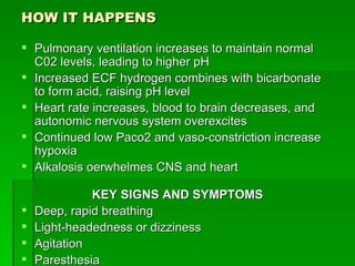
  • 98. HOW IT HAPPENS Pulmonary ventilation increases to maintain normal C02 levels, leading to higher pH Increased ECF hydrogen combines with bicarbonate to form acid, raising pH level Heart rate increases, blood to brain decreases, and autonomic nervous system overexcites Continued low Paco2 and vaso-constriction increase hypoxia Alkalosis oerwhelmes CNS and heart KEY SIGNS AND SYMPTOMS Deep, rapid breathing Light-headedness or dizziness Agitation Paresthesia
  • 99. COMPLICATIONS Cardiac arrythmias not responsive to conventional treatment Hypocalcemic tetany Seizures Periods of apnea DIAGNOSIS ABG analysis Paco 2 < 35 mm Hg; blood pH increases ECG indicates arrhythmias Toxicology screening may reveal salicylate poisoning Basic urine pH detected TREATMENT Removal of ingested toxins Treatment of fever, sepsis, CNS Oxygen administration Breathing into a paper bag to treat hyperventilation Adjustment of mechanical ventilation
  • 100. KEY NURSING ACTIONS Watch for and report changes in neurologic, neuromuscular, or cardiovascular functions Be alert for twitching and cardiac arrythmias, which may be associated with alkalemia and electrolyte imbalances.
  • 101. CHARACTERISTICS OF METABOLIC ACIDOSIS Characterized by excess acid and deficient bicarbonate Can occur with increased production or decreased clearance of nonvolatile acid, or loss of bicarbonate; can be fatal ALERT ! More prevalent in children, who are more vulnerable to acid-base imbalances. CAUSES Excessive fat metabolism in absence of usable carbohydrates Cardiac pump failure Pulmonary or hepatic disease Anemia Renal insufficiency or failure Aspirin overdose Addison’s disease Hypoaldosteronism
  • 102. HOW IT HAPPENS Excess hydrogen ions decrease blood pH and respiration increases Paco 2 fall freeshydrogen ions to bind with bicarbonate Respiration can’t compensate Kidneys try to compensate by secreting excess hydrogen into renal tubules Excess hydrogen ions in ECF diffuse into cells; cells release potassium ions Imbalance impairs neural excitability KEY SIGNS AND SYMPTOMS Headache Lethargy Drowsiness CNS depression Kussmaul’s respirations Hypotension Stupor
  • 103. COMPLICATIONS Weakness Flaccid paralysis Coma Ventricular arrhythmias Cardiac arrest Growth retardation in children Bone disorders DIAGNOSIS Arterial pH < 7.35 Paco 2 < 34 mm Hg Bicarbonate levels < 22 mEq/L Urine pH <4.5 in absence of renal disease Serum potassium > 5.5 mEq/L Glucose levels > 150 mg/dl Anion gap: ≤ 12 mEq/L indicates normal anion gap metabolic acidosis; ≥ 14 mEq/L indicates high- anion gap metabolic acidosis
  • 104. TREATMENT Address underlying cause Sodium bicarbonate I.V. I.V. lactated Ringer’s solution Mechanical ventilation Antibiotic therapy Dialysis Antidiarrheal administration KEY NURSING ACTIONS Keep sodium bicarbonate ampules handy for emergencies. Position the patient to prevent aspiration in case of vomiting. Teach diabetic patients how to test urine; encourage adherence to hypoglycemic therapy.
  • 105. CHARACTERISTICS OF METABOLIC ALKALOSIS Low acid or high bicarbonate levels cause metabolic, respiratory, and renal responses, producing characteristic symptoms Always secondary to underlying cause Prognosis good with prompt diagnosis and treatment Can lead to coma and death CAUSES Loss of acid, retention of base, or renal mechanisms associated with low serum levels of potassium and chloride Chronic vomiting NG tube drainage or lavage without adequate electrolyte replacement Fistulas Use of steroids Massive blood transfusions Cushing’s disease
  • 106. HOW IT HAPPENS Excess unbound bicarbonate raises blood pH, inhibiting respiration and raising Paco 2 Blood bicarbonate rises to > 28 mEq/L, is excreted into urine; hydrogen is retained Hydrogen in ECF drops and moves out of ICF to maintain balance; potassium moves into ICF Calcium ionization decreases; sodium moves into cells Neural impulses triggered in peripheral nervous system and CNS KEY SIGNS AND SYMPTOMS Irritability Confusion Nausea Vomiting Diarrhea
  • 107. DIAGNOSIS Blood pH > 7.45 and bicarbonate level > 26 mEq/L Paco 2 > 45 mm Hg Potassium < 3.5 mEq/L Calcium < 8.9 mg/dl Chloride < 98 mEq/L TREATMENT Cautious use of ammonium chloride I.V. or hydrochloric acid I.V. potassium chloride and normal saline Discontinue diuretics and administer supplementary potassium chloride Oral or I.V. acetazolamide KEY NURSING ACTIONS Monitor patient’s status. Dilute potassium for I.V. fluids containing potassium salts Don’t give ammonium chloride to patients with renal or hepatic disease. Watch for muscle weakness, tetany, or decreased activity. Irrigate NG tubes with isotonic saline solution.
  • 108. GASTROINTESTINAL SYSTEM KEY FACTS ABOUT THE GI SYSTEM Supplies essential nutrients Serves to digest food and eliminate waste Profoundly affects quality of life Two major components: GI tract and accessory organs Malfunction in system can produce life – threatening metabolic effects GI TRACT Hollow muscular tube from mouth to anus Oral cavity, pharynx, esophagus, stomach, small and large intestine Peristalsis propels ingested, material; sphincters prevent reflux PATHOPHYSIOLOGIC CHANGES Disorders typically nonspecific, reflect disruption in functions
  • 109. CHARACTERISTICS OF INTESTINAL OBSTRUCTION Partial or complete blockage of lumen in small or large bowel Small- bowel obstruction more common, more serious Complete obstruction can cause death within hours Most likely to occur after abdominal surgery or in persons with congenital bowel deformities CAUSES Adhesions and strangulated hernias Carcinomas Foreign bodies Compression of the bowel wall physiologic disturbances
  • 110. HOW IT HAPPEN Simple intestinal obstruction: blockage prevents intestinal contents from passing Strangulated intestinal obstruction: blood supply to obstructed section cut off, in addition to blockage of lumen Closed-loop intestinal obstruction: both ends of bowel section are occluded When obstruction occurs, fluid, air, gas collect near site Peristalsis increases temporarily, injuring intestinal mucosa, causing distention Distention blocks flow of venous blood, halts normal absorptive processes Bowel begins to secrete water, sodium, potassium into fluid pooled in lumen Small intestine obstruction results in metabolic alkalosis Lower bowel obstruction results in metabolic acidosis Obstruction may lead to ischemia, necrosis, death
  • 111. PARALYTIC ILEUS Causes Trauma, toxemia, or peritonitis Electrolyte deficiencies Vascular causes Treatment Intubation Intestinal tube Cholinergic agents ALERT ! Watch for air-fluid lock syndrome in older adults who remain recumbent for extended periods.
  • 112. KEY SIGNS AND SYMPTOMS Colicky pain Nausea and vomiting Constipation Abdominal distention Drowsiness Intense thirst Malaise Aching Dry oral mucous membranes, tongue Bowel sound, borborygmi rushes Abdominal tenderness, moderate distention
  • 113. SYMPTOM PROGRESSION ON INTESTINAL OBSTRUCTION Obstruction Fluid, gas, and air collect behind obstruction Peristalsis temporarily increases in attempt to force contents past obstruction. Distention increases at and above obstruction site. Distention impedes blood supply to bowel, halting absorption. Bowel wall swells as water, sodium, and potassium are secreted into intestine and not absorbed from it. Gas-forming bacteria collect above obstruction, increasing distention. Dehydration results because fluids aren’t absorbed into bloodstream. With no treatment, severe hypovolemia occurs. Shock Sepsis Death
  • 114. COMPLICATIONS Perforation Peritonitis Septicemia Secondary infection Metabolic alkalosis or acidosis DIAGNOSIS Progressive, colicky, abdominal pain, distention, with or without nausea and vomiting X-rays show presence and location of intestinal gas or fluid “ Stepladder” pattern emerges in small bowel on X-ray Barium enema reveals distended, air-filled colon or closed loop of sigmoid with extreme distention
  • 115. TREATMENT Correction of fluid and electrolyte imbalances Decompression of bowel Treatment of shock, peritonitis Blood replacement I.V. fluid administration Passage of NG tube Miller- Abbott or Cantor tube Surgical resection with anastomosis, colostomy, ileostomy KEY NURSING ACTIONS Monitor vital signs and observe for signs of shock. Stay alert for signs and symptoms of metabolic alkalosis or acidosis. Watch for signs and symptoms of secondary infections. Monitor urine output carefully to access renal function, circulating blood volume, and possible urine retention caused by bladder compression by the distended intestine. Look for signs of dehydration Record the amount and color of drainage from the decompression tube. Keep the patient in Fowler’s position as much as possible.
  • 116. CHARACTERISTICS OF PEPTIC ULCERS Circumscribed lesions in mucosal membrane extending below epithelium Can develop in lower esophagus, stomach, pylorus, duodenum, jejunum Erosions, commonly called ulcers but don’t extend below epithelium May be acute or chronic Chronic ulcers identified by scar tissue at base 80% of peptic ulcers: duodenal Most common in men between ages 20 and 50 Usually follow chronic course with remissions, exacerbations 5% to 10% of patients need surgery Gastric ulcers most common in middle- age and elderly men, users of NSAIDs, tobacco CAUSES H. pylori infection Use of NSAIDs Pathologic hypersecretory disorders
  • 117. HOW IT HAPPENS May result from destruction of mucosal barrier Duodenal ulcers appear to result from excessive acid production H. pylori releases toxin that destroys gastric and duodenal mucosa Epithelium’s resistance to acid digestion reduced Results in gastritis, ulcer disease Salicylates and NSAIDs inhibit secretion of prostaglandins KEY SIGNS AND SYMPTOMS Vary by type of ulcer Gastric ulcer: pain, worsens with eating; nausea and anorexia Duodenal ulcer: gnawing, dull, aching, “hungerlike” epigastric pain; relived by food or antacids; usually recurs 2 to 4 hours later
  • 118. COMPLICATIONS Hemorrhage Shock Gastric perforation Gastric outlet obstruction DIAGNOSIS Barium swallow Upper GI and small bowel series Urea breath test results reflect activity of H. pylori
  • 119. TREATMENT Antimicrobial agents Misoprostol Antacids Anticholinergic drugs Sucralfate Proton gastric acid pump inhibitor Dietary measures NG tube Gastroscopy surgery
  • 120. KEY NURSING ACTIONS Watch for adverse reactions to H 2 -receptor antagonists and omeprazole Advise any patient who uses antacids, has a history of heart disease, or follows a sodium-restricted diet to take only those antacids that contain low amounts of sodium Warn the patient to avoid steroids and NSAIDs because they irritate the gastric mucosa. AFTER GASTRIC SURGERY Keep the NG tube patent Monitor intake and output, including NG tube drainage. Replace fluids and electrolytes Monitor for possible complications.
  • 121. TREATING PEPTIC ULCER DISEASE Bile salts, aspirin, nonsteroidal anti-inflammation, alcohol, ischema Damaged mucosal barrier Increased number of parietal and chief cells Inade q uate mucosal blood supply Decreased function of mucosal cells Decreased q uality of mucus Loss of tight junctions between cells Increased sensitivity to food and other stimuli Decreased inhibition of gastric secretions Bile or pancreatic enzyme reflux from duodenum Conversion of pepsinogen to pepsin Antimicrobials Further mucosal erosion Destruction of blood vessels Bleeding Colonization by Helicobacter pylori Ulceration Mucosal injury Dietary measures Misoprostol Sucralfate Histamine-2 blockers Antacids Formation and liberation of histamine Increased acid secretion Local Vasodilatation Anticholinergic drugs Excessive vagal stimulation Stimulation of cholinergic intramural plexus, causing muscle spasm Increased capillary permeability Loss of plasma proteins Mucosal edema Loss of plasma in gastric lumen Key = treatment
  • 122. NERVOUS SYSTEM KEY FACTS ABOUT THE NERVOUS SYSTEM Coordinates and organizes functions of all body systems Has three main divisions: CNS, peripheral nervous system, ANS Neurons transmit nerve impulses through body Most neurons have several dendrites, only one axon Three types: sensory, motor, and interneurons Nervous system disorders can cause signs and symptoms in any body system; hard to diagnose
  • 123. REVIEWING THE CNS Cerebrum – frontal lobe, temporal lobe, parietal lobe, occipital lobe Cerebellum – maintains muscle tone, coordination Brain stem – pons, midbrain, medulla oblongata Primitive structures – thalamus, hypothalamus, limbic system, RAS Spinal cord – relays sensory, motor impulses REVIEWING THE PERIPHERAL NERVOUS SYSTEM Cranial nerves – 1 2 pairs: olfactory, optic, oculomotor, trochlear, trigeminal, abducens, facial, acoustic, glossopharyngeal, vagus, spinal accessory, hypoglossal Spinal nerves - 3 1 pairs with sensory and motor neurons ANS – innervates internal organs; sympathetic nervous system
  • 124. PATHOPHYSIOLOGIC CHANGES Typically involve alteration in arousal, cognition, movement, muscle tone, homeostatic mechanisms or pain Most cause more than one alteration One alteration may lead to another KEY FACTS ABOUT AROUSAL Level of consciousness or state of awareness SEVERAL MECHANISMS CAN ALTER AROUSAL Direct destruction of RAS and pathways Destruction of brain stem Compression of RAS by disease process Mechanisms may result from structural, metabolic, or psychogenic disturbances
  • 125. 6 STAGES OF ALTERED AROUSAL Usually begins with interruption or disruption in diencephalon Patient shows evidence of dullness, confusion, lethargy, and stupor Continued decreases result from midbrain dysfunction Coma results if medulla and pons are effected Patient will progress through rostal-caudal progression Levels: confusion, disorientation, lethargy, obtundation, stupor, coma 5 NEUROLOGIC FUNCTION AREAS USED TO IDENTIFY CAUSE Level of consciousness Pattern of breathing Pupillary changes Eye movement and reflex responses Motor responses
  • 126. KEY FACTS ABOUT COGNITION Ability to be aware, perceive, reason, judge, remember, use intuition Reflects higher functioning of cerebral cortex Alteration results from direct or indirect destruction May manifest as agnosia, aphasia, or dysphasia ALTERED AROUSAL STAGES Confusion Disorientation Lethargy Obtundation Stupor coma
  • 127. CHARACTERISTICS OF ALZHEIMER’S DISEASE Degenerative disorder of cerebral cortex, especially frontal lobe Accounts for more than 50% of dementia cases Found in elderly population Primary progressive dementia has poor prognosis CAUSES Exact cause unknown NEUROCHEMICAL FACTORS Deficiencies in acetylcholine, somatostatin, substance P, and norephineprine ENVIRONMENTAL FACTORS Repeated head trauma Exposure to aluminum or manganese GENETIC FACTORS Family history Down syndrome
  • 128. HOW IT HAPPENS Brain tissue exhibits neurofibrillatory tangles, neuritic pla q ues, granulovascular changes Structural changes: cortical atrophy, ventricular dilation, amyloid deposits around cortical blood vessels, and reduced brain volume KEY SIGNS AND SYMPTOMS Reflect neurologic abnormalities associates with the disease Loss of memory Flattening of affect and personality Deterioration of personal hygiene Progressive impaired communication COMPLICATIONS Injury from violent behavior Pneumonia and other infections Malnutrition
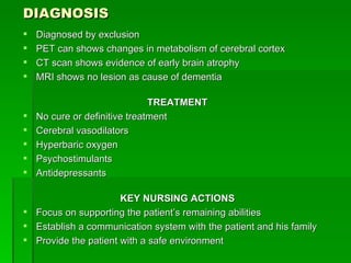
  • 129. DIAGNOSIS Diagnosed by exclusion PET can shows changes in metabolism of cerebral cortex CT scan shows evidence of early brain atrophy MRI shows no lesion as cause of dementia TREATMENT No cure or definitive treatment Cerebral vasodilators Hyperbaric oxygen Psychostimulants Antidepressants KEY NURSING ACTIONS Focus on supporting the patient’s remaining abilities Establish a communication system with the patient and his family Provide the patient with a safe environment