SlideShare a Scribd company logo
Peptic Ulcer Disease
By
Aniedu, Ugochukwu
OUTLINE
• Introduction
• Pathophysiology
• Etiology/ Risk factors
• Types of PUD
• Clinical Presentation
• Investigation/ Diagnostic test
• Complications of PUD
• Management
• Summary
• References.
INTRODUCTION
• Peptic Ulcer is a lesion in the lining
(mucosa) of the digestive tract, typically in
the stomach or duodenum, caused by the
digestive action of pepsin and stomach
acid.
Lesion may subsequently occur into the lamina
propria and submucosa to cause bleeding. –
Most of peptic ulcer occur either in the
duodenum, or in the stomach – Ulcer may also
occur in the lower esophagus due to reflexing of
gastric content – Rarely in certain areas of the
PATHOPHYSIOLOGY
Under normal conditions, a physiologic balance
exists between gastric acid secretion and
gastroduodenal mucosal defense. Mucosal injury
and, thus, peptic ulcer occur when the balance
between the aggressive factors and the
defensive mechanisms is disrupted. Aggressive
factors, such as NSAIDs, H pylori infection,
alcohol, bile salts, acid, and pepsin, can alter the
mucosal defense by allowing back diffusion of
hydrogen ions and subsequent epithelial cell
injury.
ETIOLOGY/ RISK FACTORS
• Lifestyle
– Smoking
– Acidic drinks
– Medications
• H. Pylori infection
– 90% have this bacterium
– Passed from person to
person (fecal-oral route
or oral-oral route)
• Age
– Duodenal 30-40
– Gastric over 50
• Gender
– Duodenal: are increasing
in older women
• Genetic factors
– More likely if family
member has Hx
• Other factors: stress
can worsen but not the
cause
TYPES
• GASTRIC PEPTIC ULCER
• DUODENAL PEPTIC ULCER
Gastric and Duodenal Ulcers
CLINICAL PRESENTATION
Peptic ulcer
Peptic ulcer
INVESTIGATION/ DIAGNOSTIC TEST
INVESTIGATION
• Stool examination for fecal occult blood.
• Complete blood count (CBC) for decrease
in blood cells.
DIAGNOSTIC TEST
• Esophagogastrodeuodenoscopy (EGD)
– Endoscopic procedure
• Visualizes ulcer crater
• Ability to take tissue biopsy to R/O cancer and diagnose
H. pylori
– Upper gastrointestinal series (UGI)
• Barium swallow
• X-ray that visualizes structures of the upper GI tract
– Urea Breath Testing
• Used to detect H.pylori
• Client drinks a carbon-enriched urea solution
• Exhaled carbon dioxide is then measured
In all patients with “Alarming symptoms” endoscopy
is required.
Dysphagia.
Weight loss.
Vomiting.
Anorexia.
Hematemesis or Melena
Complications of Peptic Ulcers
• Hemorrhage
– Blood vessels damaged as ulcer erodes into the muscles of
stomach or duodenal wall
– Coffee ground vomitus or occult blood in tarry stools
• Perforation
– An ulcer can erode through the entire wall
– Bacteria and partially digested food spill into
peritoneum=peritonitis
• Narrowing and obstruction (pyloric)
– Swelling and scarring can cause obstruction of food leaving
stomach=repeated vomiting
MANAGEMENT
• LIFE STYLE MODIFICATION
• HYPOSECRETORY DRUG THERAPY
• H. pylori ERADICATION THERAPY
• SURGERY
Peptic ulcer
Hyposecretory Drugs
• Proton Pump Inhibitors
– Suppress acid production
– Prilosec, Prevacid
• H2-Receptor Antagonists
– Block histamine-stimulated
gastric secretions
– Zantac, Pepcid, Axid
• Antacids
– Neutralizes acid and
prevents formation of pepsin
(Maalox, Mylanta)
– Give 2 hours after meals
and at bedtime
• Prostaglandin Analogs
– Reduce gastric acid and
enhances mucosal
resistance to injury
– Cytotec
• Mucosal barrier fortifiers
– Forms a protective coat
• Carafate/Sucralfate
– cytoprotective
H. pylori Eradication Therapy:
Indications:
Failure of medical treatment.
Development of complications
High level of gastric secretion and
combined duodenal and gastric ulcer.
Principle:
Reduce acid and pepsin
secretion.
Types of Surgical Procedures
• GASTROENTEROSTOMY
Creates a passage between
the body of stomach to small
intestines.
Allows regurgitation of alkaline
duodenal contents into the
stomach.
Keeps acid away from ulcerated
area
Types of Surgical Procedures
• VAGOTOMY
– Cuts vagus nerve
– Eliminates acid-
secretion stimulus
Types of Surgical Procedures
• PYLOROPLASTY
– Widens the pylorus
to guarantee
stomach emptying
even without vagus
nerve stimulation
Types of Surgical Procedures
• ANTRECTOMY/ SUBTOTAL GASTRECTOMY
– Lower half of stomach (antrum) makes most of the
acid
– Removing this portion (antrectomy) decreases acid
production
• SUBTOTAL GASTRECTOMY
– Removes ½ to 2/3 of stomach
• Remainder must be reattached to the rest of the
bowel
– Billroth I
– Billroth II
Billroth I
• Distal portion of the
stomach is removed
• The remainder is
anastomosed to the
duodenum
Billroth II
• The lower portion
of the stomach is
removed and the
remainder is
anastomosed to
the jejunum
Postoperative Care
– NG tube – care and management
– Monitor for post-operative complications
Post-op Complications
• Bleeding
– Occurs at the anastomosed site
– First 24 hours and post-op days
4-7
• Duodenal stump leak
– Billroth II
– Severe abdominal pain
– Bile stained drainage on
dressing
• Gastric retention
– WILL NEED TO PUT NG TUBE
BACK IN
• Dumping Syndrome.
– Prevalent with sub total gastrostomies
– Early-30 minutes after meals
– Vertigo, tachycardia, syncope, sweating,
pallor, palpitations
– Late – 90 min-3 hours after meals
• Rx: Decrease CHO intake, Eat slowly, Avoid
fluids during meals, Increase fat, Eat small,
frequent meals
• Anemia
– Rapid gastric empyting decreases
absorption of iron
• Malabsorption of fat
– Decreased acid secretions, decreased
pancreatic secretions, increased upper
GI mobility
Summary
• H. pylori is the most common cause of PUD and
is a risk factor for gastric cancer
• H Pylori eradication reduces risk of disease
recurrence
• Test-and-Treat strategy is recommended for
patients with undifferentiated dyspepsia
• Intial evaluation with endoscopy is recommended
for those with alarm symptoms or those failing
treatment
• Optimum treatment regimens are 14d multidrug
with antibiotics and acid suppressants(Triple
therapy)
REFERENCES
• http://emedicine.medscape.com/article/181753-overview#showall. Retrieved 28th
Jan,
2016
• Fendrick M, Forsch R etal. Peptic Ulcer Disease Guidleines for Clinical Care.
University of Michigan Health System May 2005
• American Gastroenterological Association medical position statement: evaluation of
dyspepsia. Gastroenterology 1998;114:579-81.
• Krogfelt K, Lehours P, Mégraud F. Diagnosis of Helicobacter pylori Infection.
Helicobacter 2005 10:s1 5
• Meurer L, Bower D. Management of Helicobacter pylori Infection. American Family
Physician Vol 65, No. 7, 2002 pp 1327-1336
• Standards of Practice Committee of the American Society for Gastrointestinal
Endoscopy; The role of endoscopy in dyspepsia. Gastrointestinal Endoscopy Vol 54,
No. 6, 2001 pp 815-817
• Vaira D, Gatta L, Ricci C, et al. Peptic ulcer and Helicobacter pylori: update on testing
and treatment. Postgrad Med 2005;117(6):17-22, 46

More Related Content

PPTX
Peptic ulcer ppt
PDF
Gastritis
PPTX
Peptic ulcer
PPT
peptic ulcer disease.PPT
PPTX
Peptic ulcer
PPTX
Peptic ulcer disease(PUD)
PPTX
Peptic ulcer disease
PPTX
Pathology of Peptic Ulcer
Peptic ulcer ppt
Gastritis
Peptic ulcer
peptic ulcer disease.PPT
Peptic ulcer
Peptic ulcer disease(PUD)
Peptic ulcer disease
Pathology of Peptic Ulcer

What's hot (20)

PDF
Inflammatory bowel disease
PPTX
Esophagitis
PPTX
Ulcerative colitis
PPTX
Liver cirrhosis
PDF
Inflammatory Bowel Disease
PPTX
Liver cirrhosis
PPT
Crohn\'s disease
PPTX
Duodenal ulcer presentation
PPTX
Ulcerative Colitis (UC)
PPTX
Cirrhosis of Liver
PPTX
Congestive cardiac failure
PPT
Malabsorption syndrome
PPTX
Renal calculi
PPTX
Cholecystitis ppt
PPTX
Portal hypertension
PPTX
PPT
Atherosclerosis ppt
PPTX
SYSTEMIC LUPUS ERYTHEMATOSUS (SLE
PPT
Gastro esophageal Reflux Disease (GERD) and its management
PPTX
Congestive heart failure CHF
Inflammatory bowel disease
Esophagitis
Ulcerative colitis
Liver cirrhosis
Inflammatory Bowel Disease
Liver cirrhosis
Crohn\'s disease
Duodenal ulcer presentation
Ulcerative Colitis (UC)
Cirrhosis of Liver
Congestive cardiac failure
Malabsorption syndrome
Renal calculi
Cholecystitis ppt
Portal hypertension
Atherosclerosis ppt
SYSTEMIC LUPUS ERYTHEMATOSUS (SLE
Gastro esophageal Reflux Disease (GERD) and its management
Congestive heart failure CHF
Ad

Viewers also liked (20)

PDF
Çakmaklı 2.El Lcd Smart Tv Alanlar 0542 541 06 06-Elsidi Tv Alanlar
PPTX
книжкові виставки
PPTX
Entenda a Importância do SPF e DKIM na entrega dos seus e-mails
PPTX
Discover open education: finding open educational resources
PPTX
LEITURA: Aspectos Teóricos e Práticos
PPTX
Prospectiva en el ambito personal
PPT
Introdução à Linguística
PDF
Infographie : Xavier Niel
PPTX
к уроку 2
PPT
Peptic Ulcer Bleeding
PPT
к уроку 3
PPT
Παρουσίαση Σεμιναρίου Εκπαίδευση μέσα από την Τέχνη (Bullying)
PPT
Peptic ulcer
DOCX
чумаков тимофей (москва)
PPTX
DPSE Ricardo Tome day 1 - 2.21.2017 - 515pm
PPTX
Molecular basis of thyroid neoplasm subhasish
PPT
Peptic ulcer
PPTX
Peptic ulcer disease
PDF
Peptic ulcer disease
PPTX
Peptic Ulcer
Çakmaklı 2.El Lcd Smart Tv Alanlar 0542 541 06 06-Elsidi Tv Alanlar
книжкові виставки
Entenda a Importância do SPF e DKIM na entrega dos seus e-mails
Discover open education: finding open educational resources
LEITURA: Aspectos Teóricos e Práticos
Prospectiva en el ambito personal
Introdução à Linguística
Infographie : Xavier Niel
к уроку 2
Peptic Ulcer Bleeding
к уроку 3
Παρουσίαση Σεμιναρίου Εκπαίδευση μέσα από την Τέχνη (Bullying)
Peptic ulcer
чумаков тимофей (москва)
DPSE Ricardo Tome day 1 - 2.21.2017 - 515pm
Molecular basis of thyroid neoplasm subhasish
Peptic ulcer
Peptic ulcer disease
Peptic Ulcer
Ad

Similar to Peptic ulcer (20)

PPT
0 Peptic Ulcer Disease GROUP 3.pp9999999999999t
PPTX
pepticyulcerulceroepticupepticulcer.pptx
PPTX
peptic ulcer mohammad abu sad 1.pptx
PPTX
Peptic Ulcer Desease (PUD), Sores in the gastrium
PPTX
Peptic ulcer disease.pptx
PPTX
Peptic Ulcer Disease
PPTX
PUD-Peptic ulcer disease presentation slides
PPTX
Peptic ulcer.pptx
PPTX
Peptic Ulcer Disease.pptx
PPTX
B1 git med1 peptic ulcer disease
PPTX
Peptic ulcer disease
PPT
Peptic ulcer disease
DOCX
Peptic ulcer
PPTX
Peptic ulcer diseases-2.pptx444444444444
PDF
Ulcer Disease.pptx.pdf
PPT
Case presentation pud
PPTX
peptic ulcer disease.pptx
PPTX
peptic ulcer
PPTX
Peptic Ulcer Disease.pptx
PPT
Peptic Ulcer Disease Dr Shatdal
0 Peptic Ulcer Disease GROUP 3.pp9999999999999t
pepticyulcerulceroepticupepticulcer.pptx
peptic ulcer mohammad abu sad 1.pptx
Peptic Ulcer Desease (PUD), Sores in the gastrium
Peptic ulcer disease.pptx
Peptic Ulcer Disease
PUD-Peptic ulcer disease presentation slides
Peptic ulcer.pptx
Peptic Ulcer Disease.pptx
B1 git med1 peptic ulcer disease
Peptic ulcer disease
Peptic ulcer
Peptic ulcer diseases-2.pptx444444444444
Ulcer Disease.pptx.pdf
Case presentation pud
peptic ulcer disease.pptx
peptic ulcer
Peptic Ulcer Disease.pptx
Peptic Ulcer Disease Dr Shatdal

Recently uploaded (20)

PDF
Therapeutic Potential of Citrus Flavonoids in Metabolic Inflammation and Ins...
PPTX
History and examination of abdomen, & pelvis .pptx
PPTX
Fundamentals of human energy transfer .pptx
PPTX
DENTAL CARIES FOR DENTISTRY STUDENT.pptx
PPTX
Chapter-1-The-Human-Body-Orientation-Edited-55-slides.pptx
PDF
Khadir.pdf Acacia catechu drug Ayurvedic medicine
PPTX
post stroke aphasia rehabilitation physician
PDF
Deadly Stampede at Yaounde’s Olembe Stadium Forensic.pdf
PPTX
1 General Principles of Radiotherapy.pptx
PDF
CT Anatomy for Radiotherapy.pdf eryuioooop
PPTX
Important Obstetric Emergency that must be recognised
PPTX
CME 2 Acute Chest Pain preentation for education
PPT
Copy-Histopathology Practical by CMDA ESUTH CHAPTER(0) - Copy.ppt
PPT
Obstructive sleep apnea in orthodontics treatment
PPT
Breast Cancer management for medicsl student.ppt
DOCX
NEET PG 2025 | Pharmacology Recall: 20 High-Yield Questions Simplified
PDF
NEET PG 2025 | 200 High-Yield Recall Topics Across All Subjects
PPTX
Electromyography (EMG) in Physiotherapy: Principles, Procedure & Clinical App...
PPTX
Slider: TOC sampling methods for cleaning validation
PDF
Human Health And Disease hggyutgghg .pdf
Therapeutic Potential of Citrus Flavonoids in Metabolic Inflammation and Ins...
History and examination of abdomen, & pelvis .pptx
Fundamentals of human energy transfer .pptx
DENTAL CARIES FOR DENTISTRY STUDENT.pptx
Chapter-1-The-Human-Body-Orientation-Edited-55-slides.pptx
Khadir.pdf Acacia catechu drug Ayurvedic medicine
post stroke aphasia rehabilitation physician
Deadly Stampede at Yaounde’s Olembe Stadium Forensic.pdf
1 General Principles of Radiotherapy.pptx
CT Anatomy for Radiotherapy.pdf eryuioooop
Important Obstetric Emergency that must be recognised
CME 2 Acute Chest Pain preentation for education
Copy-Histopathology Practical by CMDA ESUTH CHAPTER(0) - Copy.ppt
Obstructive sleep apnea in orthodontics treatment
Breast Cancer management for medicsl student.ppt
NEET PG 2025 | Pharmacology Recall: 20 High-Yield Questions Simplified
NEET PG 2025 | 200 High-Yield Recall Topics Across All Subjects
Electromyography (EMG) in Physiotherapy: Principles, Procedure & Clinical App...
Slider: TOC sampling methods for cleaning validation
Human Health And Disease hggyutgghg .pdf

Peptic ulcer

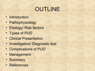
  • 2. OUTLINE • Introduction • Pathophysiology • Etiology/ Risk factors • Types of PUD • Clinical Presentation • Investigation/ Diagnostic test • Complications of PUD • Management • Summary • References.
  • 3. INTRODUCTION • Peptic Ulcer is a lesion in the lining (mucosa) of the digestive tract, typically in the stomach or duodenum, caused by the digestive action of pepsin and stomach acid.
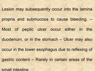
  • 4. Lesion may subsequently occur into the lamina propria and submucosa to cause bleeding. – Most of peptic ulcer occur either in the duodenum, or in the stomach – Ulcer may also occur in the lower esophagus due to reflexing of gastric content – Rarely in certain areas of the
  • 6. Under normal conditions, a physiologic balance exists between gastric acid secretion and gastroduodenal mucosal defense. Mucosal injury and, thus, peptic ulcer occur when the balance between the aggressive factors and the defensive mechanisms is disrupted. Aggressive factors, such as NSAIDs, H pylori infection, alcohol, bile salts, acid, and pepsin, can alter the mucosal defense by allowing back diffusion of hydrogen ions and subsequent epithelial cell injury.
  • 7. ETIOLOGY/ RISK FACTORS • Lifestyle – Smoking – Acidic drinks – Medications • H. Pylori infection – 90% have this bacterium – Passed from person to person (fecal-oral route or oral-oral route) • Age – Duodenal 30-40 – Gastric over 50 • Gender – Duodenal: are increasing in older women • Genetic factors – More likely if family member has Hx • Other factors: stress can worsen but not the cause
  • 8. TYPES • GASTRIC PEPTIC ULCER • DUODENAL PEPTIC ULCER
  • 14. INVESTIGATION • Stool examination for fecal occult blood. • Complete blood count (CBC) for decrease in blood cells.
  • 15. DIAGNOSTIC TEST • Esophagogastrodeuodenoscopy (EGD) – Endoscopic procedure • Visualizes ulcer crater • Ability to take tissue biopsy to R/O cancer and diagnose H. pylori – Upper gastrointestinal series (UGI) • Barium swallow • X-ray that visualizes structures of the upper GI tract – Urea Breath Testing • Used to detect H.pylori • Client drinks a carbon-enriched urea solution • Exhaled carbon dioxide is then measured
  • 16. In all patients with “Alarming symptoms” endoscopy is required. Dysphagia. Weight loss. Vomiting. Anorexia. Hematemesis or Melena
  • 17. Complications of Peptic Ulcers • Hemorrhage – Blood vessels damaged as ulcer erodes into the muscles of stomach or duodenal wall – Coffee ground vomitus or occult blood in tarry stools • Perforation – An ulcer can erode through the entire wall – Bacteria and partially digested food spill into peritoneum=peritonitis • Narrowing and obstruction (pyloric) – Swelling and scarring can cause obstruction of food leaving stomach=repeated vomiting
  • 18. MANAGEMENT • LIFE STYLE MODIFICATION • HYPOSECRETORY DRUG THERAPY • H. pylori ERADICATION THERAPY • SURGERY
  • 20. Hyposecretory Drugs • Proton Pump Inhibitors – Suppress acid production – Prilosec, Prevacid • H2-Receptor Antagonists – Block histamine-stimulated gastric secretions – Zantac, Pepcid, Axid • Antacids – Neutralizes acid and prevents formation of pepsin (Maalox, Mylanta) – Give 2 hours after meals and at bedtime • Prostaglandin Analogs – Reduce gastric acid and enhances mucosal resistance to injury – Cytotec • Mucosal barrier fortifiers – Forms a protective coat • Carafate/Sucralfate – cytoprotective
  • 22. Indications: Failure of medical treatment. Development of complications High level of gastric secretion and combined duodenal and gastric ulcer. Principle: Reduce acid and pepsin secretion.
  • 23. Types of Surgical Procedures • GASTROENTEROSTOMY Creates a passage between the body of stomach to small intestines. Allows regurgitation of alkaline duodenal contents into the stomach. Keeps acid away from ulcerated area
  • 24. Types of Surgical Procedures • VAGOTOMY – Cuts vagus nerve – Eliminates acid- secretion stimulus
  • 25. Types of Surgical Procedures • PYLOROPLASTY – Widens the pylorus to guarantee stomach emptying even without vagus nerve stimulation
  • 26. Types of Surgical Procedures • ANTRECTOMY/ SUBTOTAL GASTRECTOMY – Lower half of stomach (antrum) makes most of the acid – Removing this portion (antrectomy) decreases acid production • SUBTOTAL GASTRECTOMY – Removes ½ to 2/3 of stomach • Remainder must be reattached to the rest of the bowel – Billroth I – Billroth II
  • 27. Billroth I • Distal portion of the stomach is removed • The remainder is anastomosed to the duodenum
  • 28. Billroth II • The lower portion of the stomach is removed and the remainder is anastomosed to the jejunum
  • 29. Postoperative Care – NG tube – care and management – Monitor for post-operative complications
  • 30. Post-op Complications • Bleeding – Occurs at the anastomosed site – First 24 hours and post-op days 4-7 • Duodenal stump leak – Billroth II – Severe abdominal pain – Bile stained drainage on dressing • Gastric retention – WILL NEED TO PUT NG TUBE BACK IN • Dumping Syndrome. – Prevalent with sub total gastrostomies – Early-30 minutes after meals – Vertigo, tachycardia, syncope, sweating, pallor, palpitations – Late – 90 min-3 hours after meals • Rx: Decrease CHO intake, Eat slowly, Avoid fluids during meals, Increase fat, Eat small, frequent meals • Anemia – Rapid gastric empyting decreases absorption of iron • Malabsorption of fat – Decreased acid secretions, decreased pancreatic secretions, increased upper GI mobility
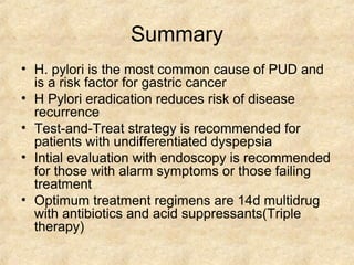
  • 31. Summary • H. pylori is the most common cause of PUD and is a risk factor for gastric cancer • H Pylori eradication reduces risk of disease recurrence • Test-and-Treat strategy is recommended for patients with undifferentiated dyspepsia • Intial evaluation with endoscopy is recommended for those with alarm symptoms or those failing treatment • Optimum treatment regimens are 14d multidrug with antibiotics and acid suppressants(Triple therapy)
  • 32. REFERENCES • http://emedicine.medscape.com/article/181753-overview#showall. Retrieved 28th Jan, 2016 • Fendrick M, Forsch R etal. Peptic Ulcer Disease Guidleines for Clinical Care. University of Michigan Health System May 2005 • American Gastroenterological Association medical position statement: evaluation of dyspepsia. Gastroenterology 1998;114:579-81. • Krogfelt K, Lehours P, Mégraud F. Diagnosis of Helicobacter pylori Infection. Helicobacter 2005 10:s1 5 • Meurer L, Bower D. Management of Helicobacter pylori Infection. American Family Physician Vol 65, No. 7, 2002 pp 1327-1336 • Standards of Practice Committee of the American Society for Gastrointestinal Endoscopy; The role of endoscopy in dyspepsia. Gastrointestinal Endoscopy Vol 54, No. 6, 2001 pp 815-817 • Vaira D, Gatta L, Ricci C, et al. Peptic ulcer and Helicobacter pylori: update on testing and treatment. Postgrad Med 2005;117(6):17-22, 46

Editor's Notes

  • #16: A barium examination of the GI tract can be used to establish a duodenal ulcer. If perforation is suspected the health care provider usually requests an upright abdomen series to demonstrate free air in the peritoneum. Do not use barium where free air is a possibility.
  • #18: Hemorrhage: With massive bleeding the patient vomits bright red or coffee ground blood. Minimal bleeding from ulcers is manifested by occult blood in a tarry stool (melena). Perforation: Gastric and duodenal ulcers can perforate or bleed. Perforation occurs when the ulcer becomes so deep that the entire thickness of the stomach or duodenum is worn away. The gastroduodenal contents may then empty into the peritoneal cavity. Symptoms of perforation are sudden, sharp pain, the abdomen is tender, rigid, and boardlike. The patient assumes the fetal position, knees to chest. Client can become acutely ill within hours. Peforation is considered a surgical emergency and can be life threatening. If this occurs the physician needs to be notified immediatley.
  • #21: Hyposecretory drugs produce a reduction in gastric acid secretions. Your proton pump inhibitors have emerged as the drug class of choice for treating clients with acid-related disorders. These drugs suppress acid production. They include Prilosec and Previcid. Your H2 receptor antagonists block histamine stimulated gastric secretions. Antacids neutralizes acid and prevents formation of pepsin. The prostoglandin analogs reduce gastric acid and enhance mucosal resistance to injury. The drug Carafate acts as a mucosal barrier by forming a protective coating
  • #24: A simple gastroenterostomy permits neutralization of gastric acid by regurgitation of alkaline duodenal contents into the stomach. The surgeon creates a passage between the body of the stomach and the small bowel. Drainage of the gastric contents diverts acid from the ulcerated area and facilitates healing.
  • #25: A vagotomy eliminates the acid-secreting stimulus to gastric cells and decreases the responsiveness of parietal cells.
  • #26: In this procedure the surgeon enlarges the pyloric stricture by incising the pylorus longitudinally and sutures the incision transversely.
  • #30: The postoperative plan of care is similar for all of the gastric operative procedures. These patients will have NG tubes in place and attached to low wall suction. Ensure the patency of these tubes. Irrigation or repositioning of the NG tube is not done after gastric surgery unless specifically ordered by the doctor.
  • #31: Dumping Syndrome: is a term that refers to a constellation of vasomotor symptoms after eating, especially following a Billiroth II procedure. This syndrome is believed to be caused because of the rapid emptying of the stomach contents into the small intestine, which shifts fluid into the gut, causing abdominal distention. Early manifestations occur within 30 minutes of eating. Symptoms include vertigo, tachycardia, syncope, pallor, sweating and palpatations. Late dumping syndrome occurs 90 minutes to 3 hours after eating, and is caused by a release of an excessive amount of insulin. The insulin release follows a rapid rise in the blood glucose level that results from the rapid entry of high carbohydrate food into the jejunum. Symptoms include dizziness, light-headedness, palpatations, diaphoresis, and confusion. Dumping syndrome is managed by dietary measures that include decreasing the amount of food taken at one time and eliminating liquids with meals.