SlideShare a Scribd company logo
CASE PRESENTATION
Presented by:
Dr. Abdullah Almazyad R1
Dr. Fatimah Alghamdi R3
Under supervision of :Dr. Abdullah Alkhudair
OBJECTIVES
Case presentation
History
Physical Examination
Differential diagnosis
Investigation and management
Patient hospital coarse
Educational topic about disease
Key point
CHIEF COMPLAINT
• Chest pain for 15 minutes followed with a sudden syncope
Any Further Questions
About Chest Pain
DIFFERENTIAL DIAGNOSES OF CHEST PAIN
• MI, Angina, Myocarditis, And Pericarditis/Dressler’s Syndrome
Cardiac
• PE, Pneumothorax/Hemothorax, Tension Pneumothorax, Pneumonia, Empyema, Pulmonary
• Neoplasm, Bronchiectasis, Pleuritis, And TB
• Pulmonary
• Esophageal: GERD, Esophageal Rupture, Spasm, Esophagitis, Ulceration, Achalasia, Neoplasm, And
• Mallory-weiss Syndrome
• Other Structures: PUD, Gastritis, Pancreatitis, And Biliary Colic
• Gastrointestinal
• Lymphoma, Thymoma
• Mediastinal
• Dissecting Aortic Aneurysm, Aortic Rupture
• Vascular
HISTORY OF PRESENTING ILLNESS
• Mr. N 52-year-old M. heavy smoker
• Presented to ER with of typical chest pain
• Pain was stared suddenly , in public place (the restaurant) while he was standing
reached maximum 10/10 in few minutes.
• Pain felt like heaviness in nature , radiating to the left shoulder and back ,
associated with diaphoresis and was aggravated by movement,
• After 15-20 of heavy chest pain M.N felt dizzy due to intense pain , people tried to
make him set and feel comfortable .
• As a result EMS were called by one of the public and took patient to ER, no CPR or
medication ware given to his way to ED.
• Patient has positive history of similar episode few days ago while sleeping , was
5/10 in severity and relieved spontaneously within few minutes
Stemi 1
PAST MEDICAL
• Mr. N is not known to have any medical illness
PAST MEDICAL HISTORY
No known cardiovascular risk factors except for smoking
No history of Hypertension
• No history of Hyperlipidaemia
• No history of Myocardial infarction
• No history of Chronic kidney disease
• No history of Atrial fibrillation
• No history of Stroke
• No history of Peripheral vascular disease
No history of Respiratory vascular disease
No history of Gastrointestinal disease:
MEDICATION HISTORY AND ALLERGY
• Patient never received :
• Anticoagulants (e.g. pulmonary embolism)
• Antiplatelets (e.g. coronary artery disease)
• Statins (e.g. coronary artery disease)
• Calcium channel blockers (e.g. hypertension)
• ACE inhibitors (e.g. hypertension)
• Antibiotics (e.g. pneumonia)
• Colchicine (e.g. pericarditis)
• No known allergy
PAST SURGICAL HISTORY
• He once was admitted for appendectomy 18 years ago , there was no
immediate or late complications
• No history of blood Transfusion
• No history of Car Accidents
FAMILY HISTORY
• No family history of premature CAD
• His father is 74 alive , he is diabetic and Hypertensive
• His mother is 72 alive , she is hypertensive only
• He has 1 brother and 3 sisters , all of them are medically free
• No history of similar episode in the family
• No history of cardiac disease
• No family history of Familial Hypercholesteremia
SOCIAL
• He is a smoker cigarettes, 250 packyears
• Sedentary lifestyle
• Works in food delivery since last year , originally was an employee in a
marketing agency ,setting most of the time
• Doesn’t follow a healthy diet, high saturated fatty diet
• No history of Recreational drug use
• No history of alcohol use/abuse
REVIEW OF SYSTEMS
• Respiratory :
• No chough , no SOB , no hemoptysis
• Gastrointestinal :
• No abdominal pain , no dysphagia , no diarrhea
• Genitourinary :
• No dysuria , no flank pain
• Neurological :
• No weakness , no dizziness , one episode of syncope for few minutes
• Musculoskeletal :
• No joint pain , no stiffness , no history of Trauma
• Hematology :
• No Bruising , no history of ease of bleeding, no history of Autoimmune disease
• Endocrine :
• No history of cold intolerance or Polyuria
PHYSICAL EXAMINATION
• Vital Signs ER
• T : 36.5 BP : 153/87 Heart Rate : 96 RR: 20 SPo2 : 99%
RA
• General examination after admission
• Conscious , Alert , Oriented to TPP , in distress due to chest pain
• Diaphoretic
• Not pale or Cyanotic
• Connected to IV Cannula
• BMI 45
HANDS AND HEENT
• Hands :
• No Nail Clubbing
• No flapping Tremor
• No Osler Nodes , Janeway lesions , or Splinter Hemorrhage
• Eyes :
• No pallor
• No Jaundice
• Nose :
• No septal Deviation , No discharge
• Ear :
• No bulging of membrane or Perforation
• Throat/Oral :
• Poor Dental Hygiene , No central Cyanosis , No Erythema
CARDIOVASCULAR
• Pulse : regular , no Radio-radial delay , no Radio-femoral delay
• Inspection :
• No scars , no prominent veins , no skin changes , no chest deformity , no visible pulsation
• JVP not raise , no cannon wave
• Palpation :
• Apex beat in 5th intercostal space , midclavicular line
• No palpable thrills , or Parasternal heaves
• No Tenderness
• Auscultation :
• First and second heart sounds are audible throughout auscultatory area
• No murmur , no S3 or S4? , no pericardial rub
• Lower Limbs : no lower limbs edema
RESPIRATORY
• Inspection :
• Chest movement was symmetrical
• Palpation :
• Trachea was central ,equal chest expansion , no Subcutaneous emphysema
• No palpable lymph nodes (anterior and posterior triangle , Supraclavicular , and axillary
region)
• Percussion :
• Bilateral Resonant all over chest
• Auscultation :
• Vesicular Breathing equal bilaterally all over chest
• No Wheezing or crackles
GASTROINTESTINAL
• Inspection :
• Abdomen is protuberant , symmetrical , Umbilicus is inverted
• No visible hernia , no visible dilated veins , no visible masses
• Palpation :
• Soft and lax , no tenderness , no palpable masses
• No Hepatomegaly or Splenomegaly
• Percussion :
• No shifting dullness
• Liver span is approximately 10 cm
• Auscultation :
• Audible heart sounds , no renal bruit
MUSCULOSKELETAL
• Joints :
• No visible joint or bone deformities
• No tenderness on movement or palpation
• No decreased ROM , swelling or erythema
NEUROLOGICAL
• GCS : 15/15
• Upper Limb
• Sensory : intact for touch and pain
• Normal power , tone and reflexes
• Lower Limb :
• Sensory : intact to touch and pian
• Normal power , tone and reflexes
SERIOUS
DIFFERENTIAL
DIAGNOSIS OF
CHEST PAIN
• Acute Coronary Syndrome
• Pulmonary Embolism
• Pneumothorax
• Aortic Dissection
• Acute Pericarditis
HOW WOULD YOU APPROACH
MR. N IN EMERGENCY DEPARTMENT
ECG AT ADMISSION
LOCALIZATION OF MI
ON ECG
STEMI ECG
• ST elevation criteria
• new ST elevation in two contiguous leads of >0.1 mV in all leads other
than leads V2-V3
• for leads V2-V3: 0.2 mV in men 40 yr, 0.25 mV in men <40 yr, or 0.15
mV in women
“TYPICAL” SEQUENTIAL CHANGES OF
EVOLVING MI
• 1. hyperacute T waves (tall, symmetric T waves) in the leads facing the infarcted
area, with or without ST elevation
• 2. ST elevation (injury pattern) in the leads facing the infarcted area usually in the first
hours post infarct
• in acute posterior MI, there is ST depression in V1-V3 (reciprocal to ST elevation in
the
• posterior leads that are not recorded in the standard 12-lead ECG) hence get a 15-
lead ECG
• 3. significant Q waves: >40 msec or >1/3 of the total QRS amplitude and present in
at least 2
• consecutive leads in the same territory (hours to days post-infarct)
• Q waves of infarction may appear in the very early stages, with or without ST changes
• non-Q wave infarction: there may be only ST or T
Stemi 1
LABS AT ADMISSION 4/11/20
• WBC : 8.61
• HGB : 17.6
• PLT : 342
• PT : 12.5
• INR : 1.1
• PTT : 35.5
• Na : 139
• K : 3.8
• Cr : 84
• Troponin : 30 , is it enough?
• on 5/11/20 it was 89000
• BNP : less then 10
LABS
• AST : 71
• ALT : 73
• ALP : 38
• HDL : 0.71
• LDL 4.11
• Triglycerides :1.76
• Cholesterol : 4.92
• Bilirubin total : 26
• Bilirubin Direct : 7.2
CXR
ECHOCARDIOGRAPHY
• LV normal in size, mildly reduced. LVEF =40%
• large sized apical, septal, anteroseptal, and anterior wall motion abnormality with
hypokinesis to akinesis of the segments
• RV is normal in size and function.
• Inadequate TR jet to assess RVSP.
• No hemodynamically significant valve disease.
• There is no pericardial effusion.
CORONARY ANGIOGRAPHY
• Was Done
• Insert vid
HOSPITAL COARSE
• During admission patient was started on IHD Medications :
• Received loading dose of Aspirin 300 mg PO , and Clopidogrel 180 mg PO
• Then kept on :
• Aspirin 100 mg PO OD
• Clopidogrel 75 mg PO OD
• Tirofiban infusion during and after cath for 18 hour
• Atorvastatin 80 mg PO OD
• Heparin Infusion until day of CABG
• Bisoprolol 2.5 mg PO OD
EDUCATIONAL REVIEW FOR STEMI
MANAGEMENT GOALS
Preventing re-occlusion
• Antiplatelet
• antithrombosis
Preventing mechanical
complication
• B-Blocker
• ACEI
• ARBs
• Aldosterone
antagonist
Preventing recurrent
Infarction
• Statin
• Cardiac rehab
Preventing sudden
cardiac death
• AICD
EDUCATIONAL REVIEW FOR ACS
MANAGEMENT AND COMPLICATION
• ACS includes the spectrum of unstable angina (UA), NSTEMI, and STEMI; this distinction aids in
providing the appropriate therapeutic intervention
1. GENERAL
MEASURES
• 1. General Measures
• ABCs: assess and correct hemodynamic status
First
• bed rest, cardiac monitoring, oxygen(if less 92)
• nitroglycerin SL followed by IV
• morphine IV
2. ANTI-PLATELET AND
ANTICOAGULATION
THERAPY
• NSTEMI
• Ticagrelor, in addition to ASA or if ASA contraindicated,
subcutaneous low molecular weight heparin or IV
unfractionated heparin (UFH)
• – LMWH preferable, except in renal failure or if CABG is
planned within 24 h
• clopidogrel used if patient ineligible for ticagrelor
• If PCI is planned: ticagrelor or prasugrel and consider IV GP
IIb/IIIa inhibitor (e.g. abciximab)
• Clopidogrel used if patient ineligible for ticagrelor and
prasugrel
• Prasugel contraindicated in those with a history of
stroke/TIA, and avoidance of or lower dose is
recommended for those >75 year old or weighing under
60 kg (TRITON-TIMI 38)
• ANTICOAGULATION OPTIONS DEPEND ON REPERFUSION
STRATEGY:
• Primary PCI: UFH during procedure; bivalirudin is a possible
alternative
• Thrombolysis: LMWH (enoxaparin) until discharge from
hospital; can use UFH as alternative because of possible rescue
PCI
• no reperfusion: LMWH (enoxaparin) until discharge from
3. B-BLOCKERS
• STEMI: contraindications include signs of heart
failure, low output states, risk of cardiogenic
shock ,heart block, asthma or airway disease;
initiate orally within 24 h of diagnosis when
indicated
• )
4. INVASIVE
STRATEGIES AND
REPERFUSION
OPTIONS STEMI
After diagnosis of STEMI is made, do not wait for results of
further investigations before implementing reperfusion therapy
THROMBOLYSIS
Goal is Door to Balloon <30 min
preferred if patient presents 12 h of symptom onset, has
contraindications to PCI, or PCI cannot be administered within 90
min
Criteria for successful THROMBOLYSIS :
• ST segment elevation resolution below half its initial magnitude
• no chest pain
• Reperfusion rhythms
• PCI
• Primary PCI: without prior thrombolytic therapy
• Rescue PCI: following failed thrombolytic,
improves mortality vs. thrombolysis with fewer intra-cranial
hemorrhages and recurrent MIs
THANK YOU

More Related Content

PPTX
Electrolyte and metabolic ECG abnormalities
PPTX
Stemi by dr.mehelina
PPT
ECG approach to arrhythmias 2017
PPT
Tachyarrhythmia
PPTX
CARDIAC ARRHYTHMIAS
PPTX
Ischemic and hemorrhagic stroke
DOCX
Hypertrophic cardiomyopathy
Electrolyte and metabolic ECG abnormalities
Stemi by dr.mehelina
ECG approach to arrhythmias 2017
Tachyarrhythmia
CARDIAC ARRHYTHMIAS
Ischemic and hemorrhagic stroke
Hypertrophic cardiomyopathy

What's hot (20)

PPTX
Altered Mental Status
PDF
Approach to palpitations
PPT
Cardiac arrhythmias
PPTX
Lvh &amp; rvh
PPT
Ecg in acute coronary syndrome and acute care
PPTX
Examination in palpitation
PPTX
ATRIAL ARRHYTHMIAS
PPTX
Sinus bradycardia
PDF
Supraventricular tachycardia
PPTX
General physical examination of CVS
PPT
Narrow complex tachycardias
PDF
PPT
Holter dmo
PPT
Syncope A Case Presentation Edited
PPTX
AORTIC STENOSIS
PPT
Pericarditis. Pericardial Disease
PPTX
Malaria ii
PPTX
STEMI Equivalent
Altered Mental Status
Approach to palpitations
Cardiac arrhythmias
Lvh &amp; rvh
Ecg in acute coronary syndrome and acute care
Examination in palpitation
ATRIAL ARRHYTHMIAS
Sinus bradycardia
Supraventricular tachycardia
General physical examination of CVS
Narrow complex tachycardias
Holter dmo
Syncope A Case Presentation Edited
AORTIC STENOSIS
Pericarditis. Pericardial Disease
Malaria ii
STEMI Equivalent
Ad

Similar to Stemi 1 (20)

PPTX
4. Ischemic Heart Disease with Hypertension-Dr.Fatema.pptx
PDF
Inferior MI with posterior extension - by. Dr. Md. Maksud Islam Shamim
PDF
refka ana ningsi isra tn.T_dr. Fahmi.pdf
DOCX
Clinical Documentation TemplateStudent Deepak Sharma .docx
DOC
Affarizal 1 st write up medicine
DOC
Affarizal 1 st write up medicine
PPSX
Approach to chest pain recording
PPT
Approach to chest pain 3 17- 2020
DOC
Affarizal 1 st write up medicine mission back up
PPTX
Acute_chest_pain.pptx educational systamic
PPTX
Case Acute coronary syndrome
PPTX
Acute Coronary Syndrome
PPTX
Aortic stenosis - case report
PPT
Clinical cardiology oration
PPT
chest pain 2015 what else you want me to write here
PPTX
CASE BASED TUTORIAL for Internal Medicine
PPTX
wub ACS.pptx
PDF
Case report- Atrial flutter
PPTX
Autopsy conference
PPTX
Acute coronary syndrome
4. Ischemic Heart Disease with Hypertension-Dr.Fatema.pptx
Inferior MI with posterior extension - by. Dr. Md. Maksud Islam Shamim
refka ana ningsi isra tn.T_dr. Fahmi.pdf
Clinical Documentation TemplateStudent Deepak Sharma .docx
Affarizal 1 st write up medicine
Approach to chest pain recording
Approach to chest pain 3 17- 2020
Affarizal 1 st write up medicine mission back up
Acute_chest_pain.pptx educational systamic
Case Acute coronary syndrome
Acute Coronary Syndrome
Aortic stenosis - case report
Clinical cardiology oration
chest pain 2015 what else you want me to write here
CASE BASED TUTORIAL for Internal Medicine
wub ACS.pptx
Case report- Atrial flutter
Autopsy conference
Acute coronary syndrome
Ad

More from Abdullah Almazyad (7)

PPTX
Ecg interpretation , Upgraded
PPT
Morning report of a Bactremia Case
PPT
Morning report - Raynaud's Phenomenon
PPTX
Rectovaginal fistula and Perineal tear
PPTX
Thalassemia
PPTX
Hepatorenal syndrome
Ecg interpretation , Upgraded
Morning report of a Bactremia Case
Morning report - Raynaud's Phenomenon
Rectovaginal fistula and Perineal tear
Thalassemia
Hepatorenal syndrome

Recently uploaded (20)

PPT
ASRH Presentation for students and teachers 2770633.ppt
PPTX
Electromyography (EMG) in Physiotherapy: Principles, Procedure & Clinical App...
PPT
Obstructive sleep apnea in orthodontics treatment
PPTX
Slider: TOC sampling methods for cleaning validation
PPT
Copy-Histopathology Practical by CMDA ESUTH CHAPTER(0) - Copy.ppt
PPTX
Fundamentals of human energy transfer .pptx
PPTX
SKIN Anatomy and physiology and associated diseases
PPTX
CEREBROVASCULAR DISORDER.POWERPOINT PRESENTATIONx
PPTX
POLYCYSTIC OVARIAN SYNDROME.pptx by Dr( med) Charles Amoateng
PDF
CT Anatomy for Radiotherapy.pdf eryuioooop
PPT
CHAPTER FIVE. '' Association in epidemiological studies and potential errors
PDF
Therapeutic Potential of Citrus Flavonoids in Metabolic Inflammation and Ins...
PDF
NEET PG 2025 | 200 High-Yield Recall Topics Across All Subjects
PPTX
Acid Base Disorders educational power point.pptx
PPTX
Pathophysiology And Clinical Features Of Peripheral Nervous System .pptx
PPTX
ACID BASE management, base deficit correction
PPTX
surgery guide for USMLE step 2-part 1.pptx
PPT
MENTAL HEALTH - NOTES.ppt for nursing students
PPTX
Neuropathic pain.ppt treatment managment
PPTX
JUVENILE NASOPHARYNGEAL ANGIOFIBROMA.pptx
ASRH Presentation for students and teachers 2770633.ppt
Electromyography (EMG) in Physiotherapy: Principles, Procedure & Clinical App...
Obstructive sleep apnea in orthodontics treatment
Slider: TOC sampling methods for cleaning validation
Copy-Histopathology Practical by CMDA ESUTH CHAPTER(0) - Copy.ppt
Fundamentals of human energy transfer .pptx
SKIN Anatomy and physiology and associated diseases
CEREBROVASCULAR DISORDER.POWERPOINT PRESENTATIONx
POLYCYSTIC OVARIAN SYNDROME.pptx by Dr( med) Charles Amoateng
CT Anatomy for Radiotherapy.pdf eryuioooop
CHAPTER FIVE. '' Association in epidemiological studies and potential errors
Therapeutic Potential of Citrus Flavonoids in Metabolic Inflammation and Ins...
NEET PG 2025 | 200 High-Yield Recall Topics Across All Subjects
Acid Base Disorders educational power point.pptx
Pathophysiology And Clinical Features Of Peripheral Nervous System .pptx
ACID BASE management, base deficit correction
surgery guide for USMLE step 2-part 1.pptx
MENTAL HEALTH - NOTES.ppt for nursing students
Neuropathic pain.ppt treatment managment
JUVENILE NASOPHARYNGEAL ANGIOFIBROMA.pptx

Stemi 1

  • 1. CASE PRESENTATION Presented by: Dr. Abdullah Almazyad R1 Dr. Fatimah Alghamdi R3 Under supervision of :Dr. Abdullah Alkhudair
  • 2. OBJECTIVES Case presentation History Physical Examination Differential diagnosis Investigation and management Patient hospital coarse Educational topic about disease Key point
  • 3. CHIEF COMPLAINT • Chest pain for 15 minutes followed with a sudden syncope
  • 5. DIFFERENTIAL DIAGNOSES OF CHEST PAIN • MI, Angina, Myocarditis, And Pericarditis/Dressler’s Syndrome Cardiac • PE, Pneumothorax/Hemothorax, Tension Pneumothorax, Pneumonia, Empyema, Pulmonary • Neoplasm, Bronchiectasis, Pleuritis, And TB • Pulmonary • Esophageal: GERD, Esophageal Rupture, Spasm, Esophagitis, Ulceration, Achalasia, Neoplasm, And • Mallory-weiss Syndrome • Other Structures: PUD, Gastritis, Pancreatitis, And Biliary Colic • Gastrointestinal • Lymphoma, Thymoma • Mediastinal • Dissecting Aortic Aneurysm, Aortic Rupture • Vascular
  • 6. HISTORY OF PRESENTING ILLNESS • Mr. N 52-year-old M. heavy smoker • Presented to ER with of typical chest pain • Pain was stared suddenly , in public place (the restaurant) while he was standing reached maximum 10/10 in few minutes. • Pain felt like heaviness in nature , radiating to the left shoulder and back , associated with diaphoresis and was aggravated by movement, • After 15-20 of heavy chest pain M.N felt dizzy due to intense pain , people tried to make him set and feel comfortable . • As a result EMS were called by one of the public and took patient to ER, no CPR or medication ware given to his way to ED. • Patient has positive history of similar episode few days ago while sleeping , was 5/10 in severity and relieved spontaneously within few minutes
  • 8. PAST MEDICAL • Mr. N is not known to have any medical illness
  • 9. PAST MEDICAL HISTORY No known cardiovascular risk factors except for smoking No history of Hypertension • No history of Hyperlipidaemia • No history of Myocardial infarction • No history of Chronic kidney disease • No history of Atrial fibrillation • No history of Stroke • No history of Peripheral vascular disease No history of Respiratory vascular disease No history of Gastrointestinal disease:
  • 10. MEDICATION HISTORY AND ALLERGY • Patient never received : • Anticoagulants (e.g. pulmonary embolism) • Antiplatelets (e.g. coronary artery disease) • Statins (e.g. coronary artery disease) • Calcium channel blockers (e.g. hypertension) • ACE inhibitors (e.g. hypertension) • Antibiotics (e.g. pneumonia) • Colchicine (e.g. pericarditis) • No known allergy
  • 11. PAST SURGICAL HISTORY • He once was admitted for appendectomy 18 years ago , there was no immediate or late complications • No history of blood Transfusion • No history of Car Accidents
  • 12. FAMILY HISTORY • No family history of premature CAD • His father is 74 alive , he is diabetic and Hypertensive • His mother is 72 alive , she is hypertensive only • He has 1 brother and 3 sisters , all of them are medically free • No history of similar episode in the family • No history of cardiac disease • No family history of Familial Hypercholesteremia
  • 13. SOCIAL • He is a smoker cigarettes, 250 packyears • Sedentary lifestyle • Works in food delivery since last year , originally was an employee in a marketing agency ,setting most of the time • Doesn’t follow a healthy diet, high saturated fatty diet • No history of Recreational drug use • No history of alcohol use/abuse
  • 14. REVIEW OF SYSTEMS • Respiratory : • No chough , no SOB , no hemoptysis • Gastrointestinal : • No abdominal pain , no dysphagia , no diarrhea • Genitourinary : • No dysuria , no flank pain • Neurological : • No weakness , no dizziness , one episode of syncope for few minutes • Musculoskeletal : • No joint pain , no stiffness , no history of Trauma
  • 15. • Hematology : • No Bruising , no history of ease of bleeding, no history of Autoimmune disease • Endocrine : • No history of cold intolerance or Polyuria
  • 16. PHYSICAL EXAMINATION • Vital Signs ER • T : 36.5 BP : 153/87 Heart Rate : 96 RR: 20 SPo2 : 99% RA • General examination after admission • Conscious , Alert , Oriented to TPP , in distress due to chest pain • Diaphoretic • Not pale or Cyanotic • Connected to IV Cannula • BMI 45
  • 17. HANDS AND HEENT • Hands : • No Nail Clubbing • No flapping Tremor • No Osler Nodes , Janeway lesions , or Splinter Hemorrhage • Eyes : • No pallor • No Jaundice • Nose : • No septal Deviation , No discharge • Ear : • No bulging of membrane or Perforation • Throat/Oral : • Poor Dental Hygiene , No central Cyanosis , No Erythema
  • 18. CARDIOVASCULAR • Pulse : regular , no Radio-radial delay , no Radio-femoral delay • Inspection : • No scars , no prominent veins , no skin changes , no chest deformity , no visible pulsation • JVP not raise , no cannon wave • Palpation : • Apex beat in 5th intercostal space , midclavicular line • No palpable thrills , or Parasternal heaves • No Tenderness • Auscultation : • First and second heart sounds are audible throughout auscultatory area • No murmur , no S3 or S4? , no pericardial rub • Lower Limbs : no lower limbs edema
  • 19. RESPIRATORY • Inspection : • Chest movement was symmetrical • Palpation : • Trachea was central ,equal chest expansion , no Subcutaneous emphysema • No palpable lymph nodes (anterior and posterior triangle , Supraclavicular , and axillary region) • Percussion : • Bilateral Resonant all over chest • Auscultation : • Vesicular Breathing equal bilaterally all over chest • No Wheezing or crackles
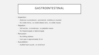
  • 20. GASTROINTESTINAL • Inspection : • Abdomen is protuberant , symmetrical , Umbilicus is inverted • No visible hernia , no visible dilated veins , no visible masses • Palpation : • Soft and lax , no tenderness , no palpable masses • No Hepatomegaly or Splenomegaly • Percussion : • No shifting dullness • Liver span is approximately 10 cm • Auscultation : • Audible heart sounds , no renal bruit
  • 21. MUSCULOSKELETAL • Joints : • No visible joint or bone deformities • No tenderness on movement or palpation • No decreased ROM , swelling or erythema
  • 22. NEUROLOGICAL • GCS : 15/15 • Upper Limb • Sensory : intact for touch and pain • Normal power , tone and reflexes • Lower Limb : • Sensory : intact to touch and pian • Normal power , tone and reflexes
  • 24. • Acute Coronary Syndrome • Pulmonary Embolism • Pneumothorax • Aortic Dissection • Acute Pericarditis
  • 25. HOW WOULD YOU APPROACH MR. N IN EMERGENCY DEPARTMENT
  • 28. STEMI ECG • ST elevation criteria • new ST elevation in two contiguous leads of >0.1 mV in all leads other than leads V2-V3 • for leads V2-V3: 0.2 mV in men 40 yr, 0.25 mV in men <40 yr, or 0.15 mV in women
  • 29. “TYPICAL” SEQUENTIAL CHANGES OF EVOLVING MI • 1. hyperacute T waves (tall, symmetric T waves) in the leads facing the infarcted area, with or without ST elevation • 2. ST elevation (injury pattern) in the leads facing the infarcted area usually in the first hours post infarct • in acute posterior MI, there is ST depression in V1-V3 (reciprocal to ST elevation in the • posterior leads that are not recorded in the standard 12-lead ECG) hence get a 15- lead ECG • 3. significant Q waves: >40 msec or >1/3 of the total QRS amplitude and present in at least 2 • consecutive leads in the same territory (hours to days post-infarct) • Q waves of infarction may appear in the very early stages, with or without ST changes • non-Q wave infarction: there may be only ST or T
  • 31. LABS AT ADMISSION 4/11/20 • WBC : 8.61 • HGB : 17.6 • PLT : 342 • PT : 12.5 • INR : 1.1 • PTT : 35.5 • Na : 139 • K : 3.8 • Cr : 84 • Troponin : 30 , is it enough? • on 5/11/20 it was 89000 • BNP : less then 10
  • 32. LABS • AST : 71 • ALT : 73 • ALP : 38 • HDL : 0.71 • LDL 4.11 • Triglycerides :1.76 • Cholesterol : 4.92 • Bilirubin total : 26 • Bilirubin Direct : 7.2
  • 33. CXR
  • 34. ECHOCARDIOGRAPHY • LV normal in size, mildly reduced. LVEF =40% • large sized apical, septal, anteroseptal, and anterior wall motion abnormality with hypokinesis to akinesis of the segments • RV is normal in size and function. • Inadequate TR jet to assess RVSP. • No hemodynamically significant valve disease. • There is no pericardial effusion.
  • 35. CORONARY ANGIOGRAPHY • Was Done • Insert vid
  • 36. HOSPITAL COARSE • During admission patient was started on IHD Medications : • Received loading dose of Aspirin 300 mg PO , and Clopidogrel 180 mg PO • Then kept on : • Aspirin 100 mg PO OD • Clopidogrel 75 mg PO OD • Tirofiban infusion during and after cath for 18 hour • Atorvastatin 80 mg PO OD • Heparin Infusion until day of CABG • Bisoprolol 2.5 mg PO OD
  • 37. EDUCATIONAL REVIEW FOR STEMI MANAGEMENT GOALS Preventing re-occlusion • Antiplatelet • antithrombosis Preventing mechanical complication • B-Blocker • ACEI • ARBs • Aldosterone antagonist Preventing recurrent Infarction • Statin • Cardiac rehab Preventing sudden cardiac death • AICD
  • 38. EDUCATIONAL REVIEW FOR ACS MANAGEMENT AND COMPLICATION • ACS includes the spectrum of unstable angina (UA), NSTEMI, and STEMI; this distinction aids in providing the appropriate therapeutic intervention
  • 39. 1. GENERAL MEASURES • 1. General Measures • ABCs: assess and correct hemodynamic status First • bed rest, cardiac monitoring, oxygen(if less 92) • nitroglycerin SL followed by IV • morphine IV
  • 40. 2. ANTI-PLATELET AND ANTICOAGULATION THERAPY • NSTEMI • Ticagrelor, in addition to ASA or if ASA contraindicated, subcutaneous low molecular weight heparin or IV unfractionated heparin (UFH) • – LMWH preferable, except in renal failure or if CABG is planned within 24 h • clopidogrel used if patient ineligible for ticagrelor • If PCI is planned: ticagrelor or prasugrel and consider IV GP IIb/IIIa inhibitor (e.g. abciximab) • Clopidogrel used if patient ineligible for ticagrelor and prasugrel • Prasugel contraindicated in those with a history of stroke/TIA, and avoidance of or lower dose is recommended for those >75 year old or weighing under 60 kg (TRITON-TIMI 38) • ANTICOAGULATION OPTIONS DEPEND ON REPERFUSION STRATEGY: • Primary PCI: UFH during procedure; bivalirudin is a possible alternative • Thrombolysis: LMWH (enoxaparin) until discharge from hospital; can use UFH as alternative because of possible rescue PCI • no reperfusion: LMWH (enoxaparin) until discharge from
  • 41. 3. B-BLOCKERS • STEMI: contraindications include signs of heart failure, low output states, risk of cardiogenic shock ,heart block, asthma or airway disease; initiate orally within 24 h of diagnosis when indicated • )
  • 42. 4. INVASIVE STRATEGIES AND REPERFUSION OPTIONS STEMI After diagnosis of STEMI is made, do not wait for results of further investigations before implementing reperfusion therapy THROMBOLYSIS Goal is Door to Balloon <30 min preferred if patient presents 12 h of symptom onset, has contraindications to PCI, or PCI cannot be administered within 90 min Criteria for successful THROMBOLYSIS : • ST segment elevation resolution below half its initial magnitude • no chest pain • Reperfusion rhythms • PCI • Primary PCI: without prior thrombolytic therapy • Rescue PCI: following failed thrombolytic, improves mortality vs. thrombolysis with fewer intra-cranial hemorrhages and recurrent MIs

Editor's Notes