INVESTIGATION OF CONCOMITANCY 
AND NONCONCOMITANCY 
RAju kAITI 
OpTOMETRIST 
DhulIkhEl hOSpITAl, kAThMANDu uNIVERSITY hOSpITAl
DDEEFFIINNIITTIIOONN OOFF 
SSTTRRAABBIISSMMUUSS 
SSttrraabbiissmmuuss oorr ssqquuiinntt iiss ddeeffiinneedd aass tthhee ddeevviiaattiioonn oorr 
mmiissaalliiggnnmmeenntt ooff tthhee eeyyeess.. 
The term strabismus is derived from a Greek word ” STRABISMOS” , 
“TO SQUINT,TO LOOK OBLIQUELY OR ASKANCE”
CCLLAASSSSIIFFIICCAATTIIOONN OOFF 
SSTTRRAABBIISSMMUUSS 
SSttrraabbiissmmuuss ccaann bbee ccllaassssiiffiieedd iinn tteerrmmss ooff:: 
1.DIRECTION OF DEVIATION 
- Hyper deviation 
- Hypo deviation 
- Divergent 
- Convergent
CCLLAASSSSIIFFIICCAATTIIOONN OOFF 
SSTTRRAABBIISSMMUUSS 
22.. CCOONNSSTTAANNCCYY 
-- ccoonnssttaanntt 
-- iinntteerrmmiitttteenntt
CCLLAASSSSIIFFIICCAATTIIOONN OOFF 
SSTTRRAABBIISSMMUUSS 
33.. CCOOMMIITTAANNCCYY 
-- ccoommiittaanntt // nnoonn--ppaarraallyyttiicc:: 
Same amplitude of deviation in all gazes 
Present in case of squint due to oculomotor imbalance 
-- iinnccoommiittaanntt // ppaarraallyyttiicc:: 
Amplitude of deviation changes with gaze 
found in paretic and restricted muscles 
Concomitant – Latin Word 
Concomitor: I Accompany (I Attend) : Duane
• Types 
1. Non-concomitant (deviation differ by 10 Pd) 
1. Non-paralytic (Special types of Strabismus 
2. Paralytic (Paralysis of Cranial Nerve/s)
Differences: 
Paralytic 
(Incomitant) 
Non-paralytic 
(Comitant) 
1 occurrence less common (15%) more common (85%) 
2 onset usually acquired & 
sudden; usually a 
sign of neurological 
or orbital disease; 
any age 
usually congenital 
3 deviation secondary deviation 
> primary deviation 
primary deviation = 
secondary deviation 
4 limitation of 
movement 
+ -
Differences:… 
5 comitance Only in late stages common 
6 diplopia present- homonymous or 
uncrossed in Esotropia; 
heteronymous or crossed 
in Exotropia 
no diplopia 
7 vertigo (nausea 
or vomiting) 
present - due to diplopia 
& false projection 
absent 
8 head posture Commonly abnormal Rarely abnormal 
9 false orientation 
(past-pointing) 
Common with recent 
paralysis 
rare 
10 H/O Head Trauma Common Uncommon
Incomitant deviation 
• Known as nonconcomitant/non comitant 
• Strabismic deviation that changes by more than 10pd 
for a given fixation distance in different directions of 
gaze 
• Misalignment of visual axis, marked by asymmetry 
between one position of gaze and another
Incomitant deviation ctd.. 
• May be congenital or acquired 
• Congenital –hydrocephalus,cerebral palsy, 
antenatal infections, birth trauma etc 
• Acquired-after 6 months of age, by 
trauma,tumour,vascular etiologies
Clinical Evaluation of Paralytic Strabismus 
1. Observation 
2. EOM Evaluation 
3. Cover Test 
4. Measurement of deviation 
5. Past Pointing 
6. Head tilt Test 
7. Diplopia/ Hess Charting 
8. Force duction/ Force Generation test 
9. Imaging Test (CT/ MRI)
Examination and Diagnosis 
1.Abnormal head posture 
• To compensate for deviation and to permit 
BSV 
• To eliminate diplopia and place eyes in most 
comfortable position 
• Three components-face turn/head tilt/chin up 
and down
• Patient position the head to reduce the need 
for affected muscle to contract 
• Head is placed in the field of action of involved 
muscle and eye moves out of field of action 
• Chin elevation for weakness of vertically acting 
muscles and in A/V pattern
Head posture: 
• Face turn -horizontal 
deviation 
• Chin elevation or 
depression –vertical 
deviation 
• Head tilt to one or the 
shoulder –torsional 
deviation
• In A/V pattern, to change the size of horizontal 
deviation in up/down gaze to get fusion and 
reduce deviation 
• Head tilt to counteract torsional and vertical 
diplopia 
• Not present in amblyopia/suppression 
• Some patients place head in opposite field to 
increase the separation between diplopic images
B.Cover Test 
• Measure primary and secondary deviation 
• Head tilt measurement with loose hand held 
prisms 
• In recently acquired paretic deviation, 
secondary angle exceeds the primary angle 
• In congenital and chronic paretic deviation-similar 
amount of deviation
B)cover test: 
1. Cover-uncover test: 
*cover test detects tropia 
*uncover test detects phoria.
Strabismus by raju
2.Alternate cover test 
measures total deviation(phoria& tropia) 
3.prismcover test: 
measures the 
actual heterotropia
Cover Test:
Cover Test:…
Cover-Uncover Test:
The Alternate Cover 
(Cross-Cover) Test:
C.Tests for Ocular Motility 
• To detect underactions and overactions 
• To differentiate paretic from mechanically 
restricted deviations 
• Check the ductions and version movements 
• Head erect with penlight as fixation target 
• Toy for children
• See limitation in eye movement that persist 
despite vigorous encouragement 
• Abduction/adduction/infraduction should 
have at least 10 mm of rotation,supraduction 
of 5-7 mm 
• Over action and underactions graded from 1 
to 4 depending on severity
Underactions denoted by – sign 
,overactions by + sign 
Each number represents a difference 
of 25% compared with normal 
movement 
- 1=slight(25%)underaction,-4=eye 
don’t extend beyond midline, 
+4=good portion of cornea 
disappears during adduction
• Ductions testing may not be as effective as 
version testing, it rules out mechanical 
restriction but don’t exclude possibility of 
paresis of EOMs 
• During ductions with paretic muscle, excessive 
innervations will flow to that eye, so needed 
movement actually be made.
Examination of ocular motility: 
1.versions: 
binocular simultaneous conjugate movement.
Strabismus by raju
Examination of squint ctd… 
1)test for ocular alignment & measurement of 
deviation. 
D)corneal light reflex test 
i)Hirschberg test
ii)krimsky test: 
measures 
the 
manifest 
deviation 
iii)Major amblyoscope: 
separate target illumination is used which can be 
moved to centre corneal light reflex.
E)dissimilar image test : 
1. Maddox rod test 
measures horizontal & vertical deviation.
2. Double Maddox rod test: 
measures the 
cyclodeviation. 
3.maddox wing test: 
measures the 
heterophoria.
Strabismus by raju
Synoptophore test : 
For assessing 
deviation 
& quantifying 
binocular vision. 
Can also detect 
suppression & 
ARC.
•Bielschowsky 
Diplopia chart 
Field of BSV 
Hess chart
Instrumentation 
• Originally to perform this test ,a black cloth 3 feet 
wide by 3 and half feet long ,marked out by a series of 
red lines subtending between them an angle of 5 
degrees used. 
• At the zero point of this coordinate system and at each 
points of interaction of the 15 and 30 degree lines with 
one another and with corresponding vertical and 
horizontal lines ,there is red dot . 
• These dots form an inner square of 8 dots and an 
outer square of 16 dots
• The patient wears red –green goggles from which his fusion 
is dissociated and holds the green flashlight 
• Fixating eyes sees from red and non-fixating sees from 
green . 
• The patient is instructed to place the green light over red 
light as much as possible. 
• The examiners marks the position indicated by the patient 
on the small card with a reduced copy of the screen . 
• The point found by the patient are connected by straight 
lines and permit the examiner to determine which ,if any 
,muscles react abnormally.
Performing Hess Screen test
Hess Screen Chart 
Additional information: 
1. Contraction of ipsilateral antagonist 
2. Overaction of contralateral synergist 
3. Inhibtion palsy of contralateral antagonist. 
Diagnosis 
- Smallest field – Affected eye 
- Field deviated maximum 
towards the centre 
- affected muscle 
Charts with sloping fields indicate 
the presence of A and v patterns
Consequences of EOM Paralysis 
Ex: LLR Palsy 
1. LMR Contract 
2. RMR Overaction 
3. RLR Under action
• The right eye is abnormal eye. The limited eye movement 
is shown by the smaller field . 
• RLR is underacting LMR is overacting . 
• Right sixth nerve palsy.
Diplopia charting
Look for 
- Gaze with maximum image separation (affected 
gaze) 
- Remote image belongs to affected eye 
- Image crossed / uncrossed
Forced duction test 
Force generation test 
Palsy Vs Paresis 
Paralytic Vs Restrictive Pathology 
Positive in restrictive pathology, 
negative 
in paralytic. 
Tug is appreciated in 
Restrictive pathology.
Force duction test 
• Also known as traction test 
• Described by wolf ,Gifford and jaensch (1900- 
1929). 
• It is performed to differentiate between the 
incomitant squint due to paralysis of EOM and 
that due to mechanical restriction of the 
ocular movement.
Force duction test 
A. No restriction is encountered when rotating 
the adducted eye into abduction . 
B. Contracture of MR muscle prevents examiner 
from the abducting the eye .
Procedures 
• Apply several drops of local anesthesia to limbal area of 
near the insertion of possibly mechanically restricted 
muscle ,which will be on the opposite side of the 
possibly paretic muscle. If the lateral rectus muscle ,the 
anesthesia would be applied near the insertion of medial 
rectus.
• The patient is then directed to look at a target 
that is held in the direction of action of the 
possible paretic muscle ,which is in the 
opposite direction of action of the possible 
mechanically restricted muscle . 
• If a RLR was the possible paretic muscle ,the 
patient would be directed to look at a target 
to the right .
• The eye is then grasped with toothless forceps near 
the limbus where the anesthesia was applied .the eye 
is grasped close to the limbus to get as little 
conjunctiva as possible in the forceps 
• An effort is then made to rotate the eye with the 
forceps in the direction of action of the possible 
paretic muscle .if the lateral rectus was the possible 
paretic muscle ,the eye would be grasped at the 
medial limbus and the rotation attempted would be 
an abduction movement .
• if the eye can be rotated with the forceps past 
the voluntary moved limit –presence of 
paretic muscle . 
• FDT is positive in cases of incomitant squint 
due to mechanical restriction and negative in 
cases of EOM PALSY.
Past Pointing 
• Described by Von Graefe 
– Anomalies of egocentric localization
Park’s Bielschowsky Head tilt Test- Park’s 
Procedure 
Useful for only Isolated Nerve Palsy 
Park’s Diagnostic Scheme 
1. Which is the hyper eye in primary gaze? 
– RE/LE 
2. Which horizontal field of gaze (right or left) does 
the hyperdeviation increase? 
3. Bielchowsky Head-Tilt Test: Toward which 
shoulder does the hyperdeviation increase?
Park’s Procedure
The 3 step Test Circling Method
Features of congenital incomitant deviation 
• Infrequent diplopia 
• Patients not aware of head posture 
• Nearly equal primary and secondary angle of 
deviation 
• Amblyopia and suppression may be present 
• Mild limitation of ocular movement
Recently acquired incomitant deviation. 
• Frequent diplopia 
• Patients aware of head posture 
• Significant difference in primary and secondary angle 
of deviation 
• Absence of Amblyopia and suppression 
• Severe limitation of ocular movement
Investigations: 
Diagnosis of strabismus is mainly clinical, if any 
neurological abnormality is suspected than we go for CT 
Scan,MRI and neurosurgical consultation.. 
Conclusion: 
Early diagnosis and management is essential for 
eliminating the effect of most difficult conditions 
-amblyopia or constant strabismus. 
Treatment under 6 years of age is ideal and 
allows better result.
References 
• Practical orthoptics (5th edition) 
– T. keith Lyle 
• Binocular vision and ocular motility 
– Gunter K. Von Noorden 
• Clinical Management of Strasbismus 
– Elizabeth E. Caloroso 
• Pediatric ophthalmology and strabismus- 
AAO section 6 
• Internet
…..THANK YOU

More Related Content

PPTX
A-V pattern strabismus
PPTX
Evaluation of squint
PPTX
Glass prescription in children
PPTX
WORTH FOUR DOT TEST.pptx
PPTX
PPT
Measuring interpupillary distance
PPTX
Slit lamp – biomicroscopy of eye
PPTX
Binocular vision and vision perception
A-V pattern strabismus
Evaluation of squint
Glass prescription in children
WORTH FOUR DOT TEST.pptx
Measuring interpupillary distance
Slit lamp – biomicroscopy of eye
Binocular vision and vision perception

What's hot (20)

PPT
fitting RGP lenses
PPTX
Ac/a ratio
PPTX
PPTX
Post surgical contact lens.pptx
PPT
Pentacam
PDF
Presbyopic contact lens description
PPTX
Test for stereopsis
PPTX
Soft contact lens complications
PPT
Contact lens verification(raju)
PPTX
Convergence insufficiency
PPTX
Orthoptic instruments
PPTX
Anisometropia
PPTX
Keratometry
PPTX
Dissociated Vertical Deviation (DVD)
PPT
Vergences of the eye
PPTX
ARC: abnormal retinal correspondence, eccentric fixation
PPT
Eccentric Fixation
PPTX
Soft Contact Lens Fitting
PPTX
Restrictive strabismus
PPT
Monovision
fitting RGP lenses
Ac/a ratio
Post surgical contact lens.pptx
Pentacam
Presbyopic contact lens description
Test for stereopsis
Soft contact lens complications
Contact lens verification(raju)
Convergence insufficiency
Orthoptic instruments
Anisometropia
Keratometry
Dissociated Vertical Deviation (DVD)
Vergences of the eye
ARC: abnormal retinal correspondence, eccentric fixation
Eccentric Fixation
Soft Contact Lens Fitting
Restrictive strabismus
Monovision
Ad

Viewers also liked (20)

PPT
Strabismus
PPTX
Ocular motility and strabismus
PPSX
Strabismus
PPT
Strabismus
PPT
Abnormal Head Posture in squint
PPTX
Motor adaptation in paretic and nonparetic strabismus
PPT
Paralytic strabismus, features and investigations of paretic strabismus
PPTX
Classification of squint
PPT
Diagnosis evaluation in strabismus
PPT
Squint/Strabismus
PPTX
Paralytic strabismus
PPTX
Hess charting
PPT
Paralytic strabismus
PPT
Occular motility
PPT
Brown's in myasthenia gravis correction: Dr Madhu Karna
PPT
Squint examination & management- simplified !!
PDF
2014 asmph strabismus & amblyopia
PPTX
Nystagmus namrata
PPTX
Binocular diplopia
Strabismus
Ocular motility and strabismus
Strabismus
Strabismus
Abnormal Head Posture in squint
Motor adaptation in paretic and nonparetic strabismus
Paralytic strabismus, features and investigations of paretic strabismus
Classification of squint
Diagnosis evaluation in strabismus
Squint/Strabismus
Paralytic strabismus
Hess charting
Paralytic strabismus
Occular motility
Brown's in myasthenia gravis correction: Dr Madhu Karna
Squint examination & management- simplified !!
2014 asmph strabismus & amblyopia
Nystagmus namrata
Binocular diplopia
Ad

Similar to Strabismus by raju (20)

PPTX
squint 4-1.pptx opthalmologist pptx squint
PPTX
squint 4-1.pptx squint opthalmologist ppt
PPT
Evaluation of squint - The Basics
PPTX
Strabismus-Clinical Examinations
PPTX
Examination of a case of strabismus
PPTX
strabismus.pptx
PPTX
Practical Clinical refraction class 3.pptx
PPTX
EXOTROPIA-FINAL PPT.pptx squint opthalmologist ppt
PDF
Role of orthoptics
PPTX
Retinoscopy
PPTX
Retinoscopy
PPT
Squint assessment
PPTX
Work up of proptosis.pptx a useful one .
PPTX
RETINOSCOPY BY DR. PUSHKAR DHIR.pptx
PPTX
Clinical examination of squint
PPTX
Assessment of ocular alignment
PPTX
Investigation in paralytic strabismus
PPTX
Approach to accommodative esotropia
PPTX
Prince investigation in paralytic strabismus
squint 4-1.pptx opthalmologist pptx squint
squint 4-1.pptx squint opthalmologist ppt
Evaluation of squint - The Basics
Strabismus-Clinical Examinations
Examination of a case of strabismus
strabismus.pptx
Practical Clinical refraction class 3.pptx
EXOTROPIA-FINAL PPT.pptx squint opthalmologist ppt
Role of orthoptics
Retinoscopy
Retinoscopy
Squint assessment
Work up of proptosis.pptx a useful one .
RETINOSCOPY BY DR. PUSHKAR DHIR.pptx
Clinical examination of squint
Assessment of ocular alignment
Investigation in paralytic strabismus
Approach to accommodative esotropia
Prince investigation in paralytic strabismus

More from Raju Kaiti (20)

PPT
Frames:types, materials and designs
PPTX
Exophthalmometry
PPT
Inconcomitant strabismus types and different tests
PPT
Principle of visual acuity charts class
PPT
Spherical, cylindrical and toric lenses
PPT
Preliminary examination
PPT
History taking
PPT
Prism & it's uses
PPT
Magnification and it's clinical uses
PPT
Polarization and it's application in Ophthalmology
PPT
Low vision introduction
PPT
Laser and it's clinical application
PPT
Coneal topography instrumentation, techniques, procedures, limitations, advan...
PPT
Color vision physiology, defects and different testing Procedures
PPT
Low vision non optical devices
PPT
Color vision and physiological processes
PPTX
Congenital nasolacrimal duct obstruction
PPT
Nightblindness and xerophthalmia
PPT
Pediatric Ophthalmic dispensing in different visual problems
PPT
RGP lens care and maintenance
Frames:types, materials and designs
Exophthalmometry
Inconcomitant strabismus types and different tests
Principle of visual acuity charts class
Spherical, cylindrical and toric lenses
Preliminary examination
History taking
Prism & it's uses
Magnification and it's clinical uses
Polarization and it's application in Ophthalmology
Low vision introduction
Laser and it's clinical application
Coneal topography instrumentation, techniques, procedures, limitations, advan...
Color vision physiology, defects and different testing Procedures
Low vision non optical devices
Color vision and physiological processes
Congenital nasolacrimal duct obstruction
Nightblindness and xerophthalmia
Pediatric Ophthalmic dispensing in different visual problems
RGP lens care and maintenance

Recently uploaded (20)

PDF
SEMEN PREPARATION TECHNIGUES FOR INTRAUTERINE INSEMINATION.pdf
PDF
Calcified coronary lesions management tips and tricks
PPT
nephrology MRCP - Member of Royal College of Physicians ppt
PPTX
CARDIOVASCULAR AND RENAL DRUGS.pptx for health study
PPTX
Wheat allergies and Disease in gastroenterology
PDF
Transcultural that can help you someday.
PPTX
Electrolyte Disturbance in Paediatric - Nitthi.pptx
PDF
OSCE SERIES ( Questions & Answers ) - Set 5.pdf
PDF
OSCE SERIES ( Questions & Answers ) - Set 3.pdf
PPTX
09. Diabetes in Pregnancy/ gestational.pptx
PPT
Dermatology for member of royalcollege.ppt
PPT
Infections Member of Royal College of Physicians.ppt
PDF
Comparison of Swim-Up and Microfluidic Sperm Sorting.pdf
PPTX
thio and propofol mechanism and uses.pptx
PPT
Rheumatology Member of Royal College of Physicians.ppt
PPTX
IMAGING EQUIPMENiiiiìiiiiiTpptxeiuueueur
PPTX
NUCLEAR-MEDICINE-Copy.pptxbabaabahahahaahha
PDF
OSCE SERIES - Set 7 ( Questions & Answers ).pdf
PDF
OSCE Series ( Questions & Answers ) - Set 6.pdf
PDF
AGE(Acute Gastroenteritis)pdf. Specific.
SEMEN PREPARATION TECHNIGUES FOR INTRAUTERINE INSEMINATION.pdf
Calcified coronary lesions management tips and tricks
nephrology MRCP - Member of Royal College of Physicians ppt
CARDIOVASCULAR AND RENAL DRUGS.pptx for health study
Wheat allergies and Disease in gastroenterology
Transcultural that can help you someday.
Electrolyte Disturbance in Paediatric - Nitthi.pptx
OSCE SERIES ( Questions & Answers ) - Set 5.pdf
OSCE SERIES ( Questions & Answers ) - Set 3.pdf
09. Diabetes in Pregnancy/ gestational.pptx
Dermatology for member of royalcollege.ppt
Infections Member of Royal College of Physicians.ppt
Comparison of Swim-Up and Microfluidic Sperm Sorting.pdf
thio and propofol mechanism and uses.pptx
Rheumatology Member of Royal College of Physicians.ppt
IMAGING EQUIPMENiiiiìiiiiiTpptxeiuueueur
NUCLEAR-MEDICINE-Copy.pptxbabaabahahahaahha
OSCE SERIES - Set 7 ( Questions & Answers ).pdf
OSCE Series ( Questions & Answers ) - Set 6.pdf
AGE(Acute Gastroenteritis)pdf. Specific.

Strabismus by raju

  • 1. INVESTIGATION OF CONCOMITANCY AND NONCONCOMITANCY RAju kAITI OpTOMETRIST DhulIkhEl hOSpITAl, kAThMANDu uNIVERSITY hOSpITAl
  • 2. DDEEFFIINNIITTIIOONN OOFF SSTTRRAABBIISSMMUUSS SSttrraabbiissmmuuss oorr ssqquuiinntt iiss ddeeffiinneedd aass tthhee ddeevviiaattiioonn oorr mmiissaalliiggnnmmeenntt ooff tthhee eeyyeess.. The term strabismus is derived from a Greek word ” STRABISMOS” , “TO SQUINT,TO LOOK OBLIQUELY OR ASKANCE”
  • 3. CCLLAASSSSIIFFIICCAATTIIOONN OOFF SSTTRRAABBIISSMMUUSS SSttrraabbiissmmuuss ccaann bbee ccllaassssiiffiieedd iinn tteerrmmss ooff:: 1.DIRECTION OF DEVIATION - Hyper deviation - Hypo deviation - Divergent - Convergent
  • 4. CCLLAASSSSIIFFIICCAATTIIOONN OOFF SSTTRRAABBIISSMMUUSS 22.. CCOONNSSTTAANNCCYY -- ccoonnssttaanntt -- iinntteerrmmiitttteenntt
  • 5. CCLLAASSSSIIFFIICCAATTIIOONN OOFF SSTTRRAABBIISSMMUUSS 33.. CCOOMMIITTAANNCCYY -- ccoommiittaanntt // nnoonn--ppaarraallyyttiicc:: Same amplitude of deviation in all gazes Present in case of squint due to oculomotor imbalance -- iinnccoommiittaanntt // ppaarraallyyttiicc:: Amplitude of deviation changes with gaze found in paretic and restricted muscles Concomitant – Latin Word Concomitor: I Accompany (I Attend) : Duane
  • 6. • Types 1. Non-concomitant (deviation differ by 10 Pd) 1. Non-paralytic (Special types of Strabismus 2. Paralytic (Paralysis of Cranial Nerve/s)
  • 7. Differences: Paralytic (Incomitant) Non-paralytic (Comitant) 1 occurrence less common (15%) more common (85%) 2 onset usually acquired & sudden; usually a sign of neurological or orbital disease; any age usually congenital 3 deviation secondary deviation > primary deviation primary deviation = secondary deviation 4 limitation of movement + -
  • 8. Differences:… 5 comitance Only in late stages common 6 diplopia present- homonymous or uncrossed in Esotropia; heteronymous or crossed in Exotropia no diplopia 7 vertigo (nausea or vomiting) present - due to diplopia & false projection absent 8 head posture Commonly abnormal Rarely abnormal 9 false orientation (past-pointing) Common with recent paralysis rare 10 H/O Head Trauma Common Uncommon
  • 9. Incomitant deviation • Known as nonconcomitant/non comitant • Strabismic deviation that changes by more than 10pd for a given fixation distance in different directions of gaze • Misalignment of visual axis, marked by asymmetry between one position of gaze and another
  • 10. Incomitant deviation ctd.. • May be congenital or acquired • Congenital –hydrocephalus,cerebral palsy, antenatal infections, birth trauma etc • Acquired-after 6 months of age, by trauma,tumour,vascular etiologies
  • 11. Clinical Evaluation of Paralytic Strabismus 1. Observation 2. EOM Evaluation 3. Cover Test 4. Measurement of deviation 5. Past Pointing 6. Head tilt Test 7. Diplopia/ Hess Charting 8. Force duction/ Force Generation test 9. Imaging Test (CT/ MRI)
  • 12. Examination and Diagnosis 1.Abnormal head posture • To compensate for deviation and to permit BSV • To eliminate diplopia and place eyes in most comfortable position • Three components-face turn/head tilt/chin up and down
  • 13. • Patient position the head to reduce the need for affected muscle to contract • Head is placed in the field of action of involved muscle and eye moves out of field of action • Chin elevation for weakness of vertically acting muscles and in A/V pattern
  • 14. Head posture: • Face turn -horizontal deviation • Chin elevation or depression –vertical deviation • Head tilt to one or the shoulder –torsional deviation
  • 15. • In A/V pattern, to change the size of horizontal deviation in up/down gaze to get fusion and reduce deviation • Head tilt to counteract torsional and vertical diplopia • Not present in amblyopia/suppression • Some patients place head in opposite field to increase the separation between diplopic images
  • 16. B.Cover Test • Measure primary and secondary deviation • Head tilt measurement with loose hand held prisms • In recently acquired paretic deviation, secondary angle exceeds the primary angle • In congenital and chronic paretic deviation-similar amount of deviation
  • 17. B)cover test: 1. Cover-uncover test: *cover test detects tropia *uncover test detects phoria.
  • 19. 2.Alternate cover test measures total deviation(phoria& tropia) 3.prismcover test: measures the actual heterotropia
  • 23. The Alternate Cover (Cross-Cover) Test:
  • 24. C.Tests for Ocular Motility • To detect underactions and overactions • To differentiate paretic from mechanically restricted deviations • Check the ductions and version movements • Head erect with penlight as fixation target • Toy for children
  • 25. • See limitation in eye movement that persist despite vigorous encouragement • Abduction/adduction/infraduction should have at least 10 mm of rotation,supraduction of 5-7 mm • Over action and underactions graded from 1 to 4 depending on severity
  • 26. Underactions denoted by – sign ,overactions by + sign Each number represents a difference of 25% compared with normal movement - 1=slight(25%)underaction,-4=eye don’t extend beyond midline, +4=good portion of cornea disappears during adduction
  • 27. • Ductions testing may not be as effective as version testing, it rules out mechanical restriction but don’t exclude possibility of paresis of EOMs • During ductions with paretic muscle, excessive innervations will flow to that eye, so needed movement actually be made.
  • 28. Examination of ocular motility: 1.versions: binocular simultaneous conjugate movement.
  • 30. Examination of squint ctd… 1)test for ocular alignment & measurement of deviation. D)corneal light reflex test i)Hirschberg test
  • 31. ii)krimsky test: measures the manifest deviation iii)Major amblyoscope: separate target illumination is used which can be moved to centre corneal light reflex.
  • 32. E)dissimilar image test : 1. Maddox rod test measures horizontal & vertical deviation.
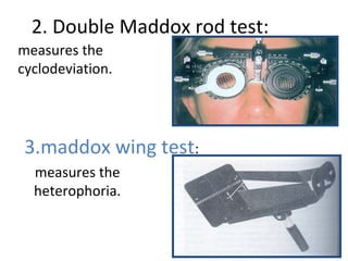
  • 33. 2. Double Maddox rod test: measures the cyclodeviation. 3.maddox wing test: measures the heterophoria.
  • 35. Synoptophore test : For assessing deviation & quantifying binocular vision. Can also detect suppression & ARC.
  • 36. •Bielschowsky Diplopia chart Field of BSV Hess chart
  • 37. Instrumentation • Originally to perform this test ,a black cloth 3 feet wide by 3 and half feet long ,marked out by a series of red lines subtending between them an angle of 5 degrees used. • At the zero point of this coordinate system and at each points of interaction of the 15 and 30 degree lines with one another and with corresponding vertical and horizontal lines ,there is red dot . • These dots form an inner square of 8 dots and an outer square of 16 dots
  • 38. • The patient wears red –green goggles from which his fusion is dissociated and holds the green flashlight • Fixating eyes sees from red and non-fixating sees from green . • The patient is instructed to place the green light over red light as much as possible. • The examiners marks the position indicated by the patient on the small card with a reduced copy of the screen . • The point found by the patient are connected by straight lines and permit the examiner to determine which ,if any ,muscles react abnormally.
  • 40. Hess Screen Chart Additional information: 1. Contraction of ipsilateral antagonist 2. Overaction of contralateral synergist 3. Inhibtion palsy of contralateral antagonist. Diagnosis - Smallest field – Affected eye - Field deviated maximum towards the centre - affected muscle Charts with sloping fields indicate the presence of A and v patterns
  • 41. Consequences of EOM Paralysis Ex: LLR Palsy 1. LMR Contract 2. RMR Overaction 3. RLR Under action
  • 42. • The right eye is abnormal eye. The limited eye movement is shown by the smaller field . • RLR is underacting LMR is overacting . • Right sixth nerve palsy.
  • 44. Look for - Gaze with maximum image separation (affected gaze) - Remote image belongs to affected eye - Image crossed / uncrossed
  • 45. Forced duction test Force generation test Palsy Vs Paresis Paralytic Vs Restrictive Pathology Positive in restrictive pathology, negative in paralytic. Tug is appreciated in Restrictive pathology.
  • 46. Force duction test • Also known as traction test • Described by wolf ,Gifford and jaensch (1900- 1929). • It is performed to differentiate between the incomitant squint due to paralysis of EOM and that due to mechanical restriction of the ocular movement.
  • 47. Force duction test A. No restriction is encountered when rotating the adducted eye into abduction . B. Contracture of MR muscle prevents examiner from the abducting the eye .
  • 48. Procedures • Apply several drops of local anesthesia to limbal area of near the insertion of possibly mechanically restricted muscle ,which will be on the opposite side of the possibly paretic muscle. If the lateral rectus muscle ,the anesthesia would be applied near the insertion of medial rectus.
  • 49. • The patient is then directed to look at a target that is held in the direction of action of the possible paretic muscle ,which is in the opposite direction of action of the possible mechanically restricted muscle . • If a RLR was the possible paretic muscle ,the patient would be directed to look at a target to the right .
  • 50. • The eye is then grasped with toothless forceps near the limbus where the anesthesia was applied .the eye is grasped close to the limbus to get as little conjunctiva as possible in the forceps • An effort is then made to rotate the eye with the forceps in the direction of action of the possible paretic muscle .if the lateral rectus was the possible paretic muscle ,the eye would be grasped at the medial limbus and the rotation attempted would be an abduction movement .
  • 51. • if the eye can be rotated with the forceps past the voluntary moved limit –presence of paretic muscle . • FDT is positive in cases of incomitant squint due to mechanical restriction and negative in cases of EOM PALSY.
  • 52. Past Pointing • Described by Von Graefe – Anomalies of egocentric localization
  • 53. Park’s Bielschowsky Head tilt Test- Park’s Procedure Useful for only Isolated Nerve Palsy Park’s Diagnostic Scheme 1. Which is the hyper eye in primary gaze? – RE/LE 2. Which horizontal field of gaze (right or left) does the hyperdeviation increase? 3. Bielchowsky Head-Tilt Test: Toward which shoulder does the hyperdeviation increase?
  • 55. The 3 step Test Circling Method
  • 56. Features of congenital incomitant deviation • Infrequent diplopia • Patients not aware of head posture • Nearly equal primary and secondary angle of deviation • Amblyopia and suppression may be present • Mild limitation of ocular movement
  • 57. Recently acquired incomitant deviation. • Frequent diplopia • Patients aware of head posture • Significant difference in primary and secondary angle of deviation • Absence of Amblyopia and suppression • Severe limitation of ocular movement
  • 58. Investigations: Diagnosis of strabismus is mainly clinical, if any neurological abnormality is suspected than we go for CT Scan,MRI and neurosurgical consultation.. Conclusion: Early diagnosis and management is essential for eliminating the effect of most difficult conditions -amblyopia or constant strabismus. Treatment under 6 years of age is ideal and allows better result.
  • 59. References • Practical orthoptics (5th edition) – T. keith Lyle • Binocular vision and ocular motility – Gunter K. Von Noorden • Clinical Management of Strasbismus – Elizabeth E. Caloroso • Pediatric ophthalmology and strabismus- AAO section 6 • Internet