SlideShare a Scribd company logo
Dr. Ajay Manickam
MS (ENT) JUNIOR RESIDENT
R.G.Kar Medical College

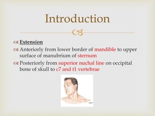
 Extension
 Anteriorly from lower border of mandible to upper
surface of manubrium of sternum
 Posteriorly from superior nuchal line on occipital
bone of skull to c7 and t1 vertebrae
Introduction

 Skin – cervical
dermatome
 Muscles – cervical
myotomes
 The branchial
apparatus
Developmental
Anatomy

 4 compartments provide longitudinal organisation
 Visceral compartment – anterior – digestive,
respiratory & endocrine glands
 Vertebral compartment – posterior – cervical
vertebrae, spinal cord, cervical nerves and muscles
 2 vascular compartments – lateral – major blood
vessels and vagus nerve
Compartments

Fascia
Superficial
Deep
1.Superficial layer
2.Middle layer
3. Deep laayer
Fascial layers

 Thin sheet of muscle
platysma, begins in
superficial fascia of thorax,
attaches to mandible and
blend with muscles of face
 Penetrated by blood vessel
that supply neck skin
 Subplatysmal flap protects
blood supply to the skin
 Facial nerve- cervical branch
Superficial fascia

 Superficial layer
 Arises from ligamentum nuchaeand spinous process
of cervical vertebrae
 Splits to enclose
trapezius,omohyoid,sternocleidomastoid,strap
muscles and parotid gland
Deep cervical fascia

 Middle layer
 Derived from superior layer of deep
cervical fascia encircles trachea,
thyroid, esophagus
 A. Investing Layer
 B. Muscular Pretracheal Layer
 C. Visceral Pretracheal Layer
 D. Prevertebral Layer
Deep cervical fascia

 Deep layer
 Arise from ligamentum
nuchaeand spinous process of
cervical vertebra
 Splits to enclose postvertebral
muscles, form layer over
vertebrae
 Floor of post triangle
 Allows pharynx to glide during
deglutition
 Extends in lower region of neck
to axilla – axillary sheath
Deep cervical fascia

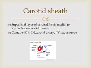
 Superficial layer of cervical fascia medial to
sternocleidomastoid muscle
 Contains 80% LN,carotid artery, IJV,vagus nerve
Carotid sheath

Triangles of neck

Fascial spaces
Between the fascial layers in the neck
are spaces that may provide conduit for
the spread of infections
They contain loose areolar fascia

 Deep Neck Spaces are described in relation to the
Hyoid bone.
 A. Entire length of the neck.
 B. Suprahyoid.
 C. Infrahyoid.
Classification of neck
spaces

 1. Superficial neck space
 2. Deep neck spaces
Retropharyngeal space
Danger space of Gillette
Pre vertebral space
Involving entire length
of neck

 Sub mental space
 Submandibular space
-Sublingual space
-Sub maxillary space
 Peri tonsillar space
 Parotid space
 Para pharyngeal space
 Masticator space
Supra-hyoid

 Pretracheal space
Infra hyoid

 Extends from base of skull to
tracheal bifurcation
 Between two parapharyngeal space
 Superior – skull base
 Anterior – musculature of pharynx
 Posteror limit – prevertebral fascia
 Communicates with – mediastinum
 It is divided into two lateral
compartments space of gillete by
fibrous raphe
Retropharyngeal space

 There are a group of inconsistent nodes in the
retropharyngeal space known as the Glands of Henle
which regresses by 5 yrs of age. Suppuration of these
nodes result in Ac. Retropharyngeal abscess and thus
commoner in children.
 There is also a constant group of nodes called the
Rouvier’s nodes which are the first nodes to enlarge
in cases of nasopharyngeal and posterior sinus
malignancies.
Retropharyngeal space

 Base of skull to diaphragm
 Located between the pre vertebral fascia and alar
fascia
 Retro pharyngeal space proper is in front of alar
fascia
 This is called danger space because of easy route of
mediastinitis
Danger space

 Potential space between cervical vertebra posteriorly and
the prevertebral fascia anteriorly
 Extends from base of skull to coccyx
 Tuberculosis of spine, penetrating traumas chief source of
infections
Prevertebral space

 Midline space between anterior bellies of digastric
muscles
 Contents – areolar tissue, lymphnode, ant jugular
vein
Submental space

 Includes submaxillary + sublingual,
divided by mylohyoid muscle
 Superficial boundary – submandibular
gland & digastric muscle
 Deep boundary – mylohyoid muscle
 Lies between mucous membrane of
floor of mouth& tongue on oneside &
superficial layer of deep cervical fascia,
from mandible to hyoid bone
 Comunicates with floor of the mouth
Submandibular space

 Between capsule of tonsil & superior constrictor
 Located lateral to the tonsils
 Infection source is mainly tonsillar crypts
 Communicates with retropharyngeal &
parapharyngeal space
Peritonsillar space

 Boundaries
The space is circumscribed by the superficial layer of the deep cervical
fascia
superior margin: external auditory canal; apex of the mastoid process
inferior margin: inferior mandibular margin (although the parotid tail can
extend further inferiorly below the angle of the mandible)
anterior margin: masticator space
 contents
parotid glands
parotid lymph nodes
facial nerve (CN VII)
external carotid artery
retromandibular vein
Fascial layer is very thick superficially , very thin on deep side of gland- burst
to parapharyngeal space- mediastinum
Parotid space

 Inverted pyramid shaped
Para pharyngeal space

 Located between superficial layer of deep
cervical fascia & muscles of mastication
 Extends from base of skull to lower border
of mandible
 Contents
muscles of mastication
ramus and body of mandible
inferior alveolar nerve,vein,artery
mandibular division of the trigeminal nerve
(V3)
enters the masticator space via the foramen
ovale
Masticator space

 Anterior and lateral to thyroid cartilage
 Contains delphian node
 Communicates – superior mediastinum
Pretracheal space

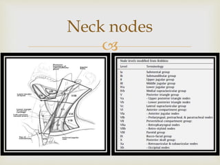
Neck nodes


Neck space infections

 Rare, but life threatening infection,that causes
progressive necrosis of the subcutaneous fat and
fascia and causes secondary necrosis of the overlying
skin.
 ETIOLOGY - Odontogenic infections
- Tonsillar infections
- As a complication of other DNSI
Necrotizing fascitis

 Cellulitis with disproportionate pain.
 Reduced skin sensation of the involved areas.
 Outer zone- Erythema
Intermediate zone- Tender ecchymosis
Central zone- Vesiculation
 Soft tissue crepitus due to gas formation.
 Hypocalcemia , Hyponatremia , Dehydration
Necrotizing fascitis

 Early correction of fluid and
electrolyte imbalance.
 I.V Penicillin and I.V Metronidazole
are the mainstays of the antimicrobial
therapy.
 Surgical debridement of all necrotic
areas is the key to successful
treatment of the patient.
 Skin grafting after wound
debridement
Necrotizing fascitis

 Life threatening
infection
 URI, tuberculous
lymphadenitis
 X-Ray soft tissue neck
lateral view, CT
 Incision & drainage
Retropharyngeal space
infections

 Children <3yDysphagia and difficulty in breathing.
 Stridor and Croupy cough maybe present,Torticollis,Bulge in the posterior
pharyngeal wall.
 The child is febrile and adopts a
peculiar posture with the neck
flexed and the head extended.
 Straightening of the cervical
spine known as Ramrod Spine
Radiographic picture of the lateral
view of neck (soft tissue) shows
widening of the prevertebral
space and even the presence of
gas shadows(air fluid levels).
Acute retropharyngeal
abscess

 Incision and Drainage of abscess is done,usually without
anaesthesia as there is risk of rupture during intubation.[the child
is kept supine with head low and mouth opened with a gag.A
vertical incision is given in the most fluctuant area.Suction should
always be available to prevent aspiration]
 Systemic Antibiotics-Broad spectrum antibiotics like Ceftriaxone
and Metronidazole may be used.
 Tracheostomy in airway obstruction
Acute retropharyngeal
abscess

 TB Spine(Pott’s Spine) where the pus collects in the
prevertebral space.
 TB of retropharyngeal lymph nodes present in the
retropharyngeal space proper.
 Post traumatic-vertebral fracture.
 Spread from Parapharyngeal abscess
Chronic retropharyngeal
abscess

 Discomfort in the throat,mild dysphagia.
 Pain is absent due to cold abscess.
 Bulge in the posterior pharyngeal wall
either centrally or laterally.
 Neck may show Tubercular lymph nodes.
 Treatment - Incision and drainage of
abscess is done through a vertical incision
along the anterior border of the
sternocleidomastoid for low abscesses, or
along its posterior border for high
abscesses.
 Full course of anti-Tubercular therapy is
given
Retropharyngeal space
infections

 Odontogenic infection – submandibular space –
submental region
 Mandibular fractures
 Cutaneous infection
 Treatment- I&D
Sub mental abscess

 Drooling, trismus,
dysphagia, stridor caused by
laryngeal edema, and
elevation of the posterior
tongue against the palate ,
fever, tachycardia.
 Aerobe, anaerobe
 Maintanence of airway
 Needle aspiration USG or CT
guided
Submandibular space
infection

 Toothache, fever, odynophagia, drooling.
 SUBLINGUAL space infection
-floor of mouth swelling.
-tongue elevation.
 SUBMAXILLARY space infection
-brawny/woody tender swelling below the chin.
 Trismus.
 Stridor- due to falling back of tongue, laryngeal edema.
 Initially there is cellulitis which is followed by abscess
formation.
Submandibular space
infection

Ludwig’s angina
Xray showing
supraglottic swelling

 Systemic antibiotics- Ceftriaxone/Cefuroxime and
Metronidazole/Clindamycin.
 Tracheostomy if airway is compromised after unsuccessful
attempts at oral/nasal intubations.
 Incision and Drainage of Abscess:
intraoral—sublingually localised infection.
extraoral—submaxillary infection.
 A transverse incision extending from one angle of mandible to
the other is made with vertical opening of midline musculature
of tongue with a blunt haemostat
Ludwig’s angina

 Quinsy
 Tonsillitis
 Odynophagia, hot
potato voice
 Complication –
ludwig’s angina,
adjacent spaces
 Needle aspiration
 I&D
Peri tonsillar space
infection

 Peritonsillar abscess is opened at the
point of maximum bulge above the
upper pole or just lateral to the point
of junctionof anterior pillar and a
horizontal line drawn through the
base of the uvula
 Interval Tonsillectomy maybe done 4 to
6 weeks after an attack of Quincy.
 Abscess/Hot Tonsillectomy are preffered
by some instead of Incision and
drainage. This has the risk of abscess
rupture during anaesthesia and
excessive bleeding at the time of
operation.
Incision & drainage

 Acute/Chronic infections of tonsils and adenoid,
bursting of the peritonsillar abscess.
 Dental infection usually from the lower last molar.
 From Bezold abscess or Petrositis.
 Infections of parotid, retropharyngeal and
submaxillary spaces.
 Penetrating injuries of neck, injection of L.A for
mandibular nerve block or for tonsillectomy.
Parapharyngeal space
infections

 More common in adults
 Infective process of upper
aerodigestive tract,
 Trismus, pyrexia, tonsil may
be medially displaced
 USG, CT, needle aspiration
under CT or USG guidance
 Small loculated –
conservatively
 Large collections – external
approach, medial to carotid
sheath, isertion of a drain
Parapharyngeal space
infections

 Incision and Drainage
 -Usually done under G.A.
 -Pre-op tracheostomy if trismus is marked.
 -Drained by a horizontal incision made 2-3 cms below
the angle of the mandible.Blunt dissection is done along
the inner surface of the medial pterygoid towards styloid
process and the abscess is evacuated and a drain is
inserted.
 [Transoral drainage should never be done due to the
danger of the great vessels which pass through this
space.]
Parapharyngeal space
infection

 Causes
 Ascent of bacterial
infection(Staphylococcus,
Streptococcus,Haemophilus) to a
dehydrated parotid via Stenson’s duct
from oral cavity.
 Suppuration of intra-parotid LNs.
 Spread of infection from the auditory
canal via the cartlaginous fissures of
Santorini or the bony foramen of Huschke.
Parotid space infection

 Symptoms
Spontaneous onset of painful parotid
enlargement followed by fever and
cellulitis which then turns into fluctuant
parotid abscess.
Pain and induration over the parotid.
Pitting edema over the parotid area
differentiates parotid abscess from
simple parotitis
Parotid massage expresses pus into the
oral cavity via the Stenson’s duct
,opposite the upper 2nd molar.
Parotid space infection

Treatment:-
Maintainence of oral hygiene, IV antibiotics
Incision and Drainage:-
 -Blair’s incision made.
 -Multiple incisions made through fascia parallel to
branches of the facial nerve.
 -Blunt dissection done to evacuate the pus.
 -Drains are placed.
Parotid space


More Related Content

PPTX
Deep Neck Spaces
PPTX
Deep neck space infections -Dr.Ashly Alexander
PPT
Neck spaces anatomy and infections
PPSX
Anatomy of Neck spaces & Infections
PPTX
Retropharyngeal space
PPTX
Deep neck space infections
PPTX
Parapharyngeal space
PPTX
Deep neck spaces and surgical anatomy
Deep Neck Spaces
Deep neck space infections -Dr.Ashly Alexander
Neck spaces anatomy and infections
Anatomy of Neck spaces & Infections
Retropharyngeal space
Deep neck space infections
Parapharyngeal space
Deep neck spaces and surgical anatomy

What's hot (20)

PPTX
Nasal Cavity and Sphenoid Sinus Anatomy
PPTX
Juvenile nasopharyngeal angiofibroma
PPTX
Surgical anatomy of Infratemporal fossa. by Dr. Aditya Tiwari
PPTX
Fisch approaches Dr Zeeshan Ahmad
PPSX
Glomus Tumour
PPTX
Anatomy of temporal bone
PPTX
surgical approaches to frontal sinus ppt
PPTX
Cortical mastoidectomy
PPTX
Surgical treatment of paraphyrngeal and retropharyngeal abscesses
PPTX
Maxillectomy and craniofacial resection
PPTX
Infratemporal fossa approaches
PPTX
1. MAXILLECTOMY.pptx
ODP
Total laryngectomy
PDF
External approaches to sinus surgery
PPTX
Spaces of middle ear and their surgical importance
PPT
Classifications in ent
PPTX
Ca maxilla
PPTX
Csf Rhinorrhea - Overview
Nasal Cavity and Sphenoid Sinus Anatomy
Juvenile nasopharyngeal angiofibroma
Surgical anatomy of Infratemporal fossa. by Dr. Aditya Tiwari
Fisch approaches Dr Zeeshan Ahmad
Glomus Tumour
Anatomy of temporal bone
surgical approaches to frontal sinus ppt
Cortical mastoidectomy
Surgical treatment of paraphyrngeal and retropharyngeal abscesses
Maxillectomy and craniofacial resection
Infratemporal fossa approaches
1. MAXILLECTOMY.pptx
Total laryngectomy
External approaches to sinus surgery
Spaces of middle ear and their surgical importance
Classifications in ent
Ca maxilla
Csf Rhinorrhea - Overview
Ad

Viewers also liked (20)

PPT
Anatomy of neck spaces and levels of cervical
PPTX
Deep neck spaces and infection ENT
PPT
CT ANATOMY OF THE NECK SPACES
PPTX
CT anatomy of Neck Spaces RV
PPTX
Surgical anatomy of deep neck spaces
PDF
Cross sectional anatomy of the neck
PPTX
Diagnostic imaging in head and neck pathology
PPTX
cervical fascia
PPTX
Presentation1.pptx, radiological anatomy of the neck.
PPT
Surgical anatomy of the head and neck /certified fixed orthodontic courses b...
PPT
Deep neck space infections
PPT
Deep neck infection
PPT
Compartments of the head and neck /certified fixed orthodontic courses by I...
PPTX
Ppt of anatomy of neck & ML imp
PPTX
Deep cervical fascia
PPTX
Odontogenic infections
PPT
MRI in neck imaging
PPTX
Layers of the neck
PPT
Space infection 2 /certified fixed orthodontic courses by Indian dental academy
PPTX
Deep cervical fascia (fascia colli)
Anatomy of neck spaces and levels of cervical
Deep neck spaces and infection ENT
CT ANATOMY OF THE NECK SPACES
CT anatomy of Neck Spaces RV
Surgical anatomy of deep neck spaces
Cross sectional anatomy of the neck
Diagnostic imaging in head and neck pathology
cervical fascia
Presentation1.pptx, radiological anatomy of the neck.
Surgical anatomy of the head and neck /certified fixed orthodontic courses b...
Deep neck space infections
Deep neck infection
Compartments of the head and neck /certified fixed orthodontic courses by I...
Ppt of anatomy of neck & ML imp
Deep cervical fascia
Odontogenic infections
MRI in neck imaging
Layers of the neck
Space infection 2 /certified fixed orthodontic courses by Indian dental academy
Deep cervical fascia (fascia colli)
Ad

Similar to SURGICAL ANATOMY OF DEEP NECK SPACES (20)

PPTX
Abscesses in Ent and HEAD, NECK region .pptx
PPTX
DEEP NECK SPACE INFECTIONS by dr Priyanka FINAL-1.pptx
PPTX
Deep space neck infection .
PPTX
Deepneck space space infections.undar.pptx
PPTX
Fascial Spaces in the Head & Neck
PPTX
DEEP NECK SPACES-1.pptx
PPT
deep neck infection EDITED.ppt and spread
PPT
Deep neck spaces are anatomical compartments in the neck, defined by the deep...
PPT
Deep neck-infection-051005 /certified fixed orthodontic courses by Indian den...
PPTX
NECK SPACE AND LYMPHATIC SYSTEM.pptx
PPTX
DEEP NECK SPACES ANATOMY.pptx
PPTX
DEEP NECK SPACE INFECTIONS - DR ADITYA GOEL (MS-ENT)
PDF
Deep Neck Spaces and Neck Infection & Abscess.pdf
PPT
Deep neck-infections /certified fixed orthodontic courses by Indian dental ac...
PPT
Anatomy
PPTX
Deep facial spaces of head and neck
PPTX
space infection in oral [Autosaved].pptx
PPTX
SPACE INFECTIONS-TYPES, DIAGNSOSIS AND MANAGEMENT
PDF
deepneckspaceinfections-dr-190619164411.pdf
PPTX
DEEP NECK SPACES.pptx
Abscesses in Ent and HEAD, NECK region .pptx
DEEP NECK SPACE INFECTIONS by dr Priyanka FINAL-1.pptx
Deep space neck infection .
Deepneck space space infections.undar.pptx
Fascial Spaces in the Head & Neck
DEEP NECK SPACES-1.pptx
deep neck infection EDITED.ppt and spread
Deep neck spaces are anatomical compartments in the neck, defined by the deep...
Deep neck-infection-051005 /certified fixed orthodontic courses by Indian den...
NECK SPACE AND LYMPHATIC SYSTEM.pptx
DEEP NECK SPACES ANATOMY.pptx
DEEP NECK SPACE INFECTIONS - DR ADITYA GOEL (MS-ENT)
Deep Neck Spaces and Neck Infection & Abscess.pdf
Deep neck-infections /certified fixed orthodontic courses by Indian dental ac...
Anatomy
Deep facial spaces of head and neck
space infection in oral [Autosaved].pptx
SPACE INFECTIONS-TYPES, DIAGNSOSIS AND MANAGEMENT
deepneckspaceinfections-dr-190619164411.pdf
DEEP NECK SPACES.pptx

More from Ajay Manickam (20)

PPTX
direct LARYNGOSCOPY.pptx
PPTX
PDF
Cutaneous horn of pinna
PPTX
Sino Nasal malignancy & Anterior skull base surgery, Endoscopy is the best ???
PPTX
NACT in Head and Neck cancer
PPTX
Nasopharyngeal cancer
PPTX
Inter group rhabdomyosarcoma study group (irsg)
PPTX
Thoracic duct injury in neck dissection
PPTX
Sentinel node biopsy in oral cancer
PPTX
Frontal sinus procedures
PPTX
MIDLINE LUMP'S DISCRETE FORTUNE
PPTX
Glomus tumors
PPT
Lasers in otology
PPTX
Otosclerosis
PPTX
Local flaps in ent
PPTX
The mechanism of hearing
PPTX
ASSESSMENT OF Deafness
PPT
Meinere'S disease
PPTX
Diseases of the external ear
PPTX
Ossiculoplasty
direct LARYNGOSCOPY.pptx
Cutaneous horn of pinna
Sino Nasal malignancy & Anterior skull base surgery, Endoscopy is the best ???
NACT in Head and Neck cancer
Nasopharyngeal cancer
Inter group rhabdomyosarcoma study group (irsg)
Thoracic duct injury in neck dissection
Sentinel node biopsy in oral cancer
Frontal sinus procedures
MIDLINE LUMP'S DISCRETE FORTUNE
Glomus tumors
Lasers in otology
Otosclerosis
Local flaps in ent
The mechanism of hearing
ASSESSMENT OF Deafness
Meinere'S disease
Diseases of the external ear
Ossiculoplasty

Recently uploaded (20)

PPTX
1 General Principles of Radiotherapy.pptx
PDF
Khadir.pdf Acacia catechu drug Ayurvedic medicine
DOC
Adobe Premiere Pro CC Crack With Serial Key Full Free Download 2025
PPTX
neonatal infection(7392992y282939y5.pptx
PPTX
Chapter-1-The-Human-Body-Orientation-Edited-55-slides.pptx
PPTX
SKIN Anatomy and physiology and associated diseases
PDF
Medical Evidence in the Criminal Justice Delivery System in.pdf
PPTX
DENTAL CARIES FOR DENTISTRY STUDENT.pptx
PPTX
Gastroschisis- Clinical Overview 18112311
PPTX
History and examination of abdomen, & pelvis .pptx
DOCX
NEET PG 2025 | Pharmacology Recall: 20 High-Yield Questions Simplified
PPTX
15.MENINGITIS AND ENCEPHALITIS-elias.pptx
PPTX
Electromyography (EMG) in Physiotherapy: Principles, Procedure & Clinical App...
PPTX
Fundamentals of human energy transfer .pptx
PDF
Handout_ NURS 220 Topic 10-Abnormal Pregnancy.pdf
PPTX
post stroke aphasia rehabilitation physician
PPTX
Important Obstetric Emergency that must be recognised
PDF
Intl J Gynecology Obste - 2021 - Melamed - FIGO International Federation o...
PPTX
NEET PG 2025 Pharmacology Recall | Real Exam Questions from 3rd August with D...
PPTX
CEREBROVASCULAR DISORDER.POWERPOINT PRESENTATIONx
1 General Principles of Radiotherapy.pptx
Khadir.pdf Acacia catechu drug Ayurvedic medicine
Adobe Premiere Pro CC Crack With Serial Key Full Free Download 2025
neonatal infection(7392992y282939y5.pptx
Chapter-1-The-Human-Body-Orientation-Edited-55-slides.pptx
SKIN Anatomy and physiology and associated diseases
Medical Evidence in the Criminal Justice Delivery System in.pdf
DENTAL CARIES FOR DENTISTRY STUDENT.pptx
Gastroschisis- Clinical Overview 18112311
History and examination of abdomen, & pelvis .pptx
NEET PG 2025 | Pharmacology Recall: 20 High-Yield Questions Simplified
15.MENINGITIS AND ENCEPHALITIS-elias.pptx
Electromyography (EMG) in Physiotherapy: Principles, Procedure & Clinical App...
Fundamentals of human energy transfer .pptx
Handout_ NURS 220 Topic 10-Abnormal Pregnancy.pdf
post stroke aphasia rehabilitation physician
Important Obstetric Emergency that must be recognised
Intl J Gynecology Obste - 2021 - Melamed - FIGO International Federation o...
NEET PG 2025 Pharmacology Recall | Real Exam Questions from 3rd August with D...
CEREBROVASCULAR DISORDER.POWERPOINT PRESENTATIONx

SURGICAL ANATOMY OF DEEP NECK SPACES

  • 1. Dr. Ajay Manickam MS (ENT) JUNIOR RESIDENT R.G.Kar Medical College
  • 2.   Extension  Anteriorly from lower border of mandible to upper surface of manubrium of sternum  Posteriorly from superior nuchal line on occipital bone of skull to c7 and t1 vertebrae Introduction
  • 3.   Skin – cervical dermatome  Muscles – cervical myotomes  The branchial apparatus Developmental Anatomy
  • 4.   4 compartments provide longitudinal organisation  Visceral compartment – anterior – digestive, respiratory & endocrine glands  Vertebral compartment – posterior – cervical vertebrae, spinal cord, cervical nerves and muscles  2 vascular compartments – lateral – major blood vessels and vagus nerve Compartments
  • 6.   Thin sheet of muscle platysma, begins in superficial fascia of thorax, attaches to mandible and blend with muscles of face  Penetrated by blood vessel that supply neck skin  Subplatysmal flap protects blood supply to the skin  Facial nerve- cervical branch Superficial fascia
  • 7.   Superficial layer  Arises from ligamentum nuchaeand spinous process of cervical vertebrae  Splits to enclose trapezius,omohyoid,sternocleidomastoid,strap muscles and parotid gland Deep cervical fascia
  • 8.   Middle layer  Derived from superior layer of deep cervical fascia encircles trachea, thyroid, esophagus  A. Investing Layer  B. Muscular Pretracheal Layer  C. Visceral Pretracheal Layer  D. Prevertebral Layer Deep cervical fascia
  • 9.   Deep layer  Arise from ligamentum nuchaeand spinous process of cervical vertebra  Splits to enclose postvertebral muscles, form layer over vertebrae  Floor of post triangle  Allows pharynx to glide during deglutition  Extends in lower region of neck to axilla – axillary sheath Deep cervical fascia
  • 10.   Superficial layer of cervical fascia medial to sternocleidomastoid muscle  Contains 80% LN,carotid artery, IJV,vagus nerve Carotid sheath
  • 12.  Fascial spaces Between the fascial layers in the neck are spaces that may provide conduit for the spread of infections They contain loose areolar fascia
  • 13.   Deep Neck Spaces are described in relation to the Hyoid bone.  A. Entire length of the neck.  B. Suprahyoid.  C. Infrahyoid. Classification of neck spaces
  • 14.   1. Superficial neck space  2. Deep neck spaces Retropharyngeal space Danger space of Gillette Pre vertebral space Involving entire length of neck
  • 15.   Sub mental space  Submandibular space -Sublingual space -Sub maxillary space  Peri tonsillar space  Parotid space  Para pharyngeal space  Masticator space Supra-hyoid
  • 17.   Extends from base of skull to tracheal bifurcation  Between two parapharyngeal space  Superior – skull base  Anterior – musculature of pharynx  Posteror limit – prevertebral fascia  Communicates with – mediastinum  It is divided into two lateral compartments space of gillete by fibrous raphe Retropharyngeal space
  • 18.   There are a group of inconsistent nodes in the retropharyngeal space known as the Glands of Henle which regresses by 5 yrs of age. Suppuration of these nodes result in Ac. Retropharyngeal abscess and thus commoner in children.  There is also a constant group of nodes called the Rouvier’s nodes which are the first nodes to enlarge in cases of nasopharyngeal and posterior sinus malignancies. Retropharyngeal space
  • 19.   Base of skull to diaphragm  Located between the pre vertebral fascia and alar fascia  Retro pharyngeal space proper is in front of alar fascia  This is called danger space because of easy route of mediastinitis Danger space
  • 20.   Potential space between cervical vertebra posteriorly and the prevertebral fascia anteriorly  Extends from base of skull to coccyx  Tuberculosis of spine, penetrating traumas chief source of infections Prevertebral space
  • 21.   Midline space between anterior bellies of digastric muscles  Contents – areolar tissue, lymphnode, ant jugular vein Submental space
  • 22.   Includes submaxillary + sublingual, divided by mylohyoid muscle  Superficial boundary – submandibular gland & digastric muscle  Deep boundary – mylohyoid muscle  Lies between mucous membrane of floor of mouth& tongue on oneside & superficial layer of deep cervical fascia, from mandible to hyoid bone  Comunicates with floor of the mouth Submandibular space
  • 23.   Between capsule of tonsil & superior constrictor  Located lateral to the tonsils  Infection source is mainly tonsillar crypts  Communicates with retropharyngeal & parapharyngeal space Peritonsillar space
  • 24.   Boundaries The space is circumscribed by the superficial layer of the deep cervical fascia superior margin: external auditory canal; apex of the mastoid process inferior margin: inferior mandibular margin (although the parotid tail can extend further inferiorly below the angle of the mandible) anterior margin: masticator space  contents parotid glands parotid lymph nodes facial nerve (CN VII) external carotid artery retromandibular vein Fascial layer is very thick superficially , very thin on deep side of gland- burst to parapharyngeal space- mediastinum Parotid space
  • 25.   Inverted pyramid shaped Para pharyngeal space
  • 26.   Located between superficial layer of deep cervical fascia & muscles of mastication  Extends from base of skull to lower border of mandible  Contents muscles of mastication ramus and body of mandible inferior alveolar nerve,vein,artery mandibular division of the trigeminal nerve (V3) enters the masticator space via the foramen ovale Masticator space
  • 27.   Anterior and lateral to thyroid cartilage  Contains delphian node  Communicates – superior mediastinum Pretracheal space
  • 29.
  • 31.   Rare, but life threatening infection,that causes progressive necrosis of the subcutaneous fat and fascia and causes secondary necrosis of the overlying skin.  ETIOLOGY - Odontogenic infections - Tonsillar infections - As a complication of other DNSI Necrotizing fascitis
  • 32.   Cellulitis with disproportionate pain.  Reduced skin sensation of the involved areas.  Outer zone- Erythema Intermediate zone- Tender ecchymosis Central zone- Vesiculation  Soft tissue crepitus due to gas formation.  Hypocalcemia , Hyponatremia , Dehydration Necrotizing fascitis
  • 33.   Early correction of fluid and electrolyte imbalance.  I.V Penicillin and I.V Metronidazole are the mainstays of the antimicrobial therapy.  Surgical debridement of all necrotic areas is the key to successful treatment of the patient.  Skin grafting after wound debridement Necrotizing fascitis
  • 34.   Life threatening infection  URI, tuberculous lymphadenitis  X-Ray soft tissue neck lateral view, CT  Incision & drainage Retropharyngeal space infections
  • 35.   Children <3yDysphagia and difficulty in breathing.  Stridor and Croupy cough maybe present,Torticollis,Bulge in the posterior pharyngeal wall.  The child is febrile and adopts a peculiar posture with the neck flexed and the head extended.  Straightening of the cervical spine known as Ramrod Spine Radiographic picture of the lateral view of neck (soft tissue) shows widening of the prevertebral space and even the presence of gas shadows(air fluid levels). Acute retropharyngeal abscess
  • 36.   Incision and Drainage of abscess is done,usually without anaesthesia as there is risk of rupture during intubation.[the child is kept supine with head low and mouth opened with a gag.A vertical incision is given in the most fluctuant area.Suction should always be available to prevent aspiration]  Systemic Antibiotics-Broad spectrum antibiotics like Ceftriaxone and Metronidazole may be used.  Tracheostomy in airway obstruction Acute retropharyngeal abscess
  • 37.   TB Spine(Pott’s Spine) where the pus collects in the prevertebral space.  TB of retropharyngeal lymph nodes present in the retropharyngeal space proper.  Post traumatic-vertebral fracture.  Spread from Parapharyngeal abscess Chronic retropharyngeal abscess
  • 38.   Discomfort in the throat,mild dysphagia.  Pain is absent due to cold abscess.  Bulge in the posterior pharyngeal wall either centrally or laterally.  Neck may show Tubercular lymph nodes.  Treatment - Incision and drainage of abscess is done through a vertical incision along the anterior border of the sternocleidomastoid for low abscesses, or along its posterior border for high abscesses.  Full course of anti-Tubercular therapy is given Retropharyngeal space infections
  • 39.   Odontogenic infection – submandibular space – submental region  Mandibular fractures  Cutaneous infection  Treatment- I&D Sub mental abscess
  • 40.   Drooling, trismus, dysphagia, stridor caused by laryngeal edema, and elevation of the posterior tongue against the palate , fever, tachycardia.  Aerobe, anaerobe  Maintanence of airway  Needle aspiration USG or CT guided Submandibular space infection
  • 41.   Toothache, fever, odynophagia, drooling.  SUBLINGUAL space infection -floor of mouth swelling. -tongue elevation.  SUBMAXILLARY space infection -brawny/woody tender swelling below the chin.  Trismus.  Stridor- due to falling back of tongue, laryngeal edema.  Initially there is cellulitis which is followed by abscess formation. Submandibular space infection
  • 43.   Systemic antibiotics- Ceftriaxone/Cefuroxime and Metronidazole/Clindamycin.  Tracheostomy if airway is compromised after unsuccessful attempts at oral/nasal intubations.  Incision and Drainage of Abscess: intraoral—sublingually localised infection. extraoral—submaxillary infection.  A transverse incision extending from one angle of mandible to the other is made with vertical opening of midline musculature of tongue with a blunt haemostat Ludwig’s angina
  • 44.   Quinsy  Tonsillitis  Odynophagia, hot potato voice  Complication – ludwig’s angina, adjacent spaces  Needle aspiration  I&D Peri tonsillar space infection
  • 45.   Peritonsillar abscess is opened at the point of maximum bulge above the upper pole or just lateral to the point of junctionof anterior pillar and a horizontal line drawn through the base of the uvula  Interval Tonsillectomy maybe done 4 to 6 weeks after an attack of Quincy.  Abscess/Hot Tonsillectomy are preffered by some instead of Incision and drainage. This has the risk of abscess rupture during anaesthesia and excessive bleeding at the time of operation. Incision & drainage
  • 46.   Acute/Chronic infections of tonsils and adenoid, bursting of the peritonsillar abscess.  Dental infection usually from the lower last molar.  From Bezold abscess or Petrositis.  Infections of parotid, retropharyngeal and submaxillary spaces.  Penetrating injuries of neck, injection of L.A for mandibular nerve block or for tonsillectomy. Parapharyngeal space infections
  • 47.   More common in adults  Infective process of upper aerodigestive tract,  Trismus, pyrexia, tonsil may be medially displaced  USG, CT, needle aspiration under CT or USG guidance  Small loculated – conservatively  Large collections – external approach, medial to carotid sheath, isertion of a drain Parapharyngeal space infections
  • 48.   Incision and Drainage  -Usually done under G.A.  -Pre-op tracheostomy if trismus is marked.  -Drained by a horizontal incision made 2-3 cms below the angle of the mandible.Blunt dissection is done along the inner surface of the medial pterygoid towards styloid process and the abscess is evacuated and a drain is inserted.  [Transoral drainage should never be done due to the danger of the great vessels which pass through this space.] Parapharyngeal space infection
  • 49.   Causes  Ascent of bacterial infection(Staphylococcus, Streptococcus,Haemophilus) to a dehydrated parotid via Stenson’s duct from oral cavity.  Suppuration of intra-parotid LNs.  Spread of infection from the auditory canal via the cartlaginous fissures of Santorini or the bony foramen of Huschke. Parotid space infection
  • 50.   Symptoms Spontaneous onset of painful parotid enlargement followed by fever and cellulitis which then turns into fluctuant parotid abscess. Pain and induration over the parotid. Pitting edema over the parotid area differentiates parotid abscess from simple parotitis Parotid massage expresses pus into the oral cavity via the Stenson’s duct ,opposite the upper 2nd molar. Parotid space infection
  • 51.  Treatment:- Maintainence of oral hygiene, IV antibiotics Incision and Drainage:-  -Blair’s incision made.  -Multiple incisions made through fascia parallel to branches of the facial nerve.  -Blunt dissection done to evacuate the pus.  -Drains are placed. Parotid space
  • 52.