SlideShare a Scribd company logo
MODERATOR – DR. SAVITHA ARUN
PRESENTER – DR. ANNIRUDH V. GUPTA
07-NOV-2023
VISUAL PATHWAY
VISUAL PATHWAY
◦ Sequential arrangement of cells and neurons from the retina to
visual cortex which helps in perception of light
◦ Both eyes (retinas) produce single 3D image of surrounding in
visual cortex
PERCEPTIONS INVOLVED IN VISUAL
PATHWAY
◦ Light sense
◦ Colour sense
◦ Contrast sensitivity
◦ Visual acuity
◦ Visual field
STRUCTURES INVOLVED
◦ Retina
◦ Optic Nerve (O.N.)
◦ Optic Chiasma (O.C.)
◦ Optic Tract (O.T.)
◦ Lateral Geniculate Body (LGB)
◦ Optic Radiation
◦ Visual Cortex
RETINA
◦ Inner most layer of eye
◦ Visual sensory part of eye
◦ Converts light energy into electrical energy ~ Transduction
◦ Histology: 9 layers of cells
◦ Densely packed with rods(dark vision) and cones (colour vision)
◦ Macula: centre of focus of visual rays
◦ Optic disc: Nerve fibres gather and exit to form OPTIC NERVE (CN II)
VISUAL PATHWAY and its related pathology
OPTIC NERVE
◦ Connects retina to optic chiasma
◦ Carries fibres from temporal and nasal side of ipsilateral eye
◦ Length: 50mm
◦ Similar to white matter tract of brain
◦ No powers of regeneration
◦ Unique features:
1. Not covered by neurellema
2. 2-10 micron diameter of each fibre
3. Covered by meninges
PARTS OF OPTIC NERVE
1. Intra-ocular part
2. Intra-orbital part
3. Intra-canalicular part
4. Intra-cranial part
INTRA-OCULAR PART
◦ Present inside the eye
◦ Length: 1mm
◦ Diameter outside sclera: 3mm (due to myelin)
◦ Average diameter: 1.5mm (no myelin inside sclera)
◦ 4 parts:
1. Surface nerve fibre layer – seen on fundus examination
2. Pre-lamellar region
3. Lamellar region – lamina cribrosa +
4. Retro-lamellar region – oligodendrocytes +
(secrete myelin)
INTRA-ORBITAL PART
◦ From intra-ocular part to optic foramina
◦ Shape: ‘S’ shaped (to accommodate eyeball movement)
◦ Length: 25mm
◦ Covered by meninges
◦ Pial sheath divide O.N. into fasciculi
OPTIC FORAMINA
◦ Nasal side of apex
◦ Where – optic nerve enters brain
- ophthalmic artery enters orbit
◦ Annulus of ZINN:
◦ Common tendinous ring
◦ Origin of recti muscles
RELATIONS OF INTRA-ORBITAL PART
◦ Inferior-medial: central retinal artery (enters the nerve 10mm away from
eyeball)
◦ Ophthalmic artery: initially lateral, moves medially from superior aspect
◦ Structures present between optic nerve and lateral rectus:
◦ Ciliary ganglion
◦ Inferior division of oculomotor nerve
◦ Nasociliary nerve
◦ Abducens nerve
◦ Sympathetic chain
◦ Clinical: Retro-orbital neuritis
◦ Optic nerve inflammation spread to muscles of eye – painful eye movements
INTRA-CANALICULAR PART
◦ Inside optic canal
◦ Length: 9mm
◦ Ophthalmic artery crosses optic nerve from medial to lateral inferiorly
◦ Medial relation: Sphenoid and ethmoid sinus
◦ Clinical – sinus infection leads to retrobulbar neuritis due to small bony separation
- injury to bony segment leads to impingement of optic nerve
(Traumatic optic neuritis)
INTRA-CRANIAL PART
◦ Length: 16mm
◦ Diameter: 4.5mm
◦ Present above level of cavernous sinus
◦ Forms optic chiasma for nasal fibre cross-over over diaphragm sella.
◦ Only pia mater present
MENINGEAL SHEATHS OF OPTIC NERVE
◦ Intra-ocular: no meningeal cover
: all the 3 layers end at sclera
◦ Intra-orbital: all 3 present
◦ Intra-canalicular: all 3 present
◦ Intra-cranial: only pia mater present
MENINGEAL SHEATH: subarachnoid and subdural space continues to brain
Clinical: increase in ICT manifests as papilledema
Splitting of dura: occurs at apex of orbit
Outer: continues with periosteum of orbit
Inner: dural sheath of optic nerve
OPTIC CHIASMA
◦ Ensheathed by pia mater
◦ Contains cerebrospinal fluid
◦ AP diameter: 8mm
◦ Lateral diameter: 12mm
◦ Posteriorly continues with optic tract
◦ Forms anterior wall of 3rd
ventricle
◦ Nasal fibres decussate at chiasma
ANATOMICAL VARIATIONS OF O.C.
Prefixed: 10%
O.C. anterior to sella tursica
Pituitary tumors affect optic tract
Normal: 80%
O.C. above sella tursica
Pituitary tumors affect optic chiasm
Postfixed: 10%
O.C. posterior to sella tursica
Pituitary tumors affect optic nerve
OPTIC TRACT
Relay centres
• To lateral geniculate body:
for visual pathway
• To superior colliculus, into pretectal nucleus
for pupillary reflex
LATERAL GENICULATE BODY (LGB)
◦ Structure in thalamus
◦ 6 layers of neurons (grey matter)
Alternating with optic fibres (white matter)
OPTIC RADIATIONS
◦ A.K.A. : Geniculo calcarian pathway.
geniculo: starts from LGB.
calcarian: ends in calcarian fissure.
◦ From LGB to visual cortex via parietal and temporal lobe.
◦ Parietal lobe : BAUMS LOOP.
◦ Temporal lobe : MEYERS LOOP.
BAUMS AND MEYERS LOOP
BAUMS LOOP:
◦ Relay into visual cortex via parital lobe
◦ Carry superior fibers i.e. inferior visual field.
◦ The RETROLENTICULAR LIMB OF INTERNAL
CAPSULE to the visual cortex.
MEYERS LOOP:
o Relay into visual cortex via temporal lobe.
o Carry inferior fibers i.e. superior visual field.
o Anterior tip of temporal horn of the lateral
ventricle into temporal lobe.
VISUAL CORTEX
◦ Receives, integrates and process visual
information.
◦ Located in occipital lobe around calcarian fissure.
◦ The area of the visual cortex that receives the
sensory input from the LGB is the primary visual
cortex, A.K.A. visual area 1 (V1), BRODMANN
AREA 17, or the striate cortex.
◦ The extra striate area includes BRODMANN AREA
18 and BRODMANN AREA 19 (A.K.A. Visual area
V2,V3,V4, and V5)
BLOOD SUPPLY OF OPTIC NERVE
INTRA ORBITAL O.N.
 Intra ocular part
• Surface nerve fiber layer
• Major: retinal arterioles capillaries
• Minor: cilioretinal artery
• Prelamilar region:
• Vessels of cilliary region
• Lamina cribrosa:
• Short posterior ciliary artery
• Arterial circle of zinn
• Retro laminar region
• Ciliary and retinal circulation
• Centripetal branches from pial plexus
 Intra orbital part
• Peraxial supply
• Ophthalmic artery, long post ciliary artery
• Lacrimal artery and centeral retinal artery
• Axial supply
• Centeral retinal artery
BLOOD SUPPLY OF OPTIC CHIASMA
• The superior aspect of chiasma is supplied
by branches from the anterior cerebral
and anterior communicating arteries.
• The inferior aspect of the chiasma is
supplied by branches from the internal
carotid artery, anterior superior
hypophyseal artery and posterior
communicating artery.
• The pial plexus supplying the optic tract receives contribution from the posterior communicating
artery, anterior choroidal artery and branches from the middle cerebral artery.
• BLOOD SUPPLY OF LATERAL GENICULATE BODY
• Posterior cerebral artery supplies the
posteromedial aspect.
• Anterior choroidal artery supplies anterolateral
aspect of LGB.
• The hilum is supplied by a rich anastomosis
from both the posterior cerebral and the
anterior choroidal arteries.
• BLOOD SUPPLY OF THE OPTIC TRACT
BLOOD SUPPLY OF VISUAL CORTEX
• The posterior cerebral artery supplies the visual
cortex through the calcarine artery along with:
• The posterotemporal
• Parietooccipital arteries.
• (CLINICAL, in the event of calcarine artery
occlusion, the macular area is spared)
• At the posterior pole, there exists a rich
anastomosis between the posterior and middle
cerebral arteries.
• The terminal branches of middle cerebral artery supply the anterior end of the calcarine
sulcus and the lateral aspect of the occipital pole.
VISUAL FIELD DEFECTS
1. Optic nerve
2. Proximal part of optic nerve
3. Central chiasma
4. Lateral chiasma (both sides)
5. Optic tract
6. Lateral Geniculate body
7. Part of optic radiations in temporal lobe
8. Part of optic radiations in parietal lobe
9. Optic radiations
10.Visual cortex sparing the macula
11.Visual cortex, only macula.
Terms in visual field
◦ Anopia
◦ Hemianopia
◦ Homonymous
◦ Heteronymous
◦ Quadrantopia
◦ Contralateral and ipsilateral
1. Optic nerve
Left Anopia
CAUSES:
• Optic atrophy
• traumatic avulsion of the optic nerve
• Indirect optic neuropathy
• Acute optic neuritis.
2. Proximal part of optic nerve
CAUSE:
• Injury to willibrand knee
(Anterior most chiasma lesion)
I/L ANOPIA WITH
C/L TEMPORAL HEMIANOPIA
3. Central chiasma
BITEMPORAL HEMIANOPIA
CAUSES:
• Suprasellar aneurysms
• Tumours of pituitary gland
• Craniopharyngioma
4. Lateral chiasma (both sides)
BINASAL HEMIANOPIA
CAUSES:
• Distension of third ventricle causing pressure.
• Atheroma of the carotids or posterior
communicating arteries.
5. Optic tract
RIGHT HOMONYMOUS HEMIANOPIA
CAUSES:
• Syphilitic meningitis or gumma
• Tuberculosis
• Tumours of optic thalamus
• Aneurysms of superior cerebellar or posterior cerebral
arteries.
6. Lateral Geniculate body
RIHT HOMONYMOUS HEMIANOPIA
7. Part of optic radiations in
temporal lobe
RIGHT SUPERIOR QUADRANTANOPIA
(A.K.A PIE IN THE SKY)
CAUSE:
• Optic radiations include vascular occlusions
• Primary and secondary tumours
• Trauma.
8. Part of optic radiations in
parietal lobe
RIGHT INFERIOR QUADRANTOPIA
(A.K.A. PIE ON THE FLOOR)
9. Optic radiations
RIGHT HOMONYMOUS HEMIANPIA
10. Visual cortex sparing the
macula
RIGHT HOMONYMOUS HEMIANOPIA
WITH MACULAR SPARING
QUESTIONS TO REMEMBER
DESCRIBE PIE ON THE FLOOR W.R.T. VISUAL PATHWAY
BLOOD SUPPLY OF VISUAL PATHWAY
DESCRIBLE LESIONS OF
OPTIC TRACT OPTIC CHIASMA OPTIC RADIATION
LABELED DIARAM OF VISUAL PATHWAY
VISUAL PATHWAY DEFECT IN PITUTAR TUMOR OR CARANIOPHYRENGIOMA
VISUAL PATHWAY and its related pathology

More Related Content

PPTX
Neuro-Ophthalmology_Dr. Bastola.pptx
PPTX
OPTIC NERVE & VISUAL PATHWAY
PPTX
ANATOMY OF VISUAL PATHWAY - DR.RUTHRA.pptx
PDF
visual pathway and lesions in different sites of visual pathway
PPTX
anatomy of visual pathway
PPTX
Anatomy OF VISUAL PATHWAY
PPTX
Anatomy of visual pathway and its lesions.
PPTX
visual pathways sivateja
Neuro-Ophthalmology_Dr. Bastola.pptx
OPTIC NERVE & VISUAL PATHWAY
ANATOMY OF VISUAL PATHWAY - DR.RUTHRA.pptx
visual pathway and lesions in different sites of visual pathway
anatomy of visual pathway
Anatomy OF VISUAL PATHWAY
Anatomy of visual pathway and its lesions.
visual pathways sivateja

Similar to VISUAL PATHWAY and its related pathology (20)

PPTX
visual pathway diseases and clinical significance pptx
PDF
Imaging in neuro ophthalmology & revisting orbital imaging.2012 (1) (1)
PPTX
Visual pathway
PPTX
Visual pathway & optic nerve tumours
PPTX
Visual pathway and lesion
PPTX
Optic nerve The nerve of sight ppt for Mch
PPTX
Visual pathway
PPTX
Visual pathway and defects
PPTX
Kamal Luitel
PPT
Anatomy of optic nerve and its clinical significance
PPTX
Optic AND OCULOMOTOR NERVE
PPTX
Optic pathway and lesions
PPTX
Anatomy of eye, oculocardiac reflex
PPTX
Visual pathway ppt
PPTX
Ncct and cect brain and orbit
PPTX
Anatomy of Optic Nerve
PDF
VISUAL PATHWAY (part 1)_watermark[1].pdf
PPTX
Anatomy for lu4
PPTX
Anatomy of Optic Nerve
PPTX
Blood supply n nerve supply of eye
visual pathway diseases and clinical significance pptx
Imaging in neuro ophthalmology & revisting orbital imaging.2012 (1) (1)
Visual pathway
Visual pathway & optic nerve tumours
Visual pathway and lesion
Optic nerve The nerve of sight ppt for Mch
Visual pathway
Visual pathway and defects
Kamal Luitel
Anatomy of optic nerve and its clinical significance
Optic AND OCULOMOTOR NERVE
Optic pathway and lesions
Anatomy of eye, oculocardiac reflex
Visual pathway ppt
Ncct and cect brain and orbit
Anatomy of Optic Nerve
VISUAL PATHWAY (part 1)_watermark[1].pdf
Anatomy for lu4
Anatomy of Optic Nerve
Blood supply n nerve supply of eye
Ad

More from snehalghate90 (9)

PPTX
Presentation on THYROID ORBITOPATHY diseases
PPTX
Presentation on Sutures In Ophthalmology
PPTX
Angle of anterior chamber (2).presentation
PPTX
TONOMETRY and its basic principles in ophthalmology
PPTX
Presentation onOCULAR ANAESTHESIA in ophthalmology
PPTX
KERATOCONUS.pptx case presentation and discussion
PPTX
bacterial_and_viral_conjunctivitis.pptx1
PPTX
REFRACTIVE SURGERY of the eye and related pathology
PPTX
extraocular muscles anatomy presentation
Presentation on THYROID ORBITOPATHY diseases
Presentation on Sutures In Ophthalmology
Angle of anterior chamber (2).presentation
TONOMETRY and its basic principles in ophthalmology
Presentation onOCULAR ANAESTHESIA in ophthalmology
KERATOCONUS.pptx case presentation and discussion
bacterial_and_viral_conjunctivitis.pptx1
REFRACTIVE SURGERY of the eye and related pathology
extraocular muscles anatomy presentation
Ad

Recently uploaded (20)

PPTX
Chapter-1-The-Human-Body-Orientation-Edited-55-slides.pptx
PPTX
post stroke aphasia rehabilitation physician
PPT
Obstructive sleep apnea in orthodontics treatment
PPTX
CHEM421 - Biochemistry (Chapter 1 - Introduction)
PPTX
Stimulation Protocols for IUI | Dr. Laxmi Shrikhande
PDF
Handout_ NURS 220 Topic 10-Abnormal Pregnancy.pdf
PPTX
Respiratory drugs, drugs acting on the respi system
PPT
HIV lecture final - student.pptfghjjkkejjhhge
PPTX
POLYCYSTIC OVARIAN SYNDROME.pptx by Dr( med) Charles Amoateng
PPTX
ACID BASE management, base deficit correction
PPTX
LUNG ABSCESS - respiratory medicine - ppt
PDF
Human Health And Disease hggyutgghg .pdf
PPTX
History and examination of abdomen, & pelvis .pptx
PPTX
anal canal anatomy with illustrations...
PPTX
CME 2 Acute Chest Pain preentation for education
PPTX
Note on Abortion.pptx for the student note
PDF
Medical Evidence in the Criminal Justice Delivery System in.pdf
PPTX
surgery guide for USMLE step 2-part 1.pptx
PPTX
ca esophagus molecula biology detailaed molecular biology of tumors of esophagus
PPT
genitourinary-cancers_1.ppt Nursing care of clients with GU cancer
Chapter-1-The-Human-Body-Orientation-Edited-55-slides.pptx
post stroke aphasia rehabilitation physician
Obstructive sleep apnea in orthodontics treatment
CHEM421 - Biochemistry (Chapter 1 - Introduction)
Stimulation Protocols for IUI | Dr. Laxmi Shrikhande
Handout_ NURS 220 Topic 10-Abnormal Pregnancy.pdf
Respiratory drugs, drugs acting on the respi system
HIV lecture final - student.pptfghjjkkejjhhge
POLYCYSTIC OVARIAN SYNDROME.pptx by Dr( med) Charles Amoateng
ACID BASE management, base deficit correction
LUNG ABSCESS - respiratory medicine - ppt
Human Health And Disease hggyutgghg .pdf
History and examination of abdomen, & pelvis .pptx
anal canal anatomy with illustrations...
CME 2 Acute Chest Pain preentation for education
Note on Abortion.pptx for the student note
Medical Evidence in the Criminal Justice Delivery System in.pdf
surgery guide for USMLE step 2-part 1.pptx
ca esophagus molecula biology detailaed molecular biology of tumors of esophagus
genitourinary-cancers_1.ppt Nursing care of clients with GU cancer

VISUAL PATHWAY and its related pathology

  • 1. MODERATOR – DR. SAVITHA ARUN PRESENTER – DR. ANNIRUDH V. GUPTA 07-NOV-2023 VISUAL PATHWAY
  • 2. VISUAL PATHWAY ◦ Sequential arrangement of cells and neurons from the retina to visual cortex which helps in perception of light ◦ Both eyes (retinas) produce single 3D image of surrounding in visual cortex
  • 3. PERCEPTIONS INVOLVED IN VISUAL PATHWAY ◦ Light sense ◦ Colour sense ◦ Contrast sensitivity ◦ Visual acuity ◦ Visual field
  • 4. STRUCTURES INVOLVED ◦ Retina ◦ Optic Nerve (O.N.) ◦ Optic Chiasma (O.C.) ◦ Optic Tract (O.T.) ◦ Lateral Geniculate Body (LGB) ◦ Optic Radiation ◦ Visual Cortex
  • 5. RETINA ◦ Inner most layer of eye ◦ Visual sensory part of eye ◦ Converts light energy into electrical energy ~ Transduction ◦ Histology: 9 layers of cells ◦ Densely packed with rods(dark vision) and cones (colour vision) ◦ Macula: centre of focus of visual rays ◦ Optic disc: Nerve fibres gather and exit to form OPTIC NERVE (CN II)
  • 7. OPTIC NERVE ◦ Connects retina to optic chiasma ◦ Carries fibres from temporal and nasal side of ipsilateral eye ◦ Length: 50mm ◦ Similar to white matter tract of brain ◦ No powers of regeneration ◦ Unique features: 1. Not covered by neurellema 2. 2-10 micron diameter of each fibre 3. Covered by meninges
  • 8. PARTS OF OPTIC NERVE 1. Intra-ocular part 2. Intra-orbital part 3. Intra-canalicular part 4. Intra-cranial part
  • 9. INTRA-OCULAR PART ◦ Present inside the eye ◦ Length: 1mm ◦ Diameter outside sclera: 3mm (due to myelin) ◦ Average diameter: 1.5mm (no myelin inside sclera) ◦ 4 parts: 1. Surface nerve fibre layer – seen on fundus examination 2. Pre-lamellar region 3. Lamellar region – lamina cribrosa + 4. Retro-lamellar region – oligodendrocytes + (secrete myelin)
  • 10. INTRA-ORBITAL PART ◦ From intra-ocular part to optic foramina ◦ Shape: ‘S’ shaped (to accommodate eyeball movement) ◦ Length: 25mm ◦ Covered by meninges ◦ Pial sheath divide O.N. into fasciculi
  • 11. OPTIC FORAMINA ◦ Nasal side of apex ◦ Where – optic nerve enters brain - ophthalmic artery enters orbit ◦ Annulus of ZINN: ◦ Common tendinous ring ◦ Origin of recti muscles
  • 12. RELATIONS OF INTRA-ORBITAL PART ◦ Inferior-medial: central retinal artery (enters the nerve 10mm away from eyeball) ◦ Ophthalmic artery: initially lateral, moves medially from superior aspect ◦ Structures present between optic nerve and lateral rectus: ◦ Ciliary ganglion ◦ Inferior division of oculomotor nerve ◦ Nasociliary nerve ◦ Abducens nerve ◦ Sympathetic chain ◦ Clinical: Retro-orbital neuritis ◦ Optic nerve inflammation spread to muscles of eye – painful eye movements
  • 13. INTRA-CANALICULAR PART ◦ Inside optic canal ◦ Length: 9mm ◦ Ophthalmic artery crosses optic nerve from medial to lateral inferiorly ◦ Medial relation: Sphenoid and ethmoid sinus ◦ Clinical – sinus infection leads to retrobulbar neuritis due to small bony separation - injury to bony segment leads to impingement of optic nerve (Traumatic optic neuritis)
  • 14. INTRA-CRANIAL PART ◦ Length: 16mm ◦ Diameter: 4.5mm ◦ Present above level of cavernous sinus ◦ Forms optic chiasma for nasal fibre cross-over over diaphragm sella. ◦ Only pia mater present
  • 15. MENINGEAL SHEATHS OF OPTIC NERVE ◦ Intra-ocular: no meningeal cover : all the 3 layers end at sclera ◦ Intra-orbital: all 3 present ◦ Intra-canalicular: all 3 present ◦ Intra-cranial: only pia mater present MENINGEAL SHEATH: subarachnoid and subdural space continues to brain Clinical: increase in ICT manifests as papilledema Splitting of dura: occurs at apex of orbit Outer: continues with periosteum of orbit Inner: dural sheath of optic nerve
  • 16. OPTIC CHIASMA ◦ Ensheathed by pia mater ◦ Contains cerebrospinal fluid ◦ AP diameter: 8mm ◦ Lateral diameter: 12mm ◦ Posteriorly continues with optic tract ◦ Forms anterior wall of 3rd ventricle ◦ Nasal fibres decussate at chiasma
  • 17. ANATOMICAL VARIATIONS OF O.C. Prefixed: 10% O.C. anterior to sella tursica Pituitary tumors affect optic tract Normal: 80% O.C. above sella tursica Pituitary tumors affect optic chiasm Postfixed: 10% O.C. posterior to sella tursica Pituitary tumors affect optic nerve
  • 18. OPTIC TRACT Relay centres • To lateral geniculate body: for visual pathway • To superior colliculus, into pretectal nucleus for pupillary reflex
  • 19. LATERAL GENICULATE BODY (LGB) ◦ Structure in thalamus ◦ 6 layers of neurons (grey matter) Alternating with optic fibres (white matter)
  • 20. OPTIC RADIATIONS ◦ A.K.A. : Geniculo calcarian pathway. geniculo: starts from LGB. calcarian: ends in calcarian fissure. ◦ From LGB to visual cortex via parietal and temporal lobe. ◦ Parietal lobe : BAUMS LOOP. ◦ Temporal lobe : MEYERS LOOP.
  • 21. BAUMS AND MEYERS LOOP BAUMS LOOP: ◦ Relay into visual cortex via parital lobe ◦ Carry superior fibers i.e. inferior visual field. ◦ The RETROLENTICULAR LIMB OF INTERNAL CAPSULE to the visual cortex. MEYERS LOOP: o Relay into visual cortex via temporal lobe. o Carry inferior fibers i.e. superior visual field. o Anterior tip of temporal horn of the lateral ventricle into temporal lobe.
  • 22. VISUAL CORTEX ◦ Receives, integrates and process visual information. ◦ Located in occipital lobe around calcarian fissure. ◦ The area of the visual cortex that receives the sensory input from the LGB is the primary visual cortex, A.K.A. visual area 1 (V1), BRODMANN AREA 17, or the striate cortex. ◦ The extra striate area includes BRODMANN AREA 18 and BRODMANN AREA 19 (A.K.A. Visual area V2,V3,V4, and V5)
  • 23. BLOOD SUPPLY OF OPTIC NERVE INTRA ORBITAL O.N.  Intra ocular part • Surface nerve fiber layer • Major: retinal arterioles capillaries • Minor: cilioretinal artery • Prelamilar region: • Vessels of cilliary region • Lamina cribrosa: • Short posterior ciliary artery • Arterial circle of zinn • Retro laminar region • Ciliary and retinal circulation • Centripetal branches from pial plexus  Intra orbital part • Peraxial supply • Ophthalmic artery, long post ciliary artery • Lacrimal artery and centeral retinal artery • Axial supply • Centeral retinal artery
  • 24. BLOOD SUPPLY OF OPTIC CHIASMA • The superior aspect of chiasma is supplied by branches from the anterior cerebral and anterior communicating arteries. • The inferior aspect of the chiasma is supplied by branches from the internal carotid artery, anterior superior hypophyseal artery and posterior communicating artery.
  • 25. • The pial plexus supplying the optic tract receives contribution from the posterior communicating artery, anterior choroidal artery and branches from the middle cerebral artery. • BLOOD SUPPLY OF LATERAL GENICULATE BODY • Posterior cerebral artery supplies the posteromedial aspect. • Anterior choroidal artery supplies anterolateral aspect of LGB. • The hilum is supplied by a rich anastomosis from both the posterior cerebral and the anterior choroidal arteries. • BLOOD SUPPLY OF THE OPTIC TRACT
  • 26. BLOOD SUPPLY OF VISUAL CORTEX • The posterior cerebral artery supplies the visual cortex through the calcarine artery along with: • The posterotemporal • Parietooccipital arteries. • (CLINICAL, in the event of calcarine artery occlusion, the macular area is spared) • At the posterior pole, there exists a rich anastomosis between the posterior and middle cerebral arteries. • The terminal branches of middle cerebral artery supply the anterior end of the calcarine sulcus and the lateral aspect of the occipital pole.
  • 27. VISUAL FIELD DEFECTS 1. Optic nerve 2. Proximal part of optic nerve 3. Central chiasma 4. Lateral chiasma (both sides) 5. Optic tract 6. Lateral Geniculate body 7. Part of optic radiations in temporal lobe 8. Part of optic radiations in parietal lobe 9. Optic radiations 10.Visual cortex sparing the macula 11.Visual cortex, only macula.
  • 28. Terms in visual field ◦ Anopia ◦ Hemianopia ◦ Homonymous ◦ Heteronymous ◦ Quadrantopia ◦ Contralateral and ipsilateral
  • 29. 1. Optic nerve Left Anopia CAUSES: • Optic atrophy • traumatic avulsion of the optic nerve • Indirect optic neuropathy • Acute optic neuritis.
  • 30. 2. Proximal part of optic nerve CAUSE: • Injury to willibrand knee (Anterior most chiasma lesion) I/L ANOPIA WITH C/L TEMPORAL HEMIANOPIA
  • 31. 3. Central chiasma BITEMPORAL HEMIANOPIA CAUSES: • Suprasellar aneurysms • Tumours of pituitary gland • Craniopharyngioma
  • 32. 4. Lateral chiasma (both sides) BINASAL HEMIANOPIA CAUSES: • Distension of third ventricle causing pressure. • Atheroma of the carotids or posterior communicating arteries.
  • 33. 5. Optic tract RIGHT HOMONYMOUS HEMIANOPIA CAUSES: • Syphilitic meningitis or gumma • Tuberculosis • Tumours of optic thalamus • Aneurysms of superior cerebellar or posterior cerebral arteries.
  • 34. 6. Lateral Geniculate body RIHT HOMONYMOUS HEMIANOPIA
  • 35. 7. Part of optic radiations in temporal lobe RIGHT SUPERIOR QUADRANTANOPIA (A.K.A PIE IN THE SKY) CAUSE: • Optic radiations include vascular occlusions • Primary and secondary tumours • Trauma.
  • 36. 8. Part of optic radiations in parietal lobe RIGHT INFERIOR QUADRANTOPIA (A.K.A. PIE ON THE FLOOR)
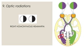
  • 37. 9. Optic radiations RIGHT HOMONYMOUS HEMIANPIA
  • 38. 10. Visual cortex sparing the macula RIGHT HOMONYMOUS HEMIANOPIA WITH MACULAR SPARING
  • 39. QUESTIONS TO REMEMBER DESCRIBE PIE ON THE FLOOR W.R.T. VISUAL PATHWAY BLOOD SUPPLY OF VISUAL PATHWAY DESCRIBLE LESIONS OF OPTIC TRACT OPTIC CHIASMA OPTIC RADIATION LABELED DIARAM OF VISUAL PATHWAY VISUAL PATHWAY DEFECT IN PITUTAR TUMOR OR CARANIOPHYRENGIOMA

Editor's Notes

  • #16: Nasal fibres (temporal visual field)
  • #22: The primary cortical region of the brain involved in…….