Accessory Structures of the Eye

Figure 17–3
Neural Tunic (Retina)

Figure 17–4c
Retina
• Rods, cones are types of photoreceptors

Figure 17–6
Rods
• Highly sensitive to light, do not discriminate colors
• Rods dominate peripheral areas of retina
• Provide low-resolution black–and–white vision in
dimly lit environments

Cones
• Sensitive to colored light
• Densely clustered in fovea
• Provide high–resolution color vision in brightly lit
environments
Photoreceptor

Figure 17–13a
• Rods and cones synapse with neurons
called bipolar cells
• Bipolar cells then synapse with neurons
called ganglion cells

Figure 17–6a
Horizontal Cells
• Where receptors synapse with bipolar cells

Amacrine Cells
• Where bipolar cells synapse with ganglion
cells
• Both facilitate or inhibit communication
between photoreceptors and ganglion cells
• Alter sensitivity of retina
Optic Disc
• Circular region just medial to fovea
• Origin of optic nerve

Figure 17–6b, c
Blind Spot

Figure 17–7
Visual Pigments
• Where light absorption
occurs
• Derivatives of rhodopsin
(opsin plus retinal)
• Retinal:
synthesized from vitamin A

Figure 17–13b
Phototransduction
1) Photon strikes retinal portion of rhodopsin
2) Opsin is activated:
– Goes from 11-cis form to 11-trans form
3) Opsin activates transducin (G protein), then
activates phosphodiesterase (PDE)
4) Cyclic-GMP (cGMP) levels decline; gated sodium
channels close
5) Dark current is reduced; neurotransmitter release
declines
In dark,
photoreceptors are
activated (stimulated)

Dark Current
In light,
photoreceptors are
inactivated (inhibited)

Figure 17–14
Bleaching
• Rhodopsin molecule breaks down into
retinal and opsin

Figure 17–15
Dark-Adapted State
• Pupil dilates
• Most pigments can be activated
• A single photon can be detected

Light-Adapted State
• Pupil constricts
• Bleaching of visual pigments occurs
• “Blinding” when go from dark to light room
Pupillary Muscle Reflexes

Figure 17–5
Color Sensitivity
• Integration of
information from
red, green, and
blue cones
• All wavelengths
reflected off an
object looks white

Figure 17–16
Color Blindness
• Inability to detect certain colors

Figure 17–17
Visual Pathway
• Begins at photoreceptors
• Ends at visual cortex of cerebral
hemispheres
• Message crosses 2 synapses before goes
toward brain:
–photoreceptor --> bipolar cell
–bipolar cell --> ganglion cell (sensory neuron)
“processing in retina”
Convergence
• Each ganglion cell
receives input from
many photoreceptors
• Therefore,
Receptive field of
ganglion cell is
monitored by many
photoreceptors
M Cells
• ganglion cells that monitor rods
• Provide information about:
–general form of object
–motion
–shadows in dim lighting

P Cells
• ganglion cells that monitor cones
• Provide information about:
–edges
–fine detail
–color
Visual Pathway
1. Axons from ganglion cells
converge on optic disc
2. Penetrate wall of eye
3. Proceed toward
diencephalon as optic
nerve (II)
4. 2 optic nerves (1 for each
eye) reach diencephalons
at optic chiasm
Figure 17–19
Optic Tracts
5. Axons from both
eyes projecting from
optic chiasm to lateral
geniculi

Optic Radiations
6. Bundles of projection
fibers from lateral
geniculates to visual
cortex
Visual Cortex
7. Info from right or left fields
of vision arrive at visual
cortex of opposite occipital
lobe:
– left half arrives at right
occipital lobe
– right half arrives at left
occipital lobe
Depth Perception
• By comparing relative
positions of objects
between left–eye and
right–eye images
But, at great distances:
- Previous familiarity
- Occlusion
- Perspective
- Motion parallax
- Shadows and light

Figure 17–19

More Related Content

PPTX
Receptors
PPT
Physiology of visual cycle
PPT
Phototransduction & Visual Pathway Mmp March 10
PPTX
Digestive system -Part II
PPTX
Special senses
PPTX
The Skin Senses (Its Parts and Sensory Functions)
PPT
NEUROPHYSIOLOGY OF VISION
PPT
Chapter 12 somatic senses and special
Receptors
Physiology of visual cycle
Phototransduction & Visual Pathway Mmp March 10
Digestive system -Part II
Special senses
The Skin Senses (Its Parts and Sensory Functions)
NEUROPHYSIOLOGY OF VISION
Chapter 12 somatic senses and special

What's hot (20)

PPTX
VITAMIN A & VISUAL CYCLE
PPTX
Sn2 reaction
PPT
PHOTOCHEMISTRY OF VISION
PPTX
Visual transduction ppt 2021
PPTX
Nose & sense of smell
PPTX
Taste ppt
PDF
Hypoxia & Cyanosis physiology of respiration 1.pdf
PPTX
The somatosensory cortex
PPT
Nervous+System+Presentation
PPT
Olfaction ppt
PPT
Anatomy of olfactory system
PPTX
Organization of the nervous system
PPTX
Dorsal column medial leminiscus
PPTX
Specialized products of Amino Acid
PPT
Olfaction
PPT
7. the sense organs
PPT
Special sense of hearing
PPT
Special Senses - Organ of Corti
PPTX
auditory pathway ppt
PPTX
Chapter 8 - Special Senses
VITAMIN A & VISUAL CYCLE
Sn2 reaction
PHOTOCHEMISTRY OF VISION
Visual transduction ppt 2021
Nose & sense of smell
Taste ppt
Hypoxia & Cyanosis physiology of respiration 1.pdf
The somatosensory cortex
Nervous+System+Presentation
Olfaction ppt
Anatomy of olfactory system
Organization of the nervous system
Dorsal column medial leminiscus
Specialized products of Amino Acid
Olfaction
7. the sense organs
Special sense of hearing
Special Senses - Organ of Corti
auditory pathway ppt
Chapter 8 - Special Senses
Ad

Viewers also liked (11)

PDF
Chapter14 peripheralns marieb
PPTX
Vitamins in animals
PPT
Visual System Structure and Function
PPT
Lecture12 4 13
PPT
Descending pathways dr_khadija (1)
PPTX
Fat soluble vitamins
PPTX
Application of Biostatistics
PPTX
Anatomy and physiology of the eye
PPTX
Blue eyes
PDF
How to Make Awesome SlideShares: Tips & Tricks
PDF
Getting Started With SlideShare
Chapter14 peripheralns marieb
Vitamins in animals
Visual System Structure and Function
Lecture12 4 13
Descending pathways dr_khadija (1)
Fat soluble vitamins
Application of Biostatistics
Anatomy and physiology of the eye
Blue eyes
How to Make Awesome SlideShares: Tips & Tricks
Getting Started With SlideShare
Ad

Similar to Lecture12 6 13 (20)

PDF
Anatomy of the Human Eye ( PDFDrive ).pdf
PDF
Ch09
PPT
Vision 300
PPT
Physio eyes-2-
PPTX
PPSX
The eye
PPT
PPT
EYE PRESENTATION Dr Sheeba.ppt
PPT
Retina preliminary
PPTX
g.neurophysiology of Vision and the eye
PPT
Vision Lucky
PPT
Physiology of Vision (1).ppt
PDF
L19. Phototransduction & retinal adaptation in three pages
PPTX
The Special Senses.pptx
PPT
PPT
Vision1
PPTX
Physiology of sense organ EYE
PPT
Pinel basics ch04
DOCX
R E T I N A - MIDTERM DOCUMENT REVIEWERS
PPTX
Sensory Systems.pptx
Anatomy of the Human Eye ( PDFDrive ).pdf
Ch09
Vision 300
Physio eyes-2-
The eye
EYE PRESENTATION Dr Sheeba.ppt
Retina preliminary
g.neurophysiology of Vision and the eye
Vision Lucky
Physiology of Vision (1).ppt
L19. Phototransduction & retinal adaptation in three pages
The Special Senses.pptx
Vision1
Physiology of sense organ EYE
Pinel basics ch04
R E T I N A - MIDTERM DOCUMENT REVIEWERS
Sensory Systems.pptx

Recently uploaded (20)

PPTX
AI IN MARKETING- PRESENTED BY ANWAR KABIR 1st June 2025.pptx
PDF
Flame analysis and combustion estimation using large language and vision assi...
PDF
Hybrid horned lizard optimization algorithm-aquila optimizer for DC motor
PDF
Consumable AI The What, Why & How for Small Teams.pdf
PPTX
2018-HIPAA-Renewal-Training for executives
PDF
From MVP to Full-Scale Product A Startup’s Software Journey.pdf
PDF
OpenACC and Open Hackathons Monthly Highlights July 2025
PDF
Five Habits of High-Impact Board Members
PDF
A review of recent deep learning applications in wood surface defect identifi...
PDF
sbt 2.0: go big (Scala Days 2025 edition)
PDF
ENT215_Completing-a-large-scale-migration-and-modernization-with-AWS.pdf
PPT
Galois Field Theory of Risk: A Perspective, Protocol, and Mathematical Backgr...
PDF
Architecture types and enterprise applications.pdf
PDF
A Late Bloomer's Guide to GenAI: Ethics, Bias, and Effective Prompting - Boha...
PPTX
Custom Battery Pack Design Considerations for Performance and Safety
PPT
Geologic Time for studying geology for geologist
PDF
1 - Historical Antecedents, Social Consideration.pdf
PPTX
Configure Apache Mutual Authentication
PPTX
Modernising the Digital Integration Hub
PPTX
Final SEM Unit 1 for mit wpu at pune .pptx
AI IN MARKETING- PRESENTED BY ANWAR KABIR 1st June 2025.pptx
Flame analysis and combustion estimation using large language and vision assi...
Hybrid horned lizard optimization algorithm-aquila optimizer for DC motor
Consumable AI The What, Why & How for Small Teams.pdf
2018-HIPAA-Renewal-Training for executives
From MVP to Full-Scale Product A Startup’s Software Journey.pdf
OpenACC and Open Hackathons Monthly Highlights July 2025
Five Habits of High-Impact Board Members
A review of recent deep learning applications in wood surface defect identifi...
sbt 2.0: go big (Scala Days 2025 edition)
ENT215_Completing-a-large-scale-migration-and-modernization-with-AWS.pdf
Galois Field Theory of Risk: A Perspective, Protocol, and Mathematical Backgr...
Architecture types and enterprise applications.pdf
A Late Bloomer's Guide to GenAI: Ethics, Bias, and Effective Prompting - Boha...
Custom Battery Pack Design Considerations for Performance and Safety
Geologic Time for studying geology for geologist
1 - Historical Antecedents, Social Consideration.pdf
Configure Apache Mutual Authentication
Modernising the Digital Integration Hub
Final SEM Unit 1 for mit wpu at pune .pptx

Lecture12 6 13

  • 1. Accessory Structures of the Eye Figure 17–3
  • 3. Retina • Rods, cones are types of photoreceptors Figure 17–6
  • 4. Rods • Highly sensitive to light, do not discriminate colors • Rods dominate peripheral areas of retina • Provide low-resolution black–and–white vision in dimly lit environments Cones • Sensitive to colored light • Densely clustered in fovea • Provide high–resolution color vision in brightly lit environments
  • 6. • Rods and cones synapse with neurons called bipolar cells • Bipolar cells then synapse with neurons called ganglion cells Figure 17–6a
  • 7. Horizontal Cells • Where receptors synapse with bipolar cells Amacrine Cells • Where bipolar cells synapse with ganglion cells • Both facilitate or inhibit communication between photoreceptors and ganglion cells • Alter sensitivity of retina
  • 8. Optic Disc • Circular region just medial to fovea • Origin of optic nerve Figure 17–6b, c
  • 10. Visual Pigments • Where light absorption occurs • Derivatives of rhodopsin (opsin plus retinal) • Retinal: synthesized from vitamin A Figure 17–13b
  • 11. Phototransduction 1) Photon strikes retinal portion of rhodopsin 2) Opsin is activated: – Goes from 11-cis form to 11-trans form 3) Opsin activates transducin (G protein), then activates phosphodiesterase (PDE) 4) Cyclic-GMP (cGMP) levels decline; gated sodium channels close 5) Dark current is reduced; neurotransmitter release declines
  • 12. In dark, photoreceptors are activated (stimulated) Dark Current In light, photoreceptors are inactivated (inhibited) Figure 17–14
  • 13. Bleaching • Rhodopsin molecule breaks down into retinal and opsin Figure 17–15
  • 14. Dark-Adapted State • Pupil dilates • Most pigments can be activated • A single photon can be detected Light-Adapted State • Pupil constricts • Bleaching of visual pigments occurs • “Blinding” when go from dark to light room
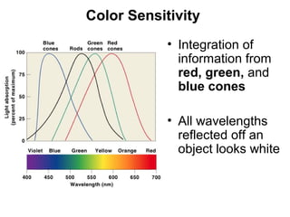
  • 16. Color Sensitivity • Integration of information from red, green, and blue cones • All wavelengths reflected off an object looks white Figure 17–16
  • 17. Color Blindness • Inability to detect certain colors Figure 17–17
  • 18. Visual Pathway • Begins at photoreceptors • Ends at visual cortex of cerebral hemispheres • Message crosses 2 synapses before goes toward brain: –photoreceptor --> bipolar cell –bipolar cell --> ganglion cell (sensory neuron) “processing in retina”
  • 19. Convergence • Each ganglion cell receives input from many photoreceptors • Therefore, Receptive field of ganglion cell is monitored by many photoreceptors
  • 20. M Cells • ganglion cells that monitor rods • Provide information about: –general form of object –motion –shadows in dim lighting P Cells • ganglion cells that monitor cones • Provide information about: –edges –fine detail –color
  • 21. Visual Pathway 1. Axons from ganglion cells converge on optic disc 2. Penetrate wall of eye 3. Proceed toward diencephalon as optic nerve (II) 4. 2 optic nerves (1 for each eye) reach diencephalons at optic chiasm Figure 17–19
  • 22. Optic Tracts 5. Axons from both eyes projecting from optic chiasm to lateral geniculi Optic Radiations 6. Bundles of projection fibers from lateral geniculates to visual cortex
  • 23. Visual Cortex 7. Info from right or left fields of vision arrive at visual cortex of opposite occipital lobe: – left half arrives at right occipital lobe – right half arrives at left occipital lobe
  • 24. Depth Perception • By comparing relative positions of objects between left–eye and right–eye images But, at great distances: - Previous familiarity - Occlusion - Perspective - Motion parallax - Shadows and light Figure 17–19