SlideShare a Scribd company logo
3. EUKARYOTES, THEIR
STRUCTURE & ELECTRON
MICROSCOPES
1.2 – Eukaryotes have a
compartmentalized
structure
WHAT IS A EUKARYOTIC CELL?
o Prokaryotes include bacteria, where as eukaryotes include algae,
protozoans, fungi, plants and animals
o Eukaryotes range in size and can exist as unicellular organisms
(protozoans) or as multicellular organism (plants/animals)
o They have a much more complicated internal structure than
prokaryotes – they are compartmentalised (they are divided up by
internal membranes into compartments). These compartments are
called organelles
o The most important of these compartments is the nucleus which
contains the cells chromosomes
o Each organelle in a eukaryotic cell has a distinctive structure and
function.
EUKARYOTIC VS PROKARYOTIC
ORGANELLES
o These are several advantages in being compartmentalised:
o Enzymes and substrates for a particular process can be much more concentrated
than if they were spread throughout the cytoplasm
o Chemical reactions that are not compatible can be separated
o Substances that could cause damage to the cell can be kept inside the organelle in
a safe compartment
o Conditions such as pH can be maintained at ideal levels within each organelle
based on their requirements
o Organelles with their contents can be moved around within the cell
PLANT AND ANIMAL CELLS
ORANGELLES
o Common organelles include the following:
o Endoplasmic reticulum, ribosomes, lysosomes, Golgi apparatus, mitochondria,
nucleus, chloroplast, vacuoles
o Many of these structures (referred to as the ‘ultrastructure’) cannot
be seen by a light microscope. Therefore our knowledge of
eukaryotes, their organelles and the functions of these have only
come about with the development of the electron microscope.
“TYPICAL” ANIMAL CELL
http://web.jjay.cuny.edu/~acarpi/NSC/images/cell.gif
ANIMAL CELL UNDER AN EM
http://waynesword.palomar.edu/images/plant3.gif
“TYPICAL” PLANT CELL
PLANT CELL UNDER AN EM
ORGANELLES – SURROUNDING
THE CELL1. Plasma (cell) Membranes
o Outer membrane of cell that controls
movement in and out of the cell
o Separates the cell contents from its
surroundings
o Double layer of phospholipids so called the
“phospholipid bilayer”
o Contains proteins throughout
o It is selectively permeable – i.e. it only allows
the passage of certain molecules into/out of
cells
o Both plant and animal cells have cell
membranes
o Most widely accepted model of this structure
is
called the “Fluid Mosaic Model”
ORGANELLES – SURROUNDING
THE CELL
2. Cell Wall
o Made of cellulose
o Most commonly found in plant cells & bacteria
o Supports & protects cells
o It is a permeable barrier – i.e. it is not selective about
what passes through it
o Because it is outside the plasma membrane it is
considered as part of the ‘extra-cellular matrix’ of the
plant cell.
o Each cell secretes its own cell wall and the walls of
adjoining cells adhere to each other via a (sticky)
middle lamella.
o The cell wall has a ‘fibrous’ appearance under very
high magnification, this makes it very porous and
allows most things through, however, it is also slightly
‘elastic’
ORGANELLES – INSIDE THE CELL
1. Nucleus
o Directs cell activities
o Separated from cytoplasm by a double nuclear
membrane (nuclear envelope)
o Contains genetic material – DNA – which is stored
as chromosomes
o Contains the nucleolus – helps in the manufacture
of ribosomes (which make proteins)
o It is pierced by pores which allow molecules to
enter and leave the nucleus (link with mRNA).
o The outer layer of the nuclear membrane is
continuous with the Endoplasmic reticulum.
ORGANELLES – INSIDE THE CELL
2. Cytoplasm/Cytosol
o Surrounded by cell membrane
o Cytoplasm refers to the jelly-like material with
organelles in it.
o If the organelles were removed, the soluble part that
would be left is called the cytosol. It consists mainly of
water with dissolved substances such as amino acids in it.
ORGANELLES – INSIDE THE CELL
3. Endoplasmic Reticulum
o Series of membranes that extend from
the nucleus throughout the cell
o Moves materials around in cell
o Smooth type: lacks ribosomes – has a
role in making fats, steroids and
membrane phospholipids
o Rough type (pictured): ribosomes
embedded in surface – has a role in
making proteins
o In an electron micrograph RER often
appear as parallel lines
ORGANELLES – INSIDE THE CELL
4. Ribosomes
o Ribosomes are the structures in the cell where the proteins are made/the sites
of protein synthesis.
o Each consists of two sections called the large and small sub-unit.
o Some ribosomes coat the outsides of the rough ER (HL - these ribosomes
synthesise proteins for use outside the cell/extracellular proteins) and others are
found ‘loose’ in the cytoplasm (HL - these ribosomes synthesis proteins for use
within the cell/intracellular proteins)
o Ribosomes in eukaryotes are larger and denser (80S) than prokaryotes (70S)
ORGANELLES – INSIDE THE CELL
5. Golgi Body
o Consists of a series of flattened vesicles called
cisternae stacked on top of each other
o It is relatively easy to recognize because the
‘ends’ of each section are slightly bulbous (like
dumb-bells)
o One side is near the RER (the cis side) and it
received products from the ER which makes its
way through the organelle to the trans side
where small vesicles bud off
o Proteins and lipids are modified and packaged
during this process and moved within the cell
and towards the plasma membrane to leave the
cell
o This organelle is especially prevalent in
glandular cells which manufacture and secrete
substances (e.g. cells in the pancreas)
ORGANELLES – INSIDE THE CELL
6. Mitochondria
o Produces energy through chemical reactions – breaking
down glucose
o Mitochondria have a two layered ‘wall’ with the inner
mitochondrial membrane forming the Cristae which increase
the surface area for the attachment of the molecules which
are associated with aerobic respiration.
o Inside the membrane is a semifluid substance called the
matrix
oRecycles and decomposes proteins, fats, and carbohydrates
o Main function is to create ATP (cellular energy) through
aerobic cellular respiration
o Not all cells have the same number of mitochondria
ORGANELLES – INSIDE THE CELL
7. Chloroplast
o Usually found in algal and plant cells
o Contains green pigment chlorophyll
o Where photosynthesis takes place – light energy is captured,
ATP is formed and oxygen is released from water
o They are found in the leaves (sometimes the stem) of plants
o Like mitochondria, chloroplasts have a two layered wall.
o Inside the chloroplasts are all the molecules associated with
the biochemistry of photosynthesis.
o There are stacks of Thylakoids (stacks called Granum) coated
in photosynthetic pigments (mainly chlorophyll) interspersed
within a cytoplasm like material called the Stroma.
o Starch granules and lipid droplets are also commonly found.
ORGANELLES – INSIDE THE CELL
8. Vacuoles
o Membrane-bound sacs for storage,
digestion, and waste removal
o Contains water solution
o Found in both plant and animal cells –
much larger in plant cells
o Help plants maintain shape
o Usually formed by the golgi body
o They enable cells to have higher surface
area to volume ratios
ORGANELLES – INSIDE THE CELL
9. Lysosomes
o Digests proteins, fats, and carbohydrates. To
do this they contain many different types of
enzymes (a type of protein)
o Also digest food, bacteria and foreign
particles
o Transports undigested material to cell
membrane for removal
o Cell breaks down if lysosome explodes
GUESS THAT
ELECTRON
MICROGRAPH!
Mitochondria
Vacuole
CHLOROPLAST
Rough Endoplasmic Reticulum
Golgi Body
Nucleus
Nucleolus
ELECTRON MICROGRAPHS
OF SPECIALISED CELLS
1.2 Application - Structure
and function of organelles
within exocrine gland cells
of the pancreas and within
palisade mesophyll cells of
the leaf.
EXOCRINE GLAND CELLS OF THE
PANCREAS
o Gland cells secrete substances – they release
them through their plasma membrane
o There are TWO types of gland cells in the
pancreas – Endocrine cells (secrete hormones
into the blood) and Exocrine cells (secrete
digestive enzymes that get carried to the small
intestine)
o Enzymes are proteins, so the exocrine gland
cells have organelles needed to synthesise
proteins in large quantities (lots of RER), process
them to make them ready for secretion (lots of
Golgi bodies), transport them to the plasma
membrane and then release them (lots of
vesicles).
o This electron micrograph of a pancreas cell
shows: mitochondrion, nucleus, golgi apparatus,
PALISADE MESOPHYLL CELLS OF
THE LEAF
o The function of the leaf if photosynthesis –
producing organic compounds from carbon
dioxide and other simple inorganic
compounds using light energy
o The cells that carry out the most
photosynthesis in the leaf are palisade
mesophyll cells. As a result they have lots of
chloroplasts and large vacuoles.
o The shape of these cells is roughly
cylindrical.
o This electron micrograph of a palisade
mesophyll cell shows: cell wall, plasma
membrane, mitochondrion, chloroplasts,
vacuole, nucleus and cytoplasm
DRAWINGS
1.2 – Skill – Drawing the
ultrastructure of
eukaryotic cells based on
electron micrographs
TIPS FOR DRAWING CELL
STRUCTURES
DRAWING EUKARYOTIC CELLS
o The ultrastructure of eukaryotic cells is very complex and it is often
best to draw only part of a cell
o The electron micrograph on the next slide shows a liver cell with
labels to identify some of the organelles that are present. Draw the
whole cell to show its ultrastructure.
3. eukaryotes, their structure & em
3. EUKARYOTES, THEIR
STRUCTURE & ELECTRON
MICROSCOPES
1.2 – Electron microscopes
have a much higher
resolution than light
microscopes
SOME TERMS
o Micrograph – a photo taken down a microscope
o Microscopes can do TWO things to enhance our perception of an
image:
1. Magnify – Magnification is an increase in the apparent size of an
object
2. Resolve – Resolution is the clarity of an object
o Microscopes that use a beam of light to see an object are called light
microscopes
o Microscopes that use a beam of electrons to see an object are
electron microscopes. There are two main types of electron
microscopes – Scanning Electron Microscopes (SEM) and Transmission
Electron Microscopes (TEM)
Dog
flea
SOME HISTORY
o Light microscopes that are made up of more than one lens are called
compound light microscopes. They are typically monocular or
binocular (have 1 or 2 eye pieces)
o The first compound microscope was developed in 1590 by Hans and
Zacharias Janssen it could magnify 3-9x
o The earliest microscopes were limited by the quality of the glass
lens used and the techniques used to create them.
o NB: Antoine van Leeuwenhoek who was the first to observe bacteria
did so because he was a lens maker and made great improvements in
the techniques used to create the lenses in microscopes
o Improved lens quality led to improvements in the quality of the
images seen – people became less sceptical of what they were seeing
SOME MORE HISTORY
o By the end of the 19th century – lens quality was no longer a
limiting factor of compound light microscopes. Ongoing research
indicated that the wavelength of light was now the main limiting
factor.
o The best light microscope cannot effectively magnify larger than
2000x. This led scientists to begin experimenting with forms of
energy other than light
o The EM was invented in 1933 by Ernst Ruska
o Images are produced using a beam of electrons – electrons that
are made to behave like light waves
o Two types:
 Transmission Electron Microscope
 Scanning Electron Microscope
RESOLUTION AND MAGNIFICATION
o In every type of microscope, magnification can be increased until a
point above which the image can no longer be focused sharply. This
is because the resolution of the microscope has been exceeded
o The resolution of a microscope depends on the wavelength of the
rays used to form the image – the shorter the wavelength the higher
the resolution
o Electrons have a much shorter wavelength than light, so electron
microscopes have a higher resolution that light microscopes – they
can therefore produce a sharp image at much higher magnifications
o TEMs – Used to view ultra-thin sections
o SEMs – Produce an image of the surface of structures
o Australia’s EM
TEM & SEM
THE EM AND DEVELOPMENTS IN
CELL THEORY
o The development of the EM has allowed scientists to study parts of
cells that are too small to be seen with a light microscope
o EM’s are now linked to computers which allows the study of sub-
cellular structures in enormous detail, providing evidence of their
functioning. Used in areas such as genetics and ecology.
o This technology has allowed modern day additions to cell theory:
4. Cells contain hereditary information which is passed on during cell
division
5. All cells have the same basic chemical composition
6. All energy flow (resulting from chemical reactions) of life occurs
within cells
THE ELECTRON MICROSCOPE
Advantages Disadvantages
• High magnification & resolution
which show an enormous amount of
detail:
 Sub-cellular structures
(organelles) seen for the first
time and in great detail (TEM) –
led to an understanding of their
functions in cells
• Specimen must be placed in a
vacuum – so must be dead. Leads
scientists to question how different
the preserved specimens are from
living tissue. Light microscope can
be either living or dead.
• Images are produced in black and
white – colour must be added after
using computer software
• Magnification of EM is 500,000x
compared to light microscope which
is 2000x
• Resolution of EM is 1nm compared
to light microscope which is 200nm
• Size
• Expense
• Maintenance (kept at a constant
temperature and pressure)
• Complex and lengthy specimen
preparation compared to light

More Related Content

PPTX
Bacteria, Animal and Plant Cells
PPTX
Parts of cell and their functions
PPTX
CELL TYPES, FUNCTIONS, AND MODIFICATION.pptx
PPT
Gas exchange in animals by madeleine
PDF
Lec 2 plant cell structures
PPT
Transport mechanism
PPTX
Types of cells notes ppt
PPTX
Cell is the Basic Unit of Life
Bacteria, Animal and Plant Cells
Parts of cell and their functions
CELL TYPES, FUNCTIONS, AND MODIFICATION.pptx
Gas exchange in animals by madeleine
Lec 2 plant cell structures
Transport mechanism
Types of cells notes ppt
Cell is the Basic Unit of Life

What's hot (20)

PPT
Integrated Science M4 Cycles in Nature
PPTX
1.4. Membrane transport
PDF
The plant cell
PPTX
Mineral resources in the philippines
PPTX
General Biology 1 - Lesson 2 Classification of Cell
PDF
Cells cell structure-cells and organisms
PPTX
Biology 1 for grade 12 SHS (cell structure)
PPT
prokayotic and eukaryotic cells.ppt
PPTX
Laboratory activity 3 (post lab)
PPTX
GAS EXCHANGE IN ANIMALS
PDF
Cell structure
PDF
Cell Theory.pdf
PPT
Basic cell types
PPTX
Chemical and nervous control
PPTX
General Biology 1- Lesson 3: Structure and Functions of Animal Tissues and Ce...
PPT
11 stem odl (1)
PPT
The cell theory
PPT
Cells structure
PPTX
Epithelium in General
PPT
Cell Transport 1
Integrated Science M4 Cycles in Nature
1.4. Membrane transport
The plant cell
Mineral resources in the philippines
General Biology 1 - Lesson 2 Classification of Cell
Cells cell structure-cells and organisms
Biology 1 for grade 12 SHS (cell structure)
prokayotic and eukaryotic cells.ppt
Laboratory activity 3 (post lab)
GAS EXCHANGE IN ANIMALS
Cell structure
Cell Theory.pdf
Basic cell types
Chemical and nervous control
General Biology 1- Lesson 3: Structure and Functions of Animal Tissues and Ce...
11 stem odl (1)
The cell theory
Cells structure
Epithelium in General
Cell Transport 1
Ad

Similar to 3. eukaryotes, their structure & em (20)

PPTX
IB Biology 1.2 Slides: Ultrastructure of Cells
PDF
Lecture 4 - Introduction to Cell Biology 23-24.pdf
PPTX
cell and its organelle
PDF
Eukaryotic cell
PPTX
Chapter 4th cell and tissues
PDF
Bio chapter 1.pdf
PDF
W2_1.2 Ultrastructure of Cell.pdf
PDF
Cell-Organelles-.pdf cell organelles
PPT
STRUCTURE OF ORGANELLES AND FUNCTIONS.ppt
PPT
(10) the cell theory 9 19
PPTX
1 Cell structure
PPTX
Cell Organelles
PDF
Lesson 3-4 Biochem.pdf
PPTX
Cell - The Unit of Life organelles about cell class 11 standard.pptx
PDF
General Introduction of Plant Cell & its Organelles by Salman Saeed Lecturer ...
PPTX
Eukaryotic Cell Structure and Functions.pptx
PPT
CELL ORGANELLES POWERPOINT PRESENTATION OKOKOK
PDF
03 Cell Organelles.pdf
PPTX
Fundamental unit of life cell
IB Biology 1.2 Slides: Ultrastructure of Cells
Lecture 4 - Introduction to Cell Biology 23-24.pdf
cell and its organelle
Eukaryotic cell
Chapter 4th cell and tissues
Bio chapter 1.pdf
W2_1.2 Ultrastructure of Cell.pdf
Cell-Organelles-.pdf cell organelles
STRUCTURE OF ORGANELLES AND FUNCTIONS.ppt
(10) the cell theory 9 19
1 Cell structure
Cell Organelles
Lesson 3-4 Biochem.pdf
Cell - The Unit of Life organelles about cell class 11 standard.pptx
General Introduction of Plant Cell & its Organelles by Salman Saeed Lecturer ...
Eukaryotic Cell Structure and Functions.pptx
CELL ORGANELLES POWERPOINT PRESENTATION OKOKOK
03 Cell Organelles.pdf
Fundamental unit of life cell
Ad

More from sophiespyrou (8)

PPTX
6. stem cells
PPTX
4. unicellular, multicellular & cell differentiation
PPTX
2. prokaryotes and their structure
PPTX
1. cell theory
PPTX
10. cell division
PPTX
9. the origin of cells
PPTX
8. membrane transport
PPTX
7. membrane structure
6. stem cells
4. unicellular, multicellular & cell differentiation
2. prokaryotes and their structure
1. cell theory
10. cell division
9. the origin of cells
8. membrane transport
7. membrane structure

Recently uploaded (20)

PDF
LNK 2025 (2).pdf MWEHEHEHEHEHEHEHEHEHEHE
PPTX
Radiologic_Anatomy_of_the_Brachial_plexus [final].pptx
PDF
What if we spent less time fighting change, and more time building what’s rig...
PPTX
UNIT III MENTAL HEALTH NURSING ASSESSMENT
PPTX
Tissue processing ( HISTOPATHOLOGICAL TECHNIQUE
PPTX
Onco Emergencies - Spinal cord compression Superior vena cava syndrome Febr...
PDF
LDMMIA Reiki Yoga Finals Review Spring Summer
PDF
Black Hat USA 2025 - Micro ICS Summit - ICS/OT Threat Landscape
PDF
IGGE1 Understanding the Self1234567891011
PDF
Paper A Mock Exam 9_ Attempt review.pdf.
PPTX
UV-Visible spectroscopy..pptx UV-Visible Spectroscopy – Electronic Transition...
PDF
Classroom Observation Tools for Teachers
PDF
RMMM.pdf make it easy to upload and study
PPTX
Digestion and Absorption of Carbohydrates, Proteina and Fats
DOC
Soft-furnishing-By-Architect-A.F.M.Mohiuddin-Akhand.doc
PPTX
Introduction to Building Materials
PPTX
Unit 4 Skeletal System.ppt.pptxopresentatiom
PPTX
CHAPTER IV. MAN AND BIOSPHERE AND ITS TOTALITY.pptx
PDF
Complications of Minimal Access Surgery at WLH
PDF
Chinmaya Tiranga quiz Grand Finale.pdf
LNK 2025 (2).pdf MWEHEHEHEHEHEHEHEHEHEHE
Radiologic_Anatomy_of_the_Brachial_plexus [final].pptx
What if we spent less time fighting change, and more time building what’s rig...
UNIT III MENTAL HEALTH NURSING ASSESSMENT
Tissue processing ( HISTOPATHOLOGICAL TECHNIQUE
Onco Emergencies - Spinal cord compression Superior vena cava syndrome Febr...
LDMMIA Reiki Yoga Finals Review Spring Summer
Black Hat USA 2025 - Micro ICS Summit - ICS/OT Threat Landscape
IGGE1 Understanding the Self1234567891011
Paper A Mock Exam 9_ Attempt review.pdf.
UV-Visible spectroscopy..pptx UV-Visible Spectroscopy – Electronic Transition...
Classroom Observation Tools for Teachers
RMMM.pdf make it easy to upload and study
Digestion and Absorption of Carbohydrates, Proteina and Fats
Soft-furnishing-By-Architect-A.F.M.Mohiuddin-Akhand.doc
Introduction to Building Materials
Unit 4 Skeletal System.ppt.pptxopresentatiom
CHAPTER IV. MAN AND BIOSPHERE AND ITS TOTALITY.pptx
Complications of Minimal Access Surgery at WLH
Chinmaya Tiranga quiz Grand Finale.pdf

3. eukaryotes, their structure & em

  • 1. 3. EUKARYOTES, THEIR STRUCTURE & ELECTRON MICROSCOPES 1.2 – Eukaryotes have a compartmentalized structure
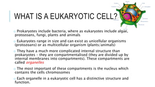
  • 2. WHAT IS A EUKARYOTIC CELL? o Prokaryotes include bacteria, where as eukaryotes include algae, protozoans, fungi, plants and animals o Eukaryotes range in size and can exist as unicellular organisms (protozoans) or as multicellular organism (plants/animals) o They have a much more complicated internal structure than prokaryotes – they are compartmentalised (they are divided up by internal membranes into compartments). These compartments are called organelles o The most important of these compartments is the nucleus which contains the cells chromosomes o Each organelle in a eukaryotic cell has a distinctive structure and function.
  • 4. ORGANELLES o These are several advantages in being compartmentalised: o Enzymes and substrates for a particular process can be much more concentrated than if they were spread throughout the cytoplasm o Chemical reactions that are not compatible can be separated o Substances that could cause damage to the cell can be kept inside the organelle in a safe compartment o Conditions such as pH can be maintained at ideal levels within each organelle based on their requirements o Organelles with their contents can be moved around within the cell
  • 6. ORANGELLES o Common organelles include the following: o Endoplasmic reticulum, ribosomes, lysosomes, Golgi apparatus, mitochondria, nucleus, chloroplast, vacuoles o Many of these structures (referred to as the ‘ultrastructure’) cannot be seen by a light microscope. Therefore our knowledge of eukaryotes, their organelles and the functions of these have only come about with the development of the electron microscope.
  • 11. ORGANELLES – SURROUNDING THE CELL1. Plasma (cell) Membranes o Outer membrane of cell that controls movement in and out of the cell o Separates the cell contents from its surroundings o Double layer of phospholipids so called the “phospholipid bilayer” o Contains proteins throughout o It is selectively permeable – i.e. it only allows the passage of certain molecules into/out of cells o Both plant and animal cells have cell membranes o Most widely accepted model of this structure is called the “Fluid Mosaic Model”
  • 12. ORGANELLES – SURROUNDING THE CELL 2. Cell Wall o Made of cellulose o Most commonly found in plant cells & bacteria o Supports & protects cells o It is a permeable barrier – i.e. it is not selective about what passes through it o Because it is outside the plasma membrane it is considered as part of the ‘extra-cellular matrix’ of the plant cell. o Each cell secretes its own cell wall and the walls of adjoining cells adhere to each other via a (sticky) middle lamella. o The cell wall has a ‘fibrous’ appearance under very high magnification, this makes it very porous and allows most things through, however, it is also slightly ‘elastic’
  • 13. ORGANELLES – INSIDE THE CELL 1. Nucleus o Directs cell activities o Separated from cytoplasm by a double nuclear membrane (nuclear envelope) o Contains genetic material – DNA – which is stored as chromosomes o Contains the nucleolus – helps in the manufacture of ribosomes (which make proteins) o It is pierced by pores which allow molecules to enter and leave the nucleus (link with mRNA). o The outer layer of the nuclear membrane is continuous with the Endoplasmic reticulum.
  • 14. ORGANELLES – INSIDE THE CELL 2. Cytoplasm/Cytosol o Surrounded by cell membrane o Cytoplasm refers to the jelly-like material with organelles in it. o If the organelles were removed, the soluble part that would be left is called the cytosol. It consists mainly of water with dissolved substances such as amino acids in it.
  • 15. ORGANELLES – INSIDE THE CELL 3. Endoplasmic Reticulum o Series of membranes that extend from the nucleus throughout the cell o Moves materials around in cell o Smooth type: lacks ribosomes – has a role in making fats, steroids and membrane phospholipids o Rough type (pictured): ribosomes embedded in surface – has a role in making proteins o In an electron micrograph RER often appear as parallel lines
  • 16. ORGANELLES – INSIDE THE CELL 4. Ribosomes o Ribosomes are the structures in the cell where the proteins are made/the sites of protein synthesis. o Each consists of two sections called the large and small sub-unit. o Some ribosomes coat the outsides of the rough ER (HL - these ribosomes synthesise proteins for use outside the cell/extracellular proteins) and others are found ‘loose’ in the cytoplasm (HL - these ribosomes synthesis proteins for use within the cell/intracellular proteins) o Ribosomes in eukaryotes are larger and denser (80S) than prokaryotes (70S)
  • 17. ORGANELLES – INSIDE THE CELL 5. Golgi Body o Consists of a series of flattened vesicles called cisternae stacked on top of each other o It is relatively easy to recognize because the ‘ends’ of each section are slightly bulbous (like dumb-bells) o One side is near the RER (the cis side) and it received products from the ER which makes its way through the organelle to the trans side where small vesicles bud off o Proteins and lipids are modified and packaged during this process and moved within the cell and towards the plasma membrane to leave the cell o This organelle is especially prevalent in glandular cells which manufacture and secrete substances (e.g. cells in the pancreas)
  • 18. ORGANELLES – INSIDE THE CELL 6. Mitochondria o Produces energy through chemical reactions – breaking down glucose o Mitochondria have a two layered ‘wall’ with the inner mitochondrial membrane forming the Cristae which increase the surface area for the attachment of the molecules which are associated with aerobic respiration. o Inside the membrane is a semifluid substance called the matrix oRecycles and decomposes proteins, fats, and carbohydrates o Main function is to create ATP (cellular energy) through aerobic cellular respiration o Not all cells have the same number of mitochondria
  • 19. ORGANELLES – INSIDE THE CELL 7. Chloroplast o Usually found in algal and plant cells o Contains green pigment chlorophyll o Where photosynthesis takes place – light energy is captured, ATP is formed and oxygen is released from water o They are found in the leaves (sometimes the stem) of plants o Like mitochondria, chloroplasts have a two layered wall. o Inside the chloroplasts are all the molecules associated with the biochemistry of photosynthesis. o There are stacks of Thylakoids (stacks called Granum) coated in photosynthetic pigments (mainly chlorophyll) interspersed within a cytoplasm like material called the Stroma. o Starch granules and lipid droplets are also commonly found.
  • 20. ORGANELLES – INSIDE THE CELL 8. Vacuoles o Membrane-bound sacs for storage, digestion, and waste removal o Contains water solution o Found in both plant and animal cells – much larger in plant cells o Help plants maintain shape o Usually formed by the golgi body o They enable cells to have higher surface area to volume ratios
  • 21. ORGANELLES – INSIDE THE CELL 9. Lysosomes o Digests proteins, fats, and carbohydrates. To do this they contain many different types of enzymes (a type of protein) o Also digest food, bacteria and foreign particles o Transports undigested material to cell membrane for removal o Cell breaks down if lysosome explodes
  • 30. ELECTRON MICROGRAPHS OF SPECIALISED CELLS 1.2 Application - Structure and function of organelles within exocrine gland cells of the pancreas and within palisade mesophyll cells of the leaf.
  • 31. EXOCRINE GLAND CELLS OF THE PANCREAS o Gland cells secrete substances – they release them through their plasma membrane o There are TWO types of gland cells in the pancreas – Endocrine cells (secrete hormones into the blood) and Exocrine cells (secrete digestive enzymes that get carried to the small intestine) o Enzymes are proteins, so the exocrine gland cells have organelles needed to synthesise proteins in large quantities (lots of RER), process them to make them ready for secretion (lots of Golgi bodies), transport them to the plasma membrane and then release them (lots of vesicles). o This electron micrograph of a pancreas cell shows: mitochondrion, nucleus, golgi apparatus,
  • 32. PALISADE MESOPHYLL CELLS OF THE LEAF o The function of the leaf if photosynthesis – producing organic compounds from carbon dioxide and other simple inorganic compounds using light energy o The cells that carry out the most photosynthesis in the leaf are palisade mesophyll cells. As a result they have lots of chloroplasts and large vacuoles. o The shape of these cells is roughly cylindrical. o This electron micrograph of a palisade mesophyll cell shows: cell wall, plasma membrane, mitochondrion, chloroplasts, vacuole, nucleus and cytoplasm
  • 33. DRAWINGS 1.2 – Skill – Drawing the ultrastructure of eukaryotic cells based on electron micrographs
  • 34. TIPS FOR DRAWING CELL STRUCTURES
  • 35. DRAWING EUKARYOTIC CELLS o The ultrastructure of eukaryotic cells is very complex and it is often best to draw only part of a cell o The electron micrograph on the next slide shows a liver cell with labels to identify some of the organelles that are present. Draw the whole cell to show its ultrastructure.
  • 37. 3. EUKARYOTES, THEIR STRUCTURE & ELECTRON MICROSCOPES 1.2 – Electron microscopes have a much higher resolution than light microscopes
  • 38. SOME TERMS o Micrograph – a photo taken down a microscope o Microscopes can do TWO things to enhance our perception of an image: 1. Magnify – Magnification is an increase in the apparent size of an object 2. Resolve – Resolution is the clarity of an object o Microscopes that use a beam of light to see an object are called light microscopes o Microscopes that use a beam of electrons to see an object are electron microscopes. There are two main types of electron microscopes – Scanning Electron Microscopes (SEM) and Transmission Electron Microscopes (TEM) Dog flea
  • 39. SOME HISTORY o Light microscopes that are made up of more than one lens are called compound light microscopes. They are typically monocular or binocular (have 1 or 2 eye pieces) o The first compound microscope was developed in 1590 by Hans and Zacharias Janssen it could magnify 3-9x o The earliest microscopes were limited by the quality of the glass lens used and the techniques used to create them. o NB: Antoine van Leeuwenhoek who was the first to observe bacteria did so because he was a lens maker and made great improvements in the techniques used to create the lenses in microscopes o Improved lens quality led to improvements in the quality of the images seen – people became less sceptical of what they were seeing
  • 40. SOME MORE HISTORY o By the end of the 19th century – lens quality was no longer a limiting factor of compound light microscopes. Ongoing research indicated that the wavelength of light was now the main limiting factor. o The best light microscope cannot effectively magnify larger than 2000x. This led scientists to begin experimenting with forms of energy other than light o The EM was invented in 1933 by Ernst Ruska o Images are produced using a beam of electrons – electrons that are made to behave like light waves o Two types:  Transmission Electron Microscope  Scanning Electron Microscope
  • 41. RESOLUTION AND MAGNIFICATION o In every type of microscope, magnification can be increased until a point above which the image can no longer be focused sharply. This is because the resolution of the microscope has been exceeded o The resolution of a microscope depends on the wavelength of the rays used to form the image – the shorter the wavelength the higher the resolution o Electrons have a much shorter wavelength than light, so electron microscopes have a higher resolution that light microscopes – they can therefore produce a sharp image at much higher magnifications o TEMs – Used to view ultra-thin sections o SEMs – Produce an image of the surface of structures o Australia’s EM
  • 43. THE EM AND DEVELOPMENTS IN CELL THEORY o The development of the EM has allowed scientists to study parts of cells that are too small to be seen with a light microscope o EM’s are now linked to computers which allows the study of sub- cellular structures in enormous detail, providing evidence of their functioning. Used in areas such as genetics and ecology. o This technology has allowed modern day additions to cell theory: 4. Cells contain hereditary information which is passed on during cell division 5. All cells have the same basic chemical composition 6. All energy flow (resulting from chemical reactions) of life occurs within cells
  • 44. THE ELECTRON MICROSCOPE Advantages Disadvantages • High magnification & resolution which show an enormous amount of detail:  Sub-cellular structures (organelles) seen for the first time and in great detail (TEM) – led to an understanding of their functions in cells • Specimen must be placed in a vacuum – so must be dead. Leads scientists to question how different the preserved specimens are from living tissue. Light microscope can be either living or dead. • Images are produced in black and white – colour must be added after using computer software • Magnification of EM is 500,000x compared to light microscope which is 2000x • Resolution of EM is 1nm compared to light microscope which is 200nm • Size • Expense • Maintenance (kept at a constant temperature and pressure) • Complex and lengthy specimen preparation compared to light