CARCINOMA ESOPHAGUS
Presentor : Dr.Gowtham
Moderator: Dr.Vikas Choudhary
Epidemiology and incidence
• Esophageal cancer is the eighth most common cancer worldwide
• Incidence of 160/100,000 in parts of South Africa and China and 540/100,000 in
Kazakhstan.
• India 8-20 / 100,000 , 6th most common in males
• Squamous cell carcinoma still accounts for most esophageal cancers diagnosed.
• M:F – 3:1 (SCC) .. …..15:1 (adeno)
• Adeno – whites …..SCC – african American
Globocan 2018
Anatomy
• The esophagus is a thin-walled, hollow tube approximately 25 cm in length.
• It is lined with stratified keratinized squamous epithelium, extending from the
cricopharyngeus muscle at the level of the cricoid cartilage superiorly to the
gastroesophageal junction inferiorly.
• The esophageal wall is composed of three layers: the mucosa, submucosa, and
muscularis propria
• The mucosal layer contains the epithelium(M1), lamina propria(M2), and muscularis
mucosae(M3).
• The epithelium is separated from the lamina propria by a basement membrane. In the
portion of the esophagus containing columnar-type epithelium, the muscularis mucosae
may consist of two layers.
• Similarly, the submucosal layer may be divided into inner (SM1), middle (SM2), and
outer(SM3) layers. The muscularis propria consists of a circular inner layer and
longitudinal outer layer.
• The adventitia (periesophageal connective tissue) lies directly on the muscularis propria.
• No serosa is present, facilitating extraesophageal spread of disease.
• AJCC 8th  4 Parts : cervical , upper thoracic ,middle thoracic ,lower thoracic
Siewert’s classification
• Type I : From >1 cm up to 5 cm above
the gastroesophageal junction (Z line),
the tumor is classified as a carcinoma
of the distal esophagus
• Type II : Within 1 cm cephalad to 2 cm
caudad to the gastroesophageal
junction.
• Type III :Tumor center is located >2
cm below the gastroesophageal
junction
AJCC 8th
• Cancers with an epicenter in the
lower thoracic esophagus or
gastroesophageal junction or within
the proximal 2 cm of the stomach
(i.e.,cardia) and extending up to the
GE junction or esophagus are staged
as a carcinoma of the esophagus.
• If the epicenter is >2 cm distal to the
gastroesophageal junction, these are
classified as stomach cancers.
Staging
• AJCC(TNM)
• Tumor
• Lymph node(N0,N1)
• Metastasis
• Most widely accepted
ELLIS (WNM)
• Wall penetration
• Lymph node(N0,N1,N2)
• Metastasis
AJCC 8TH
AJCC -8th
SCC
• Grade ,location of tumour affect
outcomes
Adenocarcinoma
• Tumour location –not a prognostic
variable
Adenosquamous – staged as squamous only
p Stage yp Stage (post neoadjuvant)
Lymphatics
• The esophagus has an extensive, longitudinal
interconnecting system of lymphatics. The
esophageal lymphatic network is primarily
located within the submucosa .However,
channels are also present within the lamina
propria, facilitating spread of even superficial
cancers of the esophagus involving the
mucosa.
• In addition to these longitudinal lymphatics,
intramural lymphatics may traverse the
muscularis propria, facilitating tumor spread to
regional lymphatic channels and
paraesophageal nodes.
• Lymph can travel the entire length of the
esophagus before draining into lymph nodes,
the entire esophagus is at potential risk for
lymphatic involvement.
• Up to 8 cm or more of “normal” tissue can
exist between gross tumor and
micrometastases “skip areas”.
• Lymphatics of the esophagus drain into nodes
that usually follow arteries,including the
inferior thyroid artery, the bronchial and
esophageal arteries, and the left gastric artery
(celiac axis).
LN Status
• Depth of tumor penetration (T stage)
affects lymph node involvement (LNI)
• Intramucosal T1 lesions (18% LNI)
• submucosal T1 lesions (55% LNI)
• T2 lesions (60% LNI)
• T3 lesions (80% LNI)
• Chance if LN <50% - conservative eso
resection and limited lymph node dissection
• LN>50% - neoadjuvant therapy followed by
resection
Natural history and pattern of spread
• Squamous cell carcinoma is characterized by
extensive local growth and proclivity to lymph node
metastases.
• Because the esophagus has no covering serosa,
direct invasion of contiguous structures may occur
early.
• Lesions in the upper esophagus can impinge on or
invade the recurrent laryngeal nerves,carotid
arteries, and trachea. If extraesophageal extension
occurs in the mediastinum, tracheoesophageal or
bronchoesophageal fistula may occur.
• Tumors in the lower one-third of the esophagus can
invade the aorta or pericardium, resulting in
mediastinitis, massive hemorrhage, or empyema.
Histology • Squamous and adeno -95% of all carcinomas
• Psuedosarcoma – spindle cell carcinoma (SCC
variant)
• Adenoca variants –Adenoid cystic
,mucoepidermoid poorer prognosis
• Small cell carcinoma – arise from argyrophilic
cells paraneoplastic syndromes !!
(ADH,hypercalcemia) , similar to SCLC
• Nonepithelial origin – very rare
• Malignant melanoms –MS 7months
• Lymphoma –mostly extension , as primary very
rare
SCC
• Squamous cell carcinomas arise from the squamous mucosa - native to the
esophagus - 70% - upper and middle thirds
• Most common type of esophageal ca in India (90%)
• Smoking and alcohol are common eitiologic factors (5 fold increase in risk)
• Combined increase risk from 25 - 100 folds
• Dietary
• Nitrosamines (pickled foods ,
smoked food)
• long term ingestion of hot liquids
• Micronutrient deficiency (Vit. A,
B12, C, E).
• Trace Element deficiency (Cobalt,
Copper & Selenium)
• Acquired
• Cigarette smoking
• Alcohol.
• Chronic esophagitis.
• Chronic Dysphagia
• Caustic ingestion
• Radiation exposure
• Plummer – vinson
syndrome
• Tylosis(40%)
• Achalasia (16 -fold)
• Esophageal strictures
and diverticula
• p53
• Premalignant conditions
Adenocarcinoma
• almost 70 % - United States and Western countries.
Etiology :
• Increasing incidence of GERD
• Western diet
• Increased use of acid-suppression medications
Histologically it is from :
• Submucosal glands of the esophagus
• Heterotopic islands of columnar epithelium
• Malignant degeneration of metaplastic columnar epithelium (Barrett’s esophagus) – 40
fold increased risk
Barret’s esophagus
• Traditionally - the presence of columnar mucosa extending at
least 3 cm into the esophagus.
• Recently - the specialized, intestinal-type epithelium
(presence of goblet cells) found in the Barrett’s mucosa is the
only tissue predisposed to malignant degeneration - the
diagnosis of BE is presently made given any length of
endoscopically identifiable columnar mucosa that is proven
on biopsy.
• 10 % of GERD pts develop – BARRETS
• Approx 1 in every 100 patient years of followup of barrets
develop adenocarcinoma (40 fold increased risk)
Clinical manifestations
• Early - asymptomatic – mimic GERD (3-4 months before diagnosis)
• Dysphagia >90% (mc)  > 2/3rds of lumen has to be obstructed (lack of serosa)
• Weight Loss (40-70%) -most common symptoms
• Vomiting/Regurgitation
• Pain.
• Cough , choking , asp.pneumonia (TEF)
• Hoarseness.(left.RLN , vocal cords)
• Dyspnoea
• In high-incidence areas where screening is practice,the most prominent early symptom is
pain on swallowing rough or dry food
• Systemic disease – jaundice ,excessive pain ,bone pain,respiratory symptoms
Perez & Brady 7E
Investigations
• Endoscopy
• CT
• PET
• MRI
• EUS
Esophagoscopy :
• Good 1st test – dysphagia & suspecting ca
esophagus
• Can differentiate intra luminal from
intramural & intrinsic from extrinsic
• Dx of esophageal ca is best made by
endoscopic biopsy
Critical points :
• Location of lesion
• Nature of lesion (polypoid etc)
• Extent & relationship to cricophayngeus ,GEJ
Apple core
appearance
:
CT
• Imp for staging.
• Chest and abdomen – Length , thickness, LN Liver
and lung mets , T4
• Accuracy  57%- T 74%- N , 83%- M
• Many unresectable tumors by CT scan are deemed
resectable at the time of surgery.
PET :
• FDG –PET:
• Evaluates Primary mass LN ,Mets
• Sensitivity and specificity slightly greater than CT
• Not reliable as single Dx tool
• Value in evaluating response to chemo and RT
• N staging imitations :periesophageal ln difficult to distinguish from primary mass , infections
• Detect distant metastasis in 20% pts
• (which was Not detected on CT )
• PET + CT – improve accuracy of detecting
• -stage III by 23%
• -stage IV by 18%
Figure Distant lymph node metastases of esophageal
cancer detected by integrated CT PET. A, Integrated CT
PET demonstrates para-aortic lymph node metastases
showing increased FDG uptake (arrowheads). B,
Corresponding CT image shows lymph nodes
(arrowheads) measuring 5 to 8 mm in diameter. Based
on size criteria, these lymph nodes may be considered
benign on CT scan
MRI
• Not done routinely
• To identify involvement of vascular & neural
• Accurately detects T4 and mets
• But overstages T & N status
EUS(Endoscopic Ultrasonography)
• More accurately assess
-periesophageal & celiac LN involvement
-transmural extent of ds.
• Limitation :
-less significant accuracy following neoadjuvant therapy d/t not
discriminate tumor from postradiation inflammation & fibrosis
• Overall accuracy for Local nodal (N) staging 75% to 80%
• Overall accuracy for Tumor staging (T) 85% to 90%
Ca esophagus 12th
EMR :
• double-channel endoscope with a soft plastic cap at its tip. The cap is placed over
the top of the lesion, suction is applied, and a snare is brought down over the top of
the lesion
• A biopsy specimen of 1 to 1.5 cm will contain mucosa and submucosa
• may also be used as a therapeutic modality for premalignant and early
malignant conditions
Minimal invasive surgical modalities for
staging:
• Includes bronchoscopy ,Thoracoscopy and Laparoscopy.
• Highly accurate in evaluating N & M Status.
Molecular considerations
• HER2/neu oncogene : overexpressed in 15% to 30% of adenocarcinomas and 5% to
13% of SCC , Both IHC and FISH to be done , associated with tumor invasion,
lymph node metastases, and poor prognosis.
• EGFR :Overexpression of EGFR on IHC testing occurs in approximately 80% of
patients with adenocarcinoma and SCC , research for targeted therapies ongoing,
poor prognosis in terms of decreased survival.
• (VEGF) expression levels: poor prognosis , targeted therapy (ramucirumab)
• COX-2:Inhibition of COX-2 activity results in enhanced radiosensitization of tumor
tissue but not normal tissue.
MANAGEMENT OF CA
ESOPHAGUS
-Continuation……
Treatment options
• Surgery
• Radiation therapy
1.External Radiotherapy
2.Intraluminal Brachytherapy
• Chemotherapy
• Multimodality treatment combining above methods
Scheme of management
Perez 7th edition
SURGERY
RESECTABLE ESOPHAGEAL CANCERS: (STAGE I –III)
UNRESECTABLE ESOPHAGEAL CANCERS:
Endoscopic methods
• Endoscopic Mucosal Resection-
• CONSIDERED as essential diagnostic, staging and therapeutic option for patient having high
grade dysplasia, superficial esophageal tumor
• Procedure – injection of fluid in submucosal layer or use the suction to trap the lesion into
cylinder followed by tissue retrieval by snare or endoscopic knife
• Complete local remission rate – 99-100%
• 5 yr survival rate – 98%
SURGICAL RESECTION
CERVICAL ESOPHAGUS
 Proximal esophageal tumors are treated with DEFINITIVE
CHEMORADIOTHERAPY
 Total esophagectomy with resection of pharynx, larynx, thyroid gland is
required (Total pharyngo-laryngo-esophagectomy) – high morbidity with loss
of function.
THORACIC ESOPHAGUS
 IVOR LEWIS OPERATION
 Abdomen opened first and stomach mobilized
 And then Right thoracotomy -Oesophagogastric anastomosis
 2 stage approach
 McKeown OPERATION
 3 incisions
 Abdominal, right posterolateral thoracotomy
and left cervical incision
TRANSHIATAL ESOPHAGECTOMY without thoracotomy
( ORRINGER’S OPERATION )
 Avoidance of thoracotomy incision
 Avoidance of mediastinitis
 Shorter Duration of operation
 Poor visualization of upper and
middle esophageal tumors
 Anastomotic leak
 Chylothorax
 Recurrent laryngeal nerve injury
DisadvantagesAdvantages
Lymph node dissection
• En Bloc resection is the best treatment with
removal of all lymph nodesEn
bloc esophagectomy involves resection of
middle and lower esophageal tumors with an
envelope of adjacent tissue that includes the
mediastinal pleura laterally, the pericardium
anteriorly, and the azygos vein and thoracic
duct posterolaterally with the surrounding
periesophageal tissue and lymph nodes
Transhiatal vs transthoracic approach
Transhiatal approach Transthoracic approach
Thoracotomy not required Required
Distal esophagus Middle or distal esophagus
Less pulmonary complications more
Anastmotic leak high low
Disease free survival 1.4yr 1.7yr
Overall survival 1.8yr 2.0yr
5 yr survival rate 34% 36%
In hospital mortality 2% 4%
• Conclusion- either the transhiatal or transthoracic procedure can be performed with
acceptable mortality and morbidity , with either technique the outcome is almost
similar
Surgery Alone as Treatment
• 5 yr survival rate  28% , compared to 10% in patients treated medically.
• Locoregional failure rate  32-45%
• Need for adjuvant therapy
• Conclusion- as relapse rate is high and long term survival is poor ,
integration of adjuvant or neoadjuvant chemoradiation approaches in
treatment is rational and indicated.
• Optimum time for surgery ? (After NACRT)
Traditional 4-6 weeks Vs >12 weeks
Perez and Brady’s 7th edition
Radiation
 Pre op RT
 Pre op CT RT
 Post op RT
 Radical RT
 Radical CT RT
 Palliative
 Brachytherapy
Role of Preoperative Radiation Therapy
• In various trials comparing surgery alone vs
preoperative radiation followed by surgery it was
found that there is no clinical benefit of using
preoperative radiation.
• No significant survival benefit
• Local control rate was improved but non
significant.
• Conclusion- preoperative radiation therapy is not
recommended
• Dosage - 41.4-50.4 Gy/ 1.8-2.0 Gy per fraction/
23-28 Fr
Role of Preoperative Chemoradiation
• It is most common approach for resectable
esophageal cancer
• Benefits
downstaging of disease
increase rate of complete resection of disease
with negative margins
eradicate occult micrometastatic disease
• Improves local control
• Absolute Survival benefit  2yr - 13%
 5yr - 6.5%
Ca esophagus 12th
Median Survival was 49 months as compared to 24
months in surgical arm – DOUBLED !!
Regimens
• Paclitaxel+carboplatin (cat-1)
Paclitaxel 50mg/m2 I V &+ Carboplatin AUC 2 Day-1 Wkly x 5 wk
• Cisplatin+ Fluorouracil (cat-1)
Cisplatin 75-100 mg /m2 D1&D29 + Fluorouracil 750-1000 mg /m2 IV infusion
over 24 hours daily on D1- 4 & D 29–32 {35day cycle} OR
Cisplatin 15 mg /m2 D1 - 5 + Flourouracil 800 mg /m2 IV infusion over 24 hours
daily on D1-5- 21days cycle x 2
• Fluorouracil+Oxaliplatin
Oxaliplatin 85 mg /m2 IV on D1 + Leucovorin 400 mg /m2 D1+ FU 400 mg /m2 IV
Push D1 and 800 mg /m2 IV continuous infusion over 24 hrs daily on D1 & D2
14 days cycle x 3 with radiation and 3 after radiation
Radiotherapy Alone as Treatment
• Indications- inoperable disease
- medical contraindication of surgery
- as palliative treatment
• Median survival – 6-12 month
• 5 yr survival - <10%
(I- 20%, II- 10%, III- 3%, IV- 0%)
• When compared to chemoradiation the 3 yr survival rate in radiation therapy alone
group was found 0%
• Conclusion- treatment with radiation therapy alone for esophageal cancer is palliative
in majority of patients and for radical treatment it should be used in conjunction with
other modalities.
Definitive Chemoradiation
Indications :-
• In squamous cell carcinoma
-cT1b-cT4a, N0-N+
- Cervical esophagus
- If patient refuses surgery
- cT4b
• In adenocarcinoma esophagus
- cT1b-cT4a, N0-N+
- If patient refuses surgery
- cT4b
Definitive Chemoradiation
• In comparison to radiation therapy alone –
 Improved median survival 14 vs 9 months
 Improved 5 yr survival rate 27 % vs none
• When compared to surgery alone in a resectable esophageal tumor no
difference in survival rate, local failure, treatment related mortality.
• 5 yr survival rate – 27%
• Radiation Dose – 50-50.4 Gy/ 1.8-2.0 Gy per fraction/25-28 Fr
CHEMOTHERAPY REGIMENS USED IN DEFINITIVE CHEMORADIATION
• Cisplatin+ Fluorouracil 5-FU (cat-1)
( Cisplatin 75-100 mg /m2 D1 + 5-FU 750-1000 mg /m2 IV infusion over 24 hours
daily on D1- 4 {28 day cycle} x 4) 2 cycles with RT F/b 2 cycles without RT
• Oxaliplatin + 5FU (cat-1)
Oxaliplatin 85 mg/m2 D1+ Leucovorin 400 mg/m2 IV D1
+ 5-FU 400 mg/m2 IV D1 + 5-FU 800 mg/m2 IV over 24 hours D1 & D2 2weekly -
3cycles with RT f/b 3cycles without RT
OR
Oxaliplatin 85 mg/m2 IV on D1,D15,D29 for 3 doses
Fluorouracil 180 mg/m2 IV Daily on D1-33
Post operative radiation therapy
• No survival advantage
• May decrease time to local recurrence particularly in cases with involved margins
• no significant decrease in local or distal failure rates
• Dosage - 45-50.4 Gy/ 1.8-2.0 Gy per fraction/ 25-28 Fr
Post operative Chemoradiation (in case of R1 ,Node positive(adeno carcinoma), pT3
,pT4a)
• When compared to surgery alone, surgery followed by chemoradiation group showed
• 5FU +Leucovorin based chemotherapy
• Significant decrease in local failure – 19% vs 29%
• Significant survival advantage – 27 vs 36 months
• Long term followup (>10yr) – continued to show survival advantage
• Conclusion- it is appropriate to advise adjuvant chemoradiation in view of improved local
control and survival
RADIOTHERAPY TECHNIQUES
PATIENT POSITIONING & IMMOBILISATION :
• Cervical /upper third :
Supine with arms by the side with straight cervical spine &
parallel to couch top + Immobilisation of jaw ,neck & upper
thorax
• Middle and lower third:
Supine with arms above their head + Vertebral column should
be as parallel to couch as possible
GEJ tumours and involving stomach fasting 2 hours before
simulation for reproducibility.
Upper Esophagus Radiation Field
• .Superior: 5 cm proximal to tumor +
supraclavicular LN + upper mediastinal
LN
Inferior: 5 cm distal to tumor
Lateral: Tumor + 2.5 –3 cm + mediastinal
LN + 2/3 of clavicle for SCF LN
Middle esophagus Radiation field
Superior: 5 cm proximal to
tumor + upper mediastinal LN
Inferior: 5 cm distal to tumor +
mediastinal LN
Lateral: tumor + 2.5–3 cm +
mediastinal LN
Lower esophagus RT
Superior: 5 cm proximal to tumor
+ mediastinal LN
Inferior: 5 cm distal to tumor +
mediastinal LN + celiac LN (until
L1–2 vertebrae)
Lateral: tumor + 2.5–3 cm +
mediastinal LN
Fields commonly used:-
• AP/PA fields followed by AP/Right posterior oblique(RPO)/Left posterior
oblique(LPO) with or without boost
• AP/PA approach followed by Right anterior oblique(RAO)/LPO fields with
or without boost
• 3-field technique (AP/PA with left lateral or oblique field)
• 45 Gy by AP/PA field followed by additional 5.4 Gy using oblique or lateral
fields –mc used.
AP/PA FIELDS AP AND TWO OBLIQUE FIELDS
Nodal basins to be covered under CTV:-
• Cervical & Upper Esophagus – lower cervical and supraclavicular to subcarinal lymph nodes + upper
paraesophageal lymph nodes
• Middle Esophagus – Complete coverage of paraesophageal lymph nodes ( individualized field design)
• Lower Esophagus – Subcarinal to left gastric and common hepatic artery/celiac lymph node inferiorly
• Adenocarcinoma – similar to lower thoracic esophagus but paraesophageal lymph nodes also to be
included
• Middle and lower paraesophageal lymph nodes should be included in Type 1 ,Type 2 tumor with T2-T4
• Splenic and common hepatic artery nodes can be spared in Type 1 T2 tumor
• GTV– primary gross tumor + nodal gross disease based on diagnostic studies
• CTV– to cover subclinical disease
 3-5 cm proximal and distal margins covers 94-100% subclinical disease.
 2 cm radial margin
• PTV– CTV + 1cm radial, 1.5cm distal , 1cm proximal margin (as per departmental protocols)
IMRT
NCCN 2019
External
Radiation
doses and OAR
constraints
• Higher doses 60-66 Gy in cervical esophagus , but there is
little evidence beyond dose of >50.4 Gy
Brachytherapy in Carcinoma
Esophagus
• Iridium 192 is used – high dose rate
brachytherapy is used most commonally
• Insertion- Transnasal or transoral route
• 1cm proximal and 1 cm distal margin is taken
based on pretreatment tumor length.
Ca Esophagus -Brachytherapy indications
Good candidates Poor candidates Contraindications
Primary tumor length ≤
10 cm length
Primary tumor length>
10 cm length
TE fistula
Tumor confined to
esophageal wall
Extra –esophageal
extension
Cervical esophagus
location
Thoracic esophagus
location
Regional
Lymphadenopathy
Stenosis which cannot
be bypassed
No nodal / systemic
metastasis
Tumor involving EGJ or
cardia
Definitive brachytherapy Palliative brachytherapy
Palliation
• 60 -80% rate of relief from dysphagia with radiation.
• MC received shedules were 20Gy/5# , 30Gy/10# , 35Gy/15# (with conc cisplatin or
FU) better relief of dysphagia in CRT group.
• Palliation by brachytherapy
• Stent placement –inferior compared to radiation /chemotherapy
Treatment sequale
• Esophagitis , dysphagia >75% , other usual symptoms  nausea , vomiting , weight
loss
• Cytopenias with 2 drug chemoregimens are more pronounced.
• Chemoradiation patients grade 3 – grade 4 toxities as high as 40%.
• Late side effects :Stricture formation 14- 30% ,stenosis 60%
• Rare – radiation pneumonitis (14% -grade 2) , cardiac toxicity 10% (pericardial
effusion , IHD , HF)
• TEF – 5-10% ( advised stenting , excision , bypass or intubation) , MS -10 weeks!
CHEMOTHERAPY
Role of Preoperative Chemotherapy
• In resectable esophageal cancer
(adenocarcinoma of esophagus and GE
junction)
• Benfits - Downstaging of disease to facilitate
resection
-Improvement of local control
-Relief of dysphagia
• The limiting thing is delay in definitive
treatment with risk of further spread of disease
specially in non responders
• Response rate – 50%
• Improve overall survival
• Absolute benefit – 2 yr survival – 7%
5 yr survival - 4%
Regimens
• Only for adenocarcinoma of thoracic esophagus- 2 cycles 21 days apart with
• Fluorouracil – 1000 mg/m2 IV continuous infusion over 24 hr daily on D1-
D4
• Cisplatin – 80 mg/m2 IV on Day 1
Postoperative Chemotherapy
• Based on various trials no survival benefit was found
• Disease free survival rate was improved speacially in patients who had R0
esection and was found nodepositive and can be considered for these patients
• No benefit in in patients with R0 resection and N0 node
Regimen
• Capecitabine and Oxaliplatin
Capecitabine 1000mg/m2 PO BID D1-14
Oxaliplatin 130mg/m2 IV D1
21 days cycle
Systemic Therapy for Recurrent or Metastatic disease
• First line therapy -Two drug regimen is preferred (lower toxicity)
• Three drug regimen reserved for medically fit patients with good
performance status
• Trastuzumab should be added in first line chemotherapy for HER 2
overexpressing metastatic adenocarcinoma
Drug -Single agents Response rate
Cisplatin 19 to 35 %
Paclitaxel 25 to 35 %
Docetaxel, 100 mg/m2 20-25%
Irinotecan 15%
Methotrexate less than 5%
Etoposide less than 5%
Ifosamide less than 5%
Carboplatin less than 5%
Regimens used as combination chemotherapy
First line therapy:-
Cisplatin and fluoropyrimidines (cat1)
• Cisplatin 75-100 mg /m2 IV D1 + Fluorouracil 750-1000 mg /m2 IV
continuous infusion over 24 hours daily on D1-4 – 28 DAY CYCLE
• Cisplatin 50 mg /m2 IV D1 + Fluorouracil 2000 mg /m2 IV continuous
infusion over 24 hours daily on D1+ Leucovorin 200 mg /m2 IV on D1 (14 day
cycle)
• Cisplatin 80 mg /m2 IV on D1+Capecitabine 1000 mg /m2 PO BID on D1-14
(21 days cycle)
Fluoropyrimidine and Oxaliplatin:-
• Oxaliplatin 85 mg /m2 IV D1 + Leucovorin 400 mg /m2 IV D1 +
Fluorouracil 400 mg /m2 IV push on D1 and 1200 mg /m2 IV continuous
infusion over 24 hr on D1&2 – (14 days cycle)
• Oxaliplatin 85 mg /m2 IV D1 + Leucovorin 200 mg /m2 IV D1 +
Fluorouracil 2600 mg /m2 IV continuous infusion over 24 hr on D1 –
(14 days cycle)
• Capecitabine 1000 mg /m2 PO BID D1-14 + Oxaliplatin 130 mg /m2 IV
D1 (-21 days cycle)
OTHER REGIMENS – DCF modifications-
 Docetaxel + cisplatin+ fluorouracil (OR) Docetaxel + oxaliplatin+ fluorouracil
(OR) Docetaxel + carboplatin+ fluorouracil
 Paclitaxel with cisplatin or carboplatin
 Epirubicin + cisplatin + fluorouracil
TRASTUZUMAB
• Used as first line therapy in recurrent or metastatic adenocarcinoma
esophagus overexpressing HER2
• When compared with 1st line combination chemotherapy, survival advantage
(13.8 vs 11.1 months) and response rate (47 vs 35%).
• Dosage – with chemotherapy
• 8 mg/kg IV loading dose on D1 of cycle 1 then 6mg/kg IV every 21 DAYS
OR
• 6mg/kg IV loading dose on D1 of cycle 1 then 4mg/kg every 14 DAYS
Second line therapy
• Ramucirumab + Paclitaxel ( Cat 1 for EGJ adenocarcinoma )
(Ramucirumab 8mg/kg IV on D1&15 + Paclitaxel 80 mg /m2 IV on D1,8,15 – 28
days cycle)
• Ramucirumab (Cat1 foe EGJ adenocarcinoma) – 8mg/kg IV on D1 – 14 days cycle
• Docetaxel – 75-100 mg /m2 IV on D1 – 21 days cycle
• Paclitaxel – 135-250 mg /m2 on D1 – 21 days cycle
• Paclitaxel – 80 mg /m2 IV on D1,8,15 cycled every 28 days
• Other regimens – irinotecan , irinotecan+fluorouracil, irinotecan +cisplatin,
docetaxel + irinotecan
Follow up and Surveillance
Tumor
classification
Therapy used Recommendations
Tis Endoscopic
resection(ER)/Ablat
ion
EGD X 6mnthly for 1-2 yr, then annually for 3 more yrs.
Esophagectomy EGD based on symptoms. If incomplete resection then EGD
every 6 mnths for 1-2 yr
T1a with or
without BE
ER/Ablation EGD every 3 mnthlyx1yr then every 4-6 mnths for 2nd yr then
annually for 3 more yrs
T1a Esophaegctomy EGD based on symptoms. If incomplete resection then
ablation f/b EGD every 3 mnthlyx1yr then every 4-6 mnths
for 2nd yr then annually for 3 more yrs
T1b , N0 ER/Ablation EGD every 3 mnthlyx1yr then every 4-6 mnths for 2nd yr then
annually for 3 more yrs. Further surveillance will depend on
relapse. PET-CT OR CT chest & abdomen yearly x 3yrs and
then if needed clinically
Follow up and Surveillance
T1b ANY N Esophagectomy PET-CT or CT chest and abdomen with contrast every 6
mnthly x 3yrs, then if clinically warranted. If incomplete
resection then ablation f/b EGD every 3 mnthlyx1yr then
every 4-6 mnths for 2nd yr then annually for 3 more yrs
Chemoradiation (non
surgical candidates)
EGD 6-12 months x 2 yrs then annually x3 yrs
PET-CT OR CECT chest and abdomen every 6-9 months x
2yrs then annually upto 5 yrs
Chemoradiation
(cadidate of salvage
esophagectomy)
EGD 6-12 months x 2 yrs then annually x3 yrs
PET-CT OR CECT chest and abdomen every 6-9 months x
2yrs then annually upto 5 yrs
T2-T4,N0-
N+,T4b
Bimodality therapy
(chemoradiation)
EGD every 3-4 mnthly x2 yr, every 6 monthly for 3rd yr , then
as clinically warranted. PET-CT OR CECT chest and abdomen
every 6-9 months x 2yrs then annually upto 5 yrs
T2-T4,N0-
N+,T4b
Trimodality therapy Imaging (PET-CT,CECT) every 4-6 monthly X 1 yr then 6-9
monthly for next 2 yr.
SUMMARY
• Often diagnosed in late stages, so poorer outcomes.
• Multimodality treatment is recommended and essential for carcinoma
esophagus.
• Pattern of failure - ~50% local failures needs improved local treatment .
• 75% succumb to distant metastasis as well , Molecular markers and newer
targeted therapies under study.
THANK YOU !!
NCCN 2019 -SCC
Ca esophagus 12th
Ca esophagus 12th
Adenocarcinoma
Ca esophagus 12th
Ca esophagus 12th
Ca esophagus 12th
Ca esophagus 12th
Ca esophagus 12th

More Related Content

PPTX
Trials in esophageal cancer.pptx
PPTX
Carcinoma esophagus
PPTX
Carcinoma gall bladder
PPT
Esophageal disorders
PPTX
CT Imaging of CA Esophagus
PPTX
jaundice ppt
PPTX
Low Anterior Resection
PPTX
Gall bladder cancer management
Trials in esophageal cancer.pptx
Carcinoma esophagus
Carcinoma gall bladder
Esophageal disorders
CT Imaging of CA Esophagus
jaundice ppt
Low Anterior Resection
Gall bladder cancer management

What's hot (20)

PPTX
Esophageal carcinoma
PPTX
Role of neoadjuvant chemoradiation in locally advanced carcinoma
PPTX
Radiotherapy in carcinoma rectum
PDF
Testis carcinoma- management- rplnd
PPTX
Retroperitoneal sarcoma
PPTX
Soft tissue sarcoma (Retroperitoneal Sarcoma)
PPTX
Peritoneal Carcinomatosis : Dr Amit Dangi
PPT
RECTUM CANCER MANAGEMENT
PPTX
chemotherapy for gastric cancer.pptx
PPTX
Cross trial esophagus updated result
PPTX
Management of ca unknown primary
PPTX
Evidence based Surgical Management of Esophageal and Gastric Cancer
PPTX
Retro peritoneal sarcoma
PPTX
APBI-Dr Kiran
PPTX
retroperitoneal sarcoma ppt_final.pptx
PPTX
D2 gastrectomy
PPT
TARGET DELINEATION OF CANCER ESOPHAGUS
PPTX
Radiotherapy lymphoma
PPTX
Total Neoadjuvant Therapy.pptx
PPTX
Locally advanced ca breast LABC
Esophageal carcinoma
Role of neoadjuvant chemoradiation in locally advanced carcinoma
Radiotherapy in carcinoma rectum
Testis carcinoma- management- rplnd
Retroperitoneal sarcoma
Soft tissue sarcoma (Retroperitoneal Sarcoma)
Peritoneal Carcinomatosis : Dr Amit Dangi
RECTUM CANCER MANAGEMENT
chemotherapy for gastric cancer.pptx
Cross trial esophagus updated result
Management of ca unknown primary
Evidence based Surgical Management of Esophageal and Gastric Cancer
Retro peritoneal sarcoma
APBI-Dr Kiran
retroperitoneal sarcoma ppt_final.pptx
D2 gastrectomy
TARGET DELINEATION OF CANCER ESOPHAGUS
Radiotherapy lymphoma
Total Neoadjuvant Therapy.pptx
Locally advanced ca breast LABC
Ad

Similar to Ca esophagus 12th (20)

PPTX
Carcinoma of esophagus
PPT
Esopageal cancer ,
PPTX
nca oesophagus.pptx
PPTX
Carcinoma Esophagus & GE jn management.pptx
PPTX
Esophageal cancer
PPT
carcinomarectum-111113085726-phpapp01 (1).ppt
PPTX
Esophageal carcinoma
PPTX
esophagealca-180508170939 (1).pptx
PPT
Carcinomarectum 111113085726-phpapp01
PPTX
Carcinoma Esophagus new.pptx
PPTX
Ca esophagus
PPTX
Ca rectum
PPTX
Carcinoma esophagus 2020
PPTX
management of early non-small cell lung cancer
PPTX
testicular tumors
PPTX
CA Pancreas- Niazi.pptx
PPTX
Esophageal ca
PPTX
esophagealca-180508170939.pptx
PDF
esophagealca-180508170939.pdf
PPTX
Oesophageal carcinoma
Carcinoma of esophagus
Esopageal cancer ,
nca oesophagus.pptx
Carcinoma Esophagus & GE jn management.pptx
Esophageal cancer
carcinomarectum-111113085726-phpapp01 (1).ppt
Esophageal carcinoma
esophagealca-180508170939 (1).pptx
Carcinomarectum 111113085726-phpapp01
Carcinoma Esophagus new.pptx
Ca esophagus
Ca rectum
Carcinoma esophagus 2020
management of early non-small cell lung cancer
testicular tumors
CA Pancreas- Niazi.pptx
Esophageal ca
esophagealca-180508170939.pptx
esophagealca-180508170939.pdf
Oesophageal carcinoma
Ad

More from Gowtham Manimaran (9)

PPTX
Retinoblastoma 7th
PPTX
Gowtham's 5th electrons
PPTX
Gowtham's 4th phsysics photons
PPTX
Gowtham's 3rd radiobiology
PPTX
Gowtham's 2nd ca cervix anatomy
PPTX
8th hyperthermia
PPTX
6th radiobiology- cell survival curves
PPTX
9th non hodgkin's
PPTX
Hyper and hypocalcemia
Retinoblastoma 7th
Gowtham's 5th electrons
Gowtham's 4th phsysics photons
Gowtham's 3rd radiobiology
Gowtham's 2nd ca cervix anatomy
8th hyperthermia
6th radiobiology- cell survival curves
9th non hodgkin's
Hyper and hypocalcemia

Recently uploaded (20)

DOCX
ORGAN SYSTEM DISORDERS Zoology Class Ass
PPTX
GAIT IN HUMAN AMD PATHOLOGICAL GAIT ...............
PPTX
Indications for Surgical Delivery...pptx
PPT
ANTI-HYPERTENSIVE PHARMACOLOGY Department.ppt
PPTX
SUMMARY OF EAR, NOSE AND THROAT DISORDERS INCLUDING DEFINITION, CAUSES, CLINI...
PDF
Diabetes mellitus - AMBOSS.pdf
PPTX
Bronchial Asthma2025 GINA Guideline.pptx
PDF
periodontaldiseasesandtreatments-200626195738.pdf
PPTX
Surgical anatomy, physiology and procedures of esophagus.pptx
PDF
Nematodes - by Sanjan PV 20-52.pdf based on all aspects
PPTX
Bacteriology and purification of water supply
PDF
communicable diseases for healthcare - Part 1.pdf
PPTX
Genetics and health: study of genes and their roles in inheritance
PPTX
A Detailed Physiology of Endocrine System.pptx
PDF
Seizures and epilepsy (neurological disorder)- AMBOSS.pdf
PDF
neonatology-for-nurses.pdfggghjjkkkkkkjhhg
PPTX
Geriatrics_(0).pptxxvvbbbbbbbnnnnnnnnnnk
PPTX
PSYCHIATRIC SEQUALAE OF HEAD INJURY.pptx
PPT
Medical Emergencies in Maxillofacial_Surgery.ppt
PDF
Integrating Traditional Medicine with Modern Engineering Solutions (www.kiu....
ORGAN SYSTEM DISORDERS Zoology Class Ass
GAIT IN HUMAN AMD PATHOLOGICAL GAIT ...............
Indications for Surgical Delivery...pptx
ANTI-HYPERTENSIVE PHARMACOLOGY Department.ppt
SUMMARY OF EAR, NOSE AND THROAT DISORDERS INCLUDING DEFINITION, CAUSES, CLINI...
Diabetes mellitus - AMBOSS.pdf
Bronchial Asthma2025 GINA Guideline.pptx
periodontaldiseasesandtreatments-200626195738.pdf
Surgical anatomy, physiology and procedures of esophagus.pptx
Nematodes - by Sanjan PV 20-52.pdf based on all aspects
Bacteriology and purification of water supply
communicable diseases for healthcare - Part 1.pdf
Genetics and health: study of genes and their roles in inheritance
A Detailed Physiology of Endocrine System.pptx
Seizures and epilepsy (neurological disorder)- AMBOSS.pdf
neonatology-for-nurses.pdfggghjjkkkkkkjhhg
Geriatrics_(0).pptxxvvbbbbbbbnnnnnnnnnnk
PSYCHIATRIC SEQUALAE OF HEAD INJURY.pptx
Medical Emergencies in Maxillofacial_Surgery.ppt
Integrating Traditional Medicine with Modern Engineering Solutions (www.kiu....

Ca esophagus 12th

  • 1. CARCINOMA ESOPHAGUS Presentor : Dr.Gowtham Moderator: Dr.Vikas Choudhary
  • 2. Epidemiology and incidence • Esophageal cancer is the eighth most common cancer worldwide • Incidence of 160/100,000 in parts of South Africa and China and 540/100,000 in Kazakhstan. • India 8-20 / 100,000 , 6th most common in males • Squamous cell carcinoma still accounts for most esophageal cancers diagnosed. • M:F – 3:1 (SCC) .. …..15:1 (adeno) • Adeno – whites …..SCC – african American Globocan 2018
  • 3. Anatomy • The esophagus is a thin-walled, hollow tube approximately 25 cm in length. • It is lined with stratified keratinized squamous epithelium, extending from the cricopharyngeus muscle at the level of the cricoid cartilage superiorly to the gastroesophageal junction inferiorly. • The esophageal wall is composed of three layers: the mucosa, submucosa, and muscularis propria • The mucosal layer contains the epithelium(M1), lamina propria(M2), and muscularis mucosae(M3). • The epithelium is separated from the lamina propria by a basement membrane. In the portion of the esophagus containing columnar-type epithelium, the muscularis mucosae may consist of two layers. • Similarly, the submucosal layer may be divided into inner (SM1), middle (SM2), and outer(SM3) layers. The muscularis propria consists of a circular inner layer and longitudinal outer layer. • The adventitia (periesophageal connective tissue) lies directly on the muscularis propria. • No serosa is present, facilitating extraesophageal spread of disease. • AJCC 8th  4 Parts : cervical , upper thoracic ,middle thoracic ,lower thoracic
  • 4. Siewert’s classification • Type I : From >1 cm up to 5 cm above the gastroesophageal junction (Z line), the tumor is classified as a carcinoma of the distal esophagus • Type II : Within 1 cm cephalad to 2 cm caudad to the gastroesophageal junction. • Type III :Tumor center is located >2 cm below the gastroesophageal junction AJCC 8th • Cancers with an epicenter in the lower thoracic esophagus or gastroesophageal junction or within the proximal 2 cm of the stomach (i.e.,cardia) and extending up to the GE junction or esophagus are staged as a carcinoma of the esophagus. • If the epicenter is >2 cm distal to the gastroesophageal junction, these are classified as stomach cancers.
  • 5. Staging • AJCC(TNM) • Tumor • Lymph node(N0,N1) • Metastasis • Most widely accepted ELLIS (WNM) • Wall penetration • Lymph node(N0,N1,N2) • Metastasis
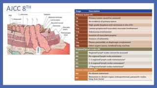
  • 7. AJCC -8th SCC • Grade ,location of tumour affect outcomes Adenocarcinoma • Tumour location –not a prognostic variable Adenosquamous – staged as squamous only
  • 8. p Stage yp Stage (post neoadjuvant)
  • 9. Lymphatics • The esophagus has an extensive, longitudinal interconnecting system of lymphatics. The esophageal lymphatic network is primarily located within the submucosa .However, channels are also present within the lamina propria, facilitating spread of even superficial cancers of the esophagus involving the mucosa. • In addition to these longitudinal lymphatics, intramural lymphatics may traverse the muscularis propria, facilitating tumor spread to regional lymphatic channels and paraesophageal nodes. • Lymph can travel the entire length of the esophagus before draining into lymph nodes, the entire esophagus is at potential risk for lymphatic involvement. • Up to 8 cm or more of “normal” tissue can exist between gross tumor and micrometastases “skip areas”. • Lymphatics of the esophagus drain into nodes that usually follow arteries,including the inferior thyroid artery, the bronchial and esophageal arteries, and the left gastric artery (celiac axis).
  • 10. LN Status • Depth of tumor penetration (T stage) affects lymph node involvement (LNI) • Intramucosal T1 lesions (18% LNI) • submucosal T1 lesions (55% LNI) • T2 lesions (60% LNI) • T3 lesions (80% LNI) • Chance if LN <50% - conservative eso resection and limited lymph node dissection • LN>50% - neoadjuvant therapy followed by resection
  • 11. Natural history and pattern of spread • Squamous cell carcinoma is characterized by extensive local growth and proclivity to lymph node metastases. • Because the esophagus has no covering serosa, direct invasion of contiguous structures may occur early. • Lesions in the upper esophagus can impinge on or invade the recurrent laryngeal nerves,carotid arteries, and trachea. If extraesophageal extension occurs in the mediastinum, tracheoesophageal or bronchoesophageal fistula may occur. • Tumors in the lower one-third of the esophagus can invade the aorta or pericardium, resulting in mediastinitis, massive hemorrhage, or empyema.
  • 12. Histology • Squamous and adeno -95% of all carcinomas • Psuedosarcoma – spindle cell carcinoma (SCC variant) • Adenoca variants –Adenoid cystic ,mucoepidermoid poorer prognosis • Small cell carcinoma – arise from argyrophilic cells paraneoplastic syndromes !! (ADH,hypercalcemia) , similar to SCLC • Nonepithelial origin – very rare • Malignant melanoms –MS 7months • Lymphoma –mostly extension , as primary very rare
  • 13. SCC • Squamous cell carcinomas arise from the squamous mucosa - native to the esophagus - 70% - upper and middle thirds • Most common type of esophageal ca in India (90%) • Smoking and alcohol are common eitiologic factors (5 fold increase in risk) • Combined increase risk from 25 - 100 folds • Dietary • Nitrosamines (pickled foods , smoked food) • long term ingestion of hot liquids • Micronutrient deficiency (Vit. A, B12, C, E). • Trace Element deficiency (Cobalt, Copper & Selenium) • Acquired • Cigarette smoking • Alcohol. • Chronic esophagitis. • Chronic Dysphagia • Caustic ingestion • Radiation exposure • Plummer – vinson syndrome • Tylosis(40%) • Achalasia (16 -fold) • Esophageal strictures and diverticula • p53 • Premalignant conditions
  • 14. Adenocarcinoma • almost 70 % - United States and Western countries. Etiology : • Increasing incidence of GERD • Western diet • Increased use of acid-suppression medications Histologically it is from : • Submucosal glands of the esophagus • Heterotopic islands of columnar epithelium • Malignant degeneration of metaplastic columnar epithelium (Barrett’s esophagus) – 40 fold increased risk
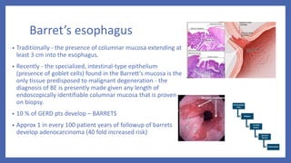
  • 15. Barret’s esophagus • Traditionally - the presence of columnar mucosa extending at least 3 cm into the esophagus. • Recently - the specialized, intestinal-type epithelium (presence of goblet cells) found in the Barrett’s mucosa is the only tissue predisposed to malignant degeneration - the diagnosis of BE is presently made given any length of endoscopically identifiable columnar mucosa that is proven on biopsy. • 10 % of GERD pts develop – BARRETS • Approx 1 in every 100 patient years of followup of barrets develop adenocarcinoma (40 fold increased risk)
  • 16. Clinical manifestations • Early - asymptomatic – mimic GERD (3-4 months before diagnosis) • Dysphagia >90% (mc)  > 2/3rds of lumen has to be obstructed (lack of serosa) • Weight Loss (40-70%) -most common symptoms • Vomiting/Regurgitation • Pain. • Cough , choking , asp.pneumonia (TEF) • Hoarseness.(left.RLN , vocal cords) • Dyspnoea • In high-incidence areas where screening is practice,the most prominent early symptom is pain on swallowing rough or dry food • Systemic disease – jaundice ,excessive pain ,bone pain,respiratory symptoms Perez & Brady 7E
  • 18. Esophagoscopy : • Good 1st test – dysphagia & suspecting ca esophagus • Can differentiate intra luminal from intramural & intrinsic from extrinsic • Dx of esophageal ca is best made by endoscopic biopsy Critical points : • Location of lesion • Nature of lesion (polypoid etc) • Extent & relationship to cricophayngeus ,GEJ Apple core appearance
  • 19. : CT • Imp for staging. • Chest and abdomen – Length , thickness, LN Liver and lung mets , T4 • Accuracy  57%- T 74%- N , 83%- M • Many unresectable tumors by CT scan are deemed resectable at the time of surgery.
  • 20. PET : • FDG –PET: • Evaluates Primary mass LN ,Mets • Sensitivity and specificity slightly greater than CT • Not reliable as single Dx tool • Value in evaluating response to chemo and RT • N staging imitations :periesophageal ln difficult to distinguish from primary mass , infections • Detect distant metastasis in 20% pts • (which was Not detected on CT ) • PET + CT – improve accuracy of detecting • -stage III by 23% • -stage IV by 18% Figure Distant lymph node metastases of esophageal cancer detected by integrated CT PET. A, Integrated CT PET demonstrates para-aortic lymph node metastases showing increased FDG uptake (arrowheads). B, Corresponding CT image shows lymph nodes (arrowheads) measuring 5 to 8 mm in diameter. Based on size criteria, these lymph nodes may be considered benign on CT scan
  • 21. MRI • Not done routinely • To identify involvement of vascular & neural • Accurately detects T4 and mets • But overstages T & N status
  • 22. EUS(Endoscopic Ultrasonography) • More accurately assess -periesophageal & celiac LN involvement -transmural extent of ds. • Limitation : -less significant accuracy following neoadjuvant therapy d/t not discriminate tumor from postradiation inflammation & fibrosis • Overall accuracy for Local nodal (N) staging 75% to 80% • Overall accuracy for Tumor staging (T) 85% to 90%
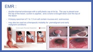
  • 24. EMR : • double-channel endoscope with a soft plastic cap at its tip. The cap is placed over the top of the lesion, suction is applied, and a snare is brought down over the top of the lesion • A biopsy specimen of 1 to 1.5 cm will contain mucosa and submucosa • may also be used as a therapeutic modality for premalignant and early malignant conditions
  • 25. Minimal invasive surgical modalities for staging: • Includes bronchoscopy ,Thoracoscopy and Laparoscopy. • Highly accurate in evaluating N & M Status.
  • 26. Molecular considerations • HER2/neu oncogene : overexpressed in 15% to 30% of adenocarcinomas and 5% to 13% of SCC , Both IHC and FISH to be done , associated with tumor invasion, lymph node metastases, and poor prognosis. • EGFR :Overexpression of EGFR on IHC testing occurs in approximately 80% of patients with adenocarcinoma and SCC , research for targeted therapies ongoing, poor prognosis in terms of decreased survival. • (VEGF) expression levels: poor prognosis , targeted therapy (ramucirumab) • COX-2:Inhibition of COX-2 activity results in enhanced radiosensitization of tumor tissue but not normal tissue.
  • 28. Treatment options • Surgery • Radiation therapy 1.External Radiotherapy 2.Intraluminal Brachytherapy • Chemotherapy • Multimodality treatment combining above methods
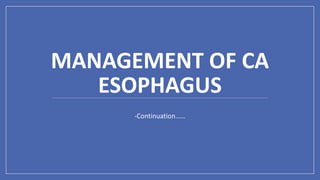
  • 30. SURGERY RESECTABLE ESOPHAGEAL CANCERS: (STAGE I –III) UNRESECTABLE ESOPHAGEAL CANCERS:
  • 31. Endoscopic methods • Endoscopic Mucosal Resection- • CONSIDERED as essential diagnostic, staging and therapeutic option for patient having high grade dysplasia, superficial esophageal tumor • Procedure – injection of fluid in submucosal layer or use the suction to trap the lesion into cylinder followed by tissue retrieval by snare or endoscopic knife • Complete local remission rate – 99-100% • 5 yr survival rate – 98%
  • 33. CERVICAL ESOPHAGUS  Proximal esophageal tumors are treated with DEFINITIVE CHEMORADIOTHERAPY  Total esophagectomy with resection of pharynx, larynx, thyroid gland is required (Total pharyngo-laryngo-esophagectomy) – high morbidity with loss of function.
  • 34. THORACIC ESOPHAGUS  IVOR LEWIS OPERATION  Abdomen opened first and stomach mobilized  And then Right thoracotomy -Oesophagogastric anastomosis  2 stage approach  McKeown OPERATION  3 incisions  Abdominal, right posterolateral thoracotomy and left cervical incision
  • 35. TRANSHIATAL ESOPHAGECTOMY without thoracotomy ( ORRINGER’S OPERATION )  Avoidance of thoracotomy incision  Avoidance of mediastinitis  Shorter Duration of operation  Poor visualization of upper and middle esophageal tumors  Anastomotic leak  Chylothorax  Recurrent laryngeal nerve injury DisadvantagesAdvantages
  • 36. Lymph node dissection • En Bloc resection is the best treatment with removal of all lymph nodesEn bloc esophagectomy involves resection of middle and lower esophageal tumors with an envelope of adjacent tissue that includes the mediastinal pleura laterally, the pericardium anteriorly, and the azygos vein and thoracic duct posterolaterally with the surrounding periesophageal tissue and lymph nodes
  • 37. Transhiatal vs transthoracic approach Transhiatal approach Transthoracic approach Thoracotomy not required Required Distal esophagus Middle or distal esophagus Less pulmonary complications more Anastmotic leak high low Disease free survival 1.4yr 1.7yr Overall survival 1.8yr 2.0yr 5 yr survival rate 34% 36% In hospital mortality 2% 4% • Conclusion- either the transhiatal or transthoracic procedure can be performed with acceptable mortality and morbidity , with either technique the outcome is almost similar
  • 38. Surgery Alone as Treatment • 5 yr survival rate  28% , compared to 10% in patients treated medically. • Locoregional failure rate  32-45% • Need for adjuvant therapy • Conclusion- as relapse rate is high and long term survival is poor , integration of adjuvant or neoadjuvant chemoradiation approaches in treatment is rational and indicated. • Optimum time for surgery ? (After NACRT) Traditional 4-6 weeks Vs >12 weeks Perez and Brady’s 7th edition
  • 39. Radiation  Pre op RT  Pre op CT RT  Post op RT  Radical RT  Radical CT RT  Palliative  Brachytherapy
  • 40. Role of Preoperative Radiation Therapy • In various trials comparing surgery alone vs preoperative radiation followed by surgery it was found that there is no clinical benefit of using preoperative radiation. • No significant survival benefit • Local control rate was improved but non significant. • Conclusion- preoperative radiation therapy is not recommended • Dosage - 41.4-50.4 Gy/ 1.8-2.0 Gy per fraction/ 23-28 Fr
  • 41. Role of Preoperative Chemoradiation • It is most common approach for resectable esophageal cancer • Benefits downstaging of disease increase rate of complete resection of disease with negative margins eradicate occult micrometastatic disease • Improves local control • Absolute Survival benefit  2yr - 13%  5yr - 6.5%
  • 43. Median Survival was 49 months as compared to 24 months in surgical arm – DOUBLED !!
  • 44. Regimens • Paclitaxel+carboplatin (cat-1) Paclitaxel 50mg/m2 I V &+ Carboplatin AUC 2 Day-1 Wkly x 5 wk • Cisplatin+ Fluorouracil (cat-1) Cisplatin 75-100 mg /m2 D1&D29 + Fluorouracil 750-1000 mg /m2 IV infusion over 24 hours daily on D1- 4 & D 29–32 {35day cycle} OR Cisplatin 15 mg /m2 D1 - 5 + Flourouracil 800 mg /m2 IV infusion over 24 hours daily on D1-5- 21days cycle x 2 • Fluorouracil+Oxaliplatin Oxaliplatin 85 mg /m2 IV on D1 + Leucovorin 400 mg /m2 D1+ FU 400 mg /m2 IV Push D1 and 800 mg /m2 IV continuous infusion over 24 hrs daily on D1 & D2 14 days cycle x 3 with radiation and 3 after radiation
  • 45. Radiotherapy Alone as Treatment • Indications- inoperable disease - medical contraindication of surgery - as palliative treatment • Median survival – 6-12 month • 5 yr survival - <10% (I- 20%, II- 10%, III- 3%, IV- 0%) • When compared to chemoradiation the 3 yr survival rate in radiation therapy alone group was found 0% • Conclusion- treatment with radiation therapy alone for esophageal cancer is palliative in majority of patients and for radical treatment it should be used in conjunction with other modalities.
  • 46. Definitive Chemoradiation Indications :- • In squamous cell carcinoma -cT1b-cT4a, N0-N+ - Cervical esophagus - If patient refuses surgery - cT4b • In adenocarcinoma esophagus - cT1b-cT4a, N0-N+ - If patient refuses surgery - cT4b
  • 47. Definitive Chemoradiation • In comparison to radiation therapy alone –  Improved median survival 14 vs 9 months  Improved 5 yr survival rate 27 % vs none • When compared to surgery alone in a resectable esophageal tumor no difference in survival rate, local failure, treatment related mortality. • 5 yr survival rate – 27% • Radiation Dose – 50-50.4 Gy/ 1.8-2.0 Gy per fraction/25-28 Fr
  • 48. CHEMOTHERAPY REGIMENS USED IN DEFINITIVE CHEMORADIATION • Cisplatin+ Fluorouracil 5-FU (cat-1) ( Cisplatin 75-100 mg /m2 D1 + 5-FU 750-1000 mg /m2 IV infusion over 24 hours daily on D1- 4 {28 day cycle} x 4) 2 cycles with RT F/b 2 cycles without RT • Oxaliplatin + 5FU (cat-1) Oxaliplatin 85 mg/m2 D1+ Leucovorin 400 mg/m2 IV D1 + 5-FU 400 mg/m2 IV D1 + 5-FU 800 mg/m2 IV over 24 hours D1 & D2 2weekly - 3cycles with RT f/b 3cycles without RT OR Oxaliplatin 85 mg/m2 IV on D1,D15,D29 for 3 doses Fluorouracil 180 mg/m2 IV Daily on D1-33
  • 49. Post operative radiation therapy • No survival advantage • May decrease time to local recurrence particularly in cases with involved margins • no significant decrease in local or distal failure rates • Dosage - 45-50.4 Gy/ 1.8-2.0 Gy per fraction/ 25-28 Fr Post operative Chemoradiation (in case of R1 ,Node positive(adeno carcinoma), pT3 ,pT4a) • When compared to surgery alone, surgery followed by chemoradiation group showed • 5FU +Leucovorin based chemotherapy • Significant decrease in local failure – 19% vs 29% • Significant survival advantage – 27 vs 36 months • Long term followup (>10yr) – continued to show survival advantage • Conclusion- it is appropriate to advise adjuvant chemoradiation in view of improved local control and survival
  • 50. RADIOTHERAPY TECHNIQUES PATIENT POSITIONING & IMMOBILISATION : • Cervical /upper third : Supine with arms by the side with straight cervical spine & parallel to couch top + Immobilisation of jaw ,neck & upper thorax • Middle and lower third: Supine with arms above their head + Vertebral column should be as parallel to couch as possible GEJ tumours and involving stomach fasting 2 hours before simulation for reproducibility.
  • 51. Upper Esophagus Radiation Field • .Superior: 5 cm proximal to tumor + supraclavicular LN + upper mediastinal LN Inferior: 5 cm distal to tumor Lateral: Tumor + 2.5 –3 cm + mediastinal LN + 2/3 of clavicle for SCF LN
  • 52. Middle esophagus Radiation field Superior: 5 cm proximal to tumor + upper mediastinal LN Inferior: 5 cm distal to tumor + mediastinal LN Lateral: tumor + 2.5–3 cm + mediastinal LN
  • 53. Lower esophagus RT Superior: 5 cm proximal to tumor + mediastinal LN Inferior: 5 cm distal to tumor + mediastinal LN + celiac LN (until L1–2 vertebrae) Lateral: tumor + 2.5–3 cm + mediastinal LN
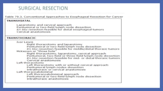
  • 54. Fields commonly used:- • AP/PA fields followed by AP/Right posterior oblique(RPO)/Left posterior oblique(LPO) with or without boost • AP/PA approach followed by Right anterior oblique(RAO)/LPO fields with or without boost • 3-field technique (AP/PA with left lateral or oblique field) • 45 Gy by AP/PA field followed by additional 5.4 Gy using oblique or lateral fields –mc used.
  • 55. AP/PA FIELDS AP AND TWO OBLIQUE FIELDS
  • 56. Nodal basins to be covered under CTV:- • Cervical & Upper Esophagus – lower cervical and supraclavicular to subcarinal lymph nodes + upper paraesophageal lymph nodes • Middle Esophagus – Complete coverage of paraesophageal lymph nodes ( individualized field design) • Lower Esophagus – Subcarinal to left gastric and common hepatic artery/celiac lymph node inferiorly • Adenocarcinoma – similar to lower thoracic esophagus but paraesophageal lymph nodes also to be included • Middle and lower paraesophageal lymph nodes should be included in Type 1 ,Type 2 tumor with T2-T4 • Splenic and common hepatic artery nodes can be spared in Type 1 T2 tumor • GTV– primary gross tumor + nodal gross disease based on diagnostic studies • CTV– to cover subclinical disease  3-5 cm proximal and distal margins covers 94-100% subclinical disease.  2 cm radial margin • PTV– CTV + 1cm radial, 1.5cm distal , 1cm proximal margin (as per departmental protocols) IMRT NCCN 2019
  • 57. External Radiation doses and OAR constraints • Higher doses 60-66 Gy in cervical esophagus , but there is little evidence beyond dose of >50.4 Gy
  • 58. Brachytherapy in Carcinoma Esophagus • Iridium 192 is used – high dose rate brachytherapy is used most commonally • Insertion- Transnasal or transoral route • 1cm proximal and 1 cm distal margin is taken based on pretreatment tumor length.
  • 59. Ca Esophagus -Brachytherapy indications Good candidates Poor candidates Contraindications Primary tumor length ≤ 10 cm length Primary tumor length> 10 cm length TE fistula Tumor confined to esophageal wall Extra –esophageal extension Cervical esophagus location Thoracic esophagus location Regional Lymphadenopathy Stenosis which cannot be bypassed No nodal / systemic metastasis Tumor involving EGJ or cardia
  • 61. Palliation • 60 -80% rate of relief from dysphagia with radiation. • MC received shedules were 20Gy/5# , 30Gy/10# , 35Gy/15# (with conc cisplatin or FU) better relief of dysphagia in CRT group. • Palliation by brachytherapy • Stent placement –inferior compared to radiation /chemotherapy
  • 62. Treatment sequale • Esophagitis , dysphagia >75% , other usual symptoms  nausea , vomiting , weight loss • Cytopenias with 2 drug chemoregimens are more pronounced. • Chemoradiation patients grade 3 – grade 4 toxities as high as 40%. • Late side effects :Stricture formation 14- 30% ,stenosis 60% • Rare – radiation pneumonitis (14% -grade 2) , cardiac toxicity 10% (pericardial effusion , IHD , HF) • TEF – 5-10% ( advised stenting , excision , bypass or intubation) , MS -10 weeks!
  • 64. Role of Preoperative Chemotherapy • In resectable esophageal cancer (adenocarcinoma of esophagus and GE junction) • Benfits - Downstaging of disease to facilitate resection -Improvement of local control -Relief of dysphagia • The limiting thing is delay in definitive treatment with risk of further spread of disease specially in non responders • Response rate – 50% • Improve overall survival • Absolute benefit – 2 yr survival – 7% 5 yr survival - 4%
  • 65. Regimens • Only for adenocarcinoma of thoracic esophagus- 2 cycles 21 days apart with • Fluorouracil – 1000 mg/m2 IV continuous infusion over 24 hr daily on D1- D4 • Cisplatin – 80 mg/m2 IV on Day 1
  • 66. Postoperative Chemotherapy • Based on various trials no survival benefit was found • Disease free survival rate was improved speacially in patients who had R0 esection and was found nodepositive and can be considered for these patients • No benefit in in patients with R0 resection and N0 node Regimen • Capecitabine and Oxaliplatin Capecitabine 1000mg/m2 PO BID D1-14 Oxaliplatin 130mg/m2 IV D1 21 days cycle
  • 67. Systemic Therapy for Recurrent or Metastatic disease • First line therapy -Two drug regimen is preferred (lower toxicity) • Three drug regimen reserved for medically fit patients with good performance status • Trastuzumab should be added in first line chemotherapy for HER 2 overexpressing metastatic adenocarcinoma Drug -Single agents Response rate Cisplatin 19 to 35 % Paclitaxel 25 to 35 % Docetaxel, 100 mg/m2 20-25% Irinotecan 15% Methotrexate less than 5% Etoposide less than 5% Ifosamide less than 5% Carboplatin less than 5%
  • 68. Regimens used as combination chemotherapy First line therapy:- Cisplatin and fluoropyrimidines (cat1) • Cisplatin 75-100 mg /m2 IV D1 + Fluorouracil 750-1000 mg /m2 IV continuous infusion over 24 hours daily on D1-4 – 28 DAY CYCLE • Cisplatin 50 mg /m2 IV D1 + Fluorouracil 2000 mg /m2 IV continuous infusion over 24 hours daily on D1+ Leucovorin 200 mg /m2 IV on D1 (14 day cycle) • Cisplatin 80 mg /m2 IV on D1+Capecitabine 1000 mg /m2 PO BID on D1-14 (21 days cycle)
  • 69. Fluoropyrimidine and Oxaliplatin:- • Oxaliplatin 85 mg /m2 IV D1 + Leucovorin 400 mg /m2 IV D1 + Fluorouracil 400 mg /m2 IV push on D1 and 1200 mg /m2 IV continuous infusion over 24 hr on D1&2 – (14 days cycle) • Oxaliplatin 85 mg /m2 IV D1 + Leucovorin 200 mg /m2 IV D1 + Fluorouracil 2600 mg /m2 IV continuous infusion over 24 hr on D1 – (14 days cycle) • Capecitabine 1000 mg /m2 PO BID D1-14 + Oxaliplatin 130 mg /m2 IV D1 (-21 days cycle) OTHER REGIMENS – DCF modifications-  Docetaxel + cisplatin+ fluorouracil (OR) Docetaxel + oxaliplatin+ fluorouracil (OR) Docetaxel + carboplatin+ fluorouracil  Paclitaxel with cisplatin or carboplatin  Epirubicin + cisplatin + fluorouracil
  • 70. TRASTUZUMAB • Used as first line therapy in recurrent or metastatic adenocarcinoma esophagus overexpressing HER2 • When compared with 1st line combination chemotherapy, survival advantage (13.8 vs 11.1 months) and response rate (47 vs 35%). • Dosage – with chemotherapy • 8 mg/kg IV loading dose on D1 of cycle 1 then 6mg/kg IV every 21 DAYS OR • 6mg/kg IV loading dose on D1 of cycle 1 then 4mg/kg every 14 DAYS
  • 71. Second line therapy • Ramucirumab + Paclitaxel ( Cat 1 for EGJ adenocarcinoma ) (Ramucirumab 8mg/kg IV on D1&15 + Paclitaxel 80 mg /m2 IV on D1,8,15 – 28 days cycle) • Ramucirumab (Cat1 foe EGJ adenocarcinoma) – 8mg/kg IV on D1 – 14 days cycle • Docetaxel – 75-100 mg /m2 IV on D1 – 21 days cycle • Paclitaxel – 135-250 mg /m2 on D1 – 21 days cycle • Paclitaxel – 80 mg /m2 IV on D1,8,15 cycled every 28 days • Other regimens – irinotecan , irinotecan+fluorouracil, irinotecan +cisplatin, docetaxel + irinotecan
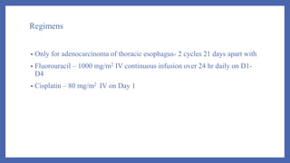
  • 72. Follow up and Surveillance Tumor classification Therapy used Recommendations Tis Endoscopic resection(ER)/Ablat ion EGD X 6mnthly for 1-2 yr, then annually for 3 more yrs. Esophagectomy EGD based on symptoms. If incomplete resection then EGD every 6 mnths for 1-2 yr T1a with or without BE ER/Ablation EGD every 3 mnthlyx1yr then every 4-6 mnths for 2nd yr then annually for 3 more yrs T1a Esophaegctomy EGD based on symptoms. If incomplete resection then ablation f/b EGD every 3 mnthlyx1yr then every 4-6 mnths for 2nd yr then annually for 3 more yrs T1b , N0 ER/Ablation EGD every 3 mnthlyx1yr then every 4-6 mnths for 2nd yr then annually for 3 more yrs. Further surveillance will depend on relapse. PET-CT OR CT chest & abdomen yearly x 3yrs and then if needed clinically
  • 73. Follow up and Surveillance T1b ANY N Esophagectomy PET-CT or CT chest and abdomen with contrast every 6 mnthly x 3yrs, then if clinically warranted. If incomplete resection then ablation f/b EGD every 3 mnthlyx1yr then every 4-6 mnths for 2nd yr then annually for 3 more yrs Chemoradiation (non surgical candidates) EGD 6-12 months x 2 yrs then annually x3 yrs PET-CT OR CECT chest and abdomen every 6-9 months x 2yrs then annually upto 5 yrs Chemoradiation (cadidate of salvage esophagectomy) EGD 6-12 months x 2 yrs then annually x3 yrs PET-CT OR CECT chest and abdomen every 6-9 months x 2yrs then annually upto 5 yrs T2-T4,N0- N+,T4b Bimodality therapy (chemoradiation) EGD every 3-4 mnthly x2 yr, every 6 monthly for 3rd yr , then as clinically warranted. PET-CT OR CECT chest and abdomen every 6-9 months x 2yrs then annually upto 5 yrs T2-T4,N0- N+,T4b Trimodality therapy Imaging (PET-CT,CECT) every 4-6 monthly X 1 yr then 6-9 monthly for next 2 yr.
  • 74. SUMMARY • Often diagnosed in late stages, so poorer outcomes. • Multimodality treatment is recommended and essential for carcinoma esophagus. • Pattern of failure - ~50% local failures needs improved local treatment . • 75% succumb to distant metastasis as well , Molecular markers and newer targeted therapies under study.