SlideShare a Scribd company logo
External Heart: Arteries that Supply
the Heart
• Coronary circulation is the functional blood
supply to the heart muscle itself (i.e. the fuel
for the heart)
• Collateral routes ensure blood delivery to
heart
• even if major vessels are occluded/clogged
Different parts of the heart are supplied by
different coronary artery segments
Heart chambers in geometric
models are subdivided into
perfusion zones using
anatomic knowledge in the
FMA ontology
CORONARY ARTERIES SUPPLYING THE
HEART WITH OXYGEN-RICH BLOOD
Right
Coronary
Artery
(RCA)
Right
Coronary
Artery
(RCA)
Left Main
Coronary
Artery (LMCA)
Left Main
Coronary
Artery (LMCA)
Left Anterior
Descending
Artery (LAD)
Left Anterior
Descending
Artery (LAD)
Posterior
Descending Artery
(PDA)
Posterior
Descending Artery
(PDA)
Left Circumflex
Coronary Artery
(LCx)
Left Circumflex
Coronary Artery
(LCx)
Arterial Coronary Circulation
External Heart: Arteries that Supply
the Heart
• Right coronary artery (in atrioventricular
groove)
– Supplies the right atrium and nearly all the
right ventricle
– marginal –supply the myocardium of the
– lateral right side of the heart
– posterior interventricular artery – supply the
– interventricular septum and adjacent portions
of the ventricular walls
External Heart: Arteries that Supply
the Heart
• Left coronary artery:
– Circumflex supplies the left atrium and the
posterior wall of the left ventricle
– Anterior interventricular artery supplies the
anterior wall of both ventricles
CERTAIN HEART WALL SEGMENTS ARE FED BY
CERTAIN CORONARY ARTERIES AS SPECIFIED BELOW:
BASE
MID
APEX
A4C
A2C
PARASTERNAL
LONG-AXIS
Key:
Red = LAD
Yellow = LCx
Green = RCA
LAD = Left Anterior Descending Coronary Artery, LCx = Left Circumflex Coronary
Artery, RCA = Right Coronary Artery
DIVIDING THE LEFT VENTRICLE INTO KEY SEGMENTS
Apical
Third
Apical
Third Mid
Third
Mid
Third
Basal
Third
Basal
Third
FURTHER DIVIDING THE LEFT VENTRICULAR WALLS
INTO 17 SPECIFIC SEGMENTS DEFINED AS:
AnteriorAnterior
AnterolateralAnterolateral
InferolateralInferolateral
InferiorInferior
InferoseptalInferoseptal
AnteroseptalAnteroseptal
ApexApex
AnteriorAnterior
AnteroseptalAnteroseptal
InferoseptalInferoseptal
AnterolateralAnterolateral
InferolateralInferolateral
InferiorInferior
AnteriorAnterior
SeptalSeptal
LateralLateral
InferiorInferior
ApexApex
BasalBasal
Mid-
Cavity
Mid-
Cavity
ApicalApical
1. Basal Anterior
2. Basal Anteroseptal
3. Basal Inferoseptal
4. Basal Inferior
5. Basal Inferolateral
6. Basal Anterolateral
7. Mid Anterior
8. Mid Anteroseptal
9. Mid Inferoseptal
10. Mid Inferior
11. Mid Inferolateral
12. Mid Anterolateral
13. Apical Anterior
14. Apical Septal
15. Apical Inferior
16. Apical Lateral
17. Apex
PUTTING IT TOGETHER: HEART WALL SEGMENTS
AND CORRESPONDING CORONARY ARTERIES
Coronary
Artery: Segments:
LAD 1, 2, 7, 8, 13, 14, 17
RCA 3, 4, 9, 10, 15
LCX 5, 6, 11, 12, 16
Coronary
Artery: Segments:
LAD 1, 2, 7, 8, 13, 14, 17
RCA 3, 4, 9, 10, 15
LCX 5, 6, 11, 12, 16
LAD = Left Anterior Descending
Artery
RCA = Right Coronary Artery
LCX = Left Circumflex Artery
LAD = Left Anterior Descending
Artery
RCA = Right Coronary Artery
LCX = Left Circumflex Artery
AnteriorAnterior
AnterolateralAnterolateral
InferolateralInferolateral
InferiorInferior
InferoseptalInferoseptal
AnteroseptalAnteroseptal
ApexApex
AnteriorAnterior
AnteroseptalAnteroseptal
InferoseptalInferoseptal
AnterolateralAnterolateral
InferolateralInferolateral
InferiorInferior
AnteriorAnterior
SeptalSeptal
LateralLateral
InferiorInferior
ApexApex
ANOTHER PERSPECTIVE – 17 SEGMENT
MODEL OF LEFT VENTRICLE
1. Basal Anterior
2. Basal Anteroseptal
3. Basal Inferoseptal
4. Basal Inferior
5. Basal Inferolateral
6. Basal Anterolateral
7. Mid Anterior
8. Mid Anteroseptal
9. Mid Inferoseptal
10. Mid Inferior
11. Mid Inferolateral
12. Mid Anterolateral
13. Apical Anterior
14. Apical Septal
15. Apical Inferior
16. Apical Lateral
17. Apex
SAMPLE – SEGMENTAL SCORING
Ortiz-Perez, J. T. et al. J Am Coll Cardiol Img 2008;1:282-293
Representative Segmentation and Pattern of Contrast
HE Over 17-Segments of the LAD, RCA, and LCX
Arteries
Ortiz-Perez, J. T. et al. J Am Coll Cardiol Img 2008;1:282-293
HEART ANATOMY – COVERINGS OF THE HEART
• Coverings of the Heart: Anatomy
• Pericardium – a double-walled sac around the
• heart composed of:
• A superficial fibrous pericardium
• A deep two-layer serous pericardium
• The parietal layer lines the internal surface
• of the fibrous pericardium
• The visceral layer or epicardium lines the
• surface of the heart
• They are separated by the fluid-filled
• pericardial cavity
• Coverings of the Heart: Physiology
• The pericardium:
• Protects the heart
COVERINGS OF THE HEART:
PHYSIOLOGY
• The pericardium:
– Protects and anchors the heart
– Prevents overfilling of the heart with blood
– Allows for the heart to work in a relatively
– friction-free environment
PERICARDIUM OF THE HEART
• Pericardium (3 layers)
• 1) Outer-fibrous pericardium
– Serous pericardium
• 2) parietal
• 3) visceral (epicardium)
• Pericardial Cavity
– between layers of serous pericardium
– serous fluid
– lubricate heart while beating
Pericardial Layers of the Heart
Heart Wall – Layers of the Heart
• Epicardium – visceral layer of the serous
• pericardium
• Myocardium – cardiac muscle layer forming
• the bulk of the heart
• Fibrous skeleton of the heart – crisscrossing,
• interlacing layer of connective tissue
• Endocardium – endothelial layer of the inner
• myocardial surface
• Cardiac Muscle Bundles
• External
Cardiac Muscle 1000X
intercalated disc
striations nucleus
Purkinje fibers 40X
EXTERNAL FEATURES OF THE HEART
• Interventricular sulcus
• Coronal/Coronary sulcus
• Auricles of atria
• Apex
• Base
• Coronary vessels
• Ligamentum Arteriosum
External Heart: Major Vessels of
the Heart (Anterior View)
• External Heart: Major Vessels of
the Heart (Anterior View)
• Vessels returning blood to the heart include:
– Superior and inferior vena cava
– Right and left pulmonary veins
• Vessels conveying blood away from the heart:
– Pulmonary trunk, which splits into right and
– left pulmonary arteries
– Ascending aorta (three branches) –
– brachiocephalic, left common carotid, and
– subclavian arteries
External Heart: Veins that Drain the
Heart
External Heart: Major Vessels of
the Heart (Posterior View)
Vessels returning blood to the heart include:
•Right and left pulmonary veins
• Superior and inferior vena cava
• Vessels conveying blood away from the heart
include:
• Aorta
• Right and left pulmonary arteries
FLOW OF BLOOD
• O2-poor blood (S+I VC, Coronary Sinus) enters Rt Atrium
• Travels through Tricuspid Valve into Rt Ventricle
• Pumped out through Pulmonary Semilunar Valve into
Pulmonary trunk (branches into Pulmonary Arteries) and to
lungs
•After circulating through lungs, O2-rich blood returns to the
heart through 4 Pulmonary veins
• The O2-rich blood enters the Left Atrium
• Travels through Bicuspid/Mitral Valve into Left Ventricle
• Pumped out through Aortic Semilunar Valve into Aorta to be
distributed to rest of body by descending aorta and branches
of aortic arch
Pathway of Blood Through the
Heart and Lungs
Right atrium > tricuspid valve > right ventricle >
pulmonary semilunar valve > pulmonary arteries >
Lungs > pulmonary veins > left atrium >
Mitral valve > left ventricle > Left ventricle >
aortic semilunar valve > aorta > systemic circulation
THE PATHWAY OF THE HEART
Cardiovascular Flow of Blood
• HeartArteries(conducting-distributing)
ArteriolesCapillaries of tissues
• At Capillaries O2 is delivered and CO2 picked
up
•CapillariesVenulesVeinsHeart
DETAILED IMAGES OF THE HEART’S
ANATOMY – CHORDAE TENDONAE
THE CARDIAC CYCLE
Semilunar Valve Function
Cardiac Intrinsic Conduction
IMPORTANT - Various
nodes and intrinsic Heart
Rate Generated:
• SA Node – 60 – 100
Beats/min
• AV Node – 40 – 60
Beats/min
• Bundle of His – 40 – 60
Beats/min
•Purkinje Fibers – Last
resort – 20 – 40
Beats/min
IMPORTANT - Various
nodes and intrinsic Heart
Rate Generated:
• SA Node – 60 – 100
Beats/min
• AV Node – 40 – 60
Beats/min
• Bundle of His – 40 – 60
Beats/min
•Purkinje Fibers – Last
resort – 20 – 40
Beats/min
Heart Excitation Related to ECG
atrial excitation begins
Impulse delayed
at AV node
Impulse passes to
heart apex; ventricular
excitation begins
Ventricular excitation
complete
SA node AV node Purkinje
fibers
Bundle
branches
Electrocardiography
Electrical activity is recorded by
electrocardiogram (ECG)
• P wave corresponds to depolarization of SA
•node
•QRS complex corresponds to ventricular
•depolarization
•T wave corresponds to ventricular
repolarization
•Atrial repolarization record is masked by the
•larger QRS complex
Electrocardiography
Electrocardiography
PR interval
•Atrial depolarization and contraction
•QT interval
•Ventricular depolarization, contraction and
repolarization
•PR segment
•Atrial contraction
•ST segment
•Ventricular contraction
•ECG Tracings
•Heart
ECG Tracings
Heart
Cardiac Cycle
Cardiac cycle refers to all
events associated
with blood flow through
the heart
•Systole – contraction of heart muscle
•Diastole – relaxation of heart muscle
Phases of the Cardiac Cycle
• Isovolumetric relaxation – early diastole
• Ventricles relax
• Backflow of blood in aorta and pulmonary trunk closes semilunar valves
• Dicrotic notch – brief rise in aortic pressure caused by backflow of blood. This
backflow
• causes the valve to close and creates a slight
pressure rebound
Phases of the Cardiac Cycle
THE CARDIAC CYCLE
Cardiodynamics
Cardiac output (CO) : the amount of blood
pumped by each ventricle in one minute
•Cardiac output equals heart rate times stroke
volume
Cardiac output (ml/min) =
Heart Rate (HR) (beats/min) X
Stroke Volume (SV) (ml/beat)
Cardiodynamics
Heart rate (HR) : number of heart beats in a
minute
•Stroke volume (SV) – amount of blood
ejected from the ventricles with each beat
•SV = EDV - ESV
Cardiac Output:
Example
CO (ml/min) = HR (75 beats/min) x
SV (70 ml/beat)
•CO = 5250 ml/min (5.25 L/min)
Cardiodynamics
Ventricular pressure increases forcing blood
through the semilunar valves: ventricular
Ejection
•End-systolic volume (ESV)
•Amount of blood that remains in the
ventricles after the contraction and closing of
the semilunar valves
Factors Affecting stroke volume
(EDV-ESV)
EDV (end diastolic volume) is affected by
•Venous return - amount of blood returning to
the heart or blood flow during filling time
•High venous return= high EDV
•Slow heartbeat and exercise increase venous
return to the heart, increasing SV
Factors Affecting stroke volume
(EDV-ESV)
Filling time -duration of ventricular diastole
•Depends on the heart rate
•Blood loss and extremely rapid heartbeat
decrease SV
•The longer the filling time the higher the
EDV will be
Factors Affecting stroke volume
(EDV-ESV)
Preload
• Stretchiness of the ventricles during
Diastole
• Directly proportional to the EDV
• Frank-Starling principle (“more in = more out”)
or increased EDV=increased SV
Factors Affecting stroke volume
(EDV-ESV)
ESV (end systolic volume). It is influenced by:
•Contractility
• Force produced during a contraction
• Positive inotropic (increase Calcium entry)
• Increased sympathetic stimuli
• Certain hormones, some drugs
• Increase SV by decreasing ESV
Factors Affecting stroke volume
(EDV-ESV)
Afterload
•The pressure that must be overcome for the
ventricles to eject blood (back pressure
exerted by blood in the large arteries leaving
the heart)
• Increased afterload will increase ESV and
decrease SV
• Increased by factors that restricts arterial blood
flow
PRELOAD AND AFTERLOAD DEPICTED
STRESS PERFUSION IMAGES
Stress-
Perfusion
Rest-
Perfusion
Baseline
LV contrast
uptake
RV contrast
uptake
Myocardial
contrast uptake
Viability and
coronary
angiography
Echo class 2_05262015
Walls of the ventricles:
Left wall is thicker!
Echo class 2_05262015
Find:
1. Walls of the ventricles
2. Auricles
3. Inner walls of the atria
4. Fossa ovalis
5. Trabeculae carnae
6. Atrioventricular valve
(a) "Bicuspid valve"
(b) "Tricuspid valve"
7. Chordae tendonae
8. Papillary muscles
9. Aortic & pulmonary valves
Coronary arteries are the FIRST
branches of the aorta!
1. Coronary arteries
(a) Left coronary artery
(b) Right coronary artery
(c) Interventricular branches
(d) Right marginal branch
2. Cardiac veins
Heart in VENTRAL
view.
(You see mostly
right ventricle!)
Heart in DORSAL view.
(You see mostly left
ventricle.)
Intrinsic regulation of heart beat
1. Sinoatrial node is
PACEMAKER OF
HEART, and beginning
of process. Geenrates
periodic impulses that
initiate contraction of
right atrium.
2. Signal then runs to
Atrioventricular node.
Message is passed along
a track of Purkinje fibers
called the...
3. Atrioventricular bundle.
Atrioventricular bundle
then splits into right and
left limbs/branches that
pass to individual inner
ventricular walls on right
and left.
Echo class 2_05262015
Maximum Heartrate Calculation
(Suggested) MAXIMUM HEARTRATE can be calculated by the
formula:
208 - (0.7)(your age) = normal maximum
heartrate.
Heartrate
Resting heartrate average is variable
depending on ages, sex, weight, etc.
MAXIMUM HEARTRATE used to be
calculated by the formula:
220 - your age = normal maximum
heartrate.
(This is now known to be oversimplified
and incorrect.)

More Related Content

PPTX
Stress echocardiography
PPTX
Echo assessment of lv systolic function and swma
PDF
CONTRAST ECHOCARDIOGRAPHY
PPTX
Aortic stenosis - Echocardiography
PPTX
Echo assessment of RV function
PDF
Echo assessment of cardiomyopathy pdf
PPTX
cath Lab Hemoduhynamic
PPTX
PRESSURE MEASUREMENT by Cardiac catheterisation_Dr Amol Patil.pptx
Stress echocardiography
Echo assessment of lv systolic function and swma
CONTRAST ECHOCARDIOGRAPHY
Aortic stenosis - Echocardiography
Echo assessment of RV function
Echo assessment of cardiomyopathy pdf
cath Lab Hemoduhynamic
PRESSURE MEASUREMENT by Cardiac catheterisation_Dr Amol Patil.pptx

What's hot (20)

PPTX
Lv systolic function
PPT
Ecg criteria of chamber enlargement
PPTX
Echo Mitral Stenosis
PPTX
Aortic stenosis Echo
PPTX
Echocardiography in mitral stenosis
PPTX
Echocardiographic evaluation of mitral regurgitation
PPTX
Basics of echo & principles of doppler echocardiography
PDF
Contrast echocardiography
PPTX
Left ventricular diastolic dysfunction in echocardiography
PDF
Echo in cardiomyopathies part 1
PPTX
Right heart catheters
PPTX
FFR(fractional flow reserve)
PPTX
Left ventricular pressure tracings
PPTX
hemodynamic in cath lab: aortic stenosis and hocm
PPTX
BMV,PTMC,BALLOON MITRAL VALVOTOMY, BAL, VIRBHAN BALAI, DR VIRBHAN
PPSX
Right heart catheterization
PDF
Echocardiographic Evaluation of LV Diastolic Function
PPTX
Repolarization ST wave Abnormalities
PPTX
Cardiac catheters
Lv systolic function
Ecg criteria of chamber enlargement
Echo Mitral Stenosis
Aortic stenosis Echo
Echocardiography in mitral stenosis
Echocardiographic evaluation of mitral regurgitation
Basics of echo & principles of doppler echocardiography
Contrast echocardiography
Left ventricular diastolic dysfunction in echocardiography
Echo in cardiomyopathies part 1
Right heart catheters
FFR(fractional flow reserve)
Left ventricular pressure tracings
hemodynamic in cath lab: aortic stenosis and hocm
BMV,PTMC,BALLOON MITRAL VALVOTOMY, BAL, VIRBHAN BALAI, DR VIRBHAN
Right heart catheterization
Echocardiographic Evaluation of LV Diastolic Function
Repolarization ST wave Abnormalities
Cardiac catheters
Ad

Viewers also liked (20)

PDF
Physiology
PPT
L7 circulatory routes
PPT
33. cardiovascular 5-1-08-09
PPTX
Cvs physio
PPT
Circulatory system
PPT
Blood circulation
ODP
Amp Slideshow
PPTX
Coronary circuln 07 02-2012
PPTX
coronary circulation
PPTX
Circulation through special regions 1
PPTX
Coronary arteries
PPTX
Coronary Circulation
PPTX
Regulation of Coronary Blood Flow
PPTX
Coronary Arteries
PPTX
Coronary circulation
PPTX
CORONARY BLOOD FLOW
PPTX
Coronary circulation 14 10-14
PPT
DRAW A NEAT DIAGRAM - EXCRETARY SYSTEM
PPTX
Anatomy of the Cardiovascular system
PPT
CORONARY CIRCULATION
Physiology
L7 circulatory routes
33. cardiovascular 5-1-08-09
Cvs physio
Circulatory system
Blood circulation
Amp Slideshow
Coronary circuln 07 02-2012
coronary circulation
Circulation through special regions 1
Coronary arteries
Coronary Circulation
Regulation of Coronary Blood Flow
Coronary Arteries
Coronary circulation
CORONARY BLOOD FLOW
Coronary circulation 14 10-14
DRAW A NEAT DIAGRAM - EXCRETARY SYSTEM
Anatomy of the Cardiovascular system
CORONARY CIRCULATION
Ad

Similar to Echo class 2_05262015 (20)

PPT
Cardiac anatomy and physiology
PPT
CARDIOVASCULAR SYSTEM - ANATOMY & PHYSIOLOGY
PDF
cardiovascular anatomy and physiology
PPTX
cardiac physiology.pptx cardiac physiology
PPTX
Anatomy of the heart lecture.pptx
PPTX
Cardiac anatomy and physiology
PPTX
Anatomy of THE CIRCULATORY SYSTEM hand out.pptx
PPT
Cardiovascularsystem 110221045748-phpapp02
PDF
Cardiovascular anatomy, nerves and other structures
PPTX
THE HEART.pptx
PPTX
CARDIO VASCULAR SYSTEM THE HEART
PPT
Human heart
PPTX
The Human Body's Cardiovascular System.ppt
PDF
thorax 2,12.pdf anatomy simplified just for u easy
PPTX
cardiovascular disease.pptx
PPT
Presentation on anatomy of heart ,coronary circulation ,mi and ecg interpreta...
PPTX
The heart
PPTX
Cardiovasular system
PDF
Anatomy & PHYSIOLOGY Cardiovascular System
PPT
THE CARDIOVASCULAR SYSTEM (heart anatomy)
Cardiac anatomy and physiology
CARDIOVASCULAR SYSTEM - ANATOMY & PHYSIOLOGY
cardiovascular anatomy and physiology
cardiac physiology.pptx cardiac physiology
Anatomy of the heart lecture.pptx
Cardiac anatomy and physiology
Anatomy of THE CIRCULATORY SYSTEM hand out.pptx
Cardiovascularsystem 110221045748-phpapp02
Cardiovascular anatomy, nerves and other structures
THE HEART.pptx
CARDIO VASCULAR SYSTEM THE HEART
Human heart
The Human Body's Cardiovascular System.ppt
thorax 2,12.pdf anatomy simplified just for u easy
cardiovascular disease.pptx
Presentation on anatomy of heart ,coronary circulation ,mi and ecg interpreta...
The heart
Cardiovasular system
Anatomy & PHYSIOLOGY Cardiovascular System
THE CARDIOVASCULAR SYSTEM (heart anatomy)

More from Clifford Thornton (8)

PDF
Vascular devices llc_implanted_vessel_clearing_modules_&_system_02-21-2021
PDF
Global innovation magazine_issue10
PPT
Thornton clifford sample_consulting
PPT
Zurn drill bit_informational_deck_cmt_12-03-2017
PPTX
Thornton clifford m_future_of_cardiology_09-01-2017_v4
PPTX
Clifford thornton spe_600_research_project_final
PPTX
Thornton lesson plan_1_indian_nations_presentation_10-18-2016_v2
PPTX
Intro to echo_class_05262015
Vascular devices llc_implanted_vessel_clearing_modules_&_system_02-21-2021
Global innovation magazine_issue10
Thornton clifford sample_consulting
Zurn drill bit_informational_deck_cmt_12-03-2017
Thornton clifford m_future_of_cardiology_09-01-2017_v4
Clifford thornton spe_600_research_project_final
Thornton lesson plan_1_indian_nations_presentation_10-18-2016_v2
Intro to echo_class_05262015

Recently uploaded (20)

PPTX
Uterus anatomy embryology, and clinical aspects
PPTX
DENTAL CARIES FOR DENTISTRY STUDENT.pptx
PPTX
Chapter-1-The-Human-Body-Orientation-Edited-55-slides.pptx
PPT
ASRH Presentation for students and teachers 2770633.ppt
PDF
Medical Evidence in the Criminal Justice Delivery System in.pdf
PPT
Obstructive sleep apnea in orthodontics treatment
DOCX
RUHS II MBBS Microbiology Paper-II with Answer Key | 6th August 2025 (New Sch...
PPTX
Fundamentals of human energy transfer .pptx
PPTX
POLYCYSTIC OVARIAN SYNDROME.pptx by Dr( med) Charles Amoateng
PPTX
Slider: TOC sampling methods for cleaning validation
PPTX
post stroke aphasia rehabilitation physician
PPTX
Electromyography (EMG) in Physiotherapy: Principles, Procedure & Clinical App...
PPTX
JUVENILE NASOPHARYNGEAL ANGIOFIBROMA.pptx
PPTX
Pathophysiology And Clinical Features Of Peripheral Nervous System .pptx
PPTX
Respiratory drugs, drugs acting on the respi system
PPTX
CME 2 Acute Chest Pain preentation for education
PPTX
Gastroschisis- Clinical Overview 18112311
PPTX
Neurotransmitter, Types of neurotransmitters,Neurotransmitter function, Neur...
PPTX
Patholysiology of MAFLD/MASLD and Role of GLP 1 agonist in obesity and cardio...
PDF
Handout_ NURS 220 Topic 10-Abnormal Pregnancy.pdf
Uterus anatomy embryology, and clinical aspects
DENTAL CARIES FOR DENTISTRY STUDENT.pptx
Chapter-1-The-Human-Body-Orientation-Edited-55-slides.pptx
ASRH Presentation for students and teachers 2770633.ppt
Medical Evidence in the Criminal Justice Delivery System in.pdf
Obstructive sleep apnea in orthodontics treatment
RUHS II MBBS Microbiology Paper-II with Answer Key | 6th August 2025 (New Sch...
Fundamentals of human energy transfer .pptx
POLYCYSTIC OVARIAN SYNDROME.pptx by Dr( med) Charles Amoateng
Slider: TOC sampling methods for cleaning validation
post stroke aphasia rehabilitation physician
Electromyography (EMG) in Physiotherapy: Principles, Procedure & Clinical App...
JUVENILE NASOPHARYNGEAL ANGIOFIBROMA.pptx
Pathophysiology And Clinical Features Of Peripheral Nervous System .pptx
Respiratory drugs, drugs acting on the respi system
CME 2 Acute Chest Pain preentation for education
Gastroschisis- Clinical Overview 18112311
Neurotransmitter, Types of neurotransmitters,Neurotransmitter function, Neur...
Patholysiology of MAFLD/MASLD and Role of GLP 1 agonist in obesity and cardio...
Handout_ NURS 220 Topic 10-Abnormal Pregnancy.pdf

Echo class 2_05262015

  • 1. External Heart: Arteries that Supply the Heart • Coronary circulation is the functional blood supply to the heart muscle itself (i.e. the fuel for the heart) • Collateral routes ensure blood delivery to heart • even if major vessels are occluded/clogged
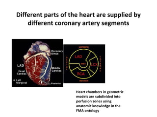
  • 2. Different parts of the heart are supplied by different coronary artery segments Heart chambers in geometric models are subdivided into perfusion zones using anatomic knowledge in the FMA ontology
  • 3. CORONARY ARTERIES SUPPLYING THE HEART WITH OXYGEN-RICH BLOOD Right Coronary Artery (RCA) Right Coronary Artery (RCA) Left Main Coronary Artery (LMCA) Left Main Coronary Artery (LMCA) Left Anterior Descending Artery (LAD) Left Anterior Descending Artery (LAD) Posterior Descending Artery (PDA) Posterior Descending Artery (PDA) Left Circumflex Coronary Artery (LCx) Left Circumflex Coronary Artery (LCx)
  • 5. External Heart: Arteries that Supply the Heart • Right coronary artery (in atrioventricular groove) – Supplies the right atrium and nearly all the right ventricle – marginal –supply the myocardium of the – lateral right side of the heart – posterior interventricular artery – supply the – interventricular septum and adjacent portions of the ventricular walls
  • 6. External Heart: Arteries that Supply the Heart • Left coronary artery: – Circumflex supplies the left atrium and the posterior wall of the left ventricle – Anterior interventricular artery supplies the anterior wall of both ventricles
  • 7. CERTAIN HEART WALL SEGMENTS ARE FED BY CERTAIN CORONARY ARTERIES AS SPECIFIED BELOW: BASE MID APEX A4C A2C PARASTERNAL LONG-AXIS Key: Red = LAD Yellow = LCx Green = RCA LAD = Left Anterior Descending Coronary Artery, LCx = Left Circumflex Coronary Artery, RCA = Right Coronary Artery
  • 8. DIVIDING THE LEFT VENTRICLE INTO KEY SEGMENTS Apical Third Apical Third Mid Third Mid Third Basal Third Basal Third
  • 9. FURTHER DIVIDING THE LEFT VENTRICULAR WALLS INTO 17 SPECIFIC SEGMENTS DEFINED AS: AnteriorAnterior AnterolateralAnterolateral InferolateralInferolateral InferiorInferior InferoseptalInferoseptal AnteroseptalAnteroseptal ApexApex AnteriorAnterior AnteroseptalAnteroseptal InferoseptalInferoseptal AnterolateralAnterolateral InferolateralInferolateral InferiorInferior AnteriorAnterior SeptalSeptal LateralLateral InferiorInferior ApexApex BasalBasal Mid- Cavity Mid- Cavity ApicalApical 1. Basal Anterior 2. Basal Anteroseptal 3. Basal Inferoseptal 4. Basal Inferior 5. Basal Inferolateral 6. Basal Anterolateral 7. Mid Anterior 8. Mid Anteroseptal 9. Mid Inferoseptal 10. Mid Inferior 11. Mid Inferolateral 12. Mid Anterolateral 13. Apical Anterior 14. Apical Septal 15. Apical Inferior 16. Apical Lateral 17. Apex 1. Basal Anterior 2. Basal Anteroseptal 3. Basal Inferoseptal 4. Basal Inferior 5. Basal Inferolateral 6. Basal Anterolateral 7. Mid Anterior 8. Mid Anteroseptal 9. Mid Inferoseptal 10. Mid Inferior 11. Mid Inferolateral 12. Mid Anterolateral 13. Apical Anterior 14. Apical Septal 15. Apical Inferior 16. Apical Lateral 17. Apex
  • 10. PUTTING IT TOGETHER: HEART WALL SEGMENTS AND CORRESPONDING CORONARY ARTERIES Coronary Artery: Segments: LAD 1, 2, 7, 8, 13, 14, 17 RCA 3, 4, 9, 10, 15 LCX 5, 6, 11, 12, 16 Coronary Artery: Segments: LAD 1, 2, 7, 8, 13, 14, 17 RCA 3, 4, 9, 10, 15 LCX 5, 6, 11, 12, 16 LAD = Left Anterior Descending Artery RCA = Right Coronary Artery LCX = Left Circumflex Artery LAD = Left Anterior Descending Artery RCA = Right Coronary Artery LCX = Left Circumflex Artery AnteriorAnterior AnterolateralAnterolateral InferolateralInferolateral InferiorInferior InferoseptalInferoseptal AnteroseptalAnteroseptal ApexApex AnteriorAnterior AnteroseptalAnteroseptal InferoseptalInferoseptal AnterolateralAnterolateral InferolateralInferolateral InferiorInferior AnteriorAnterior SeptalSeptal LateralLateral InferiorInferior ApexApex
  • 11. ANOTHER PERSPECTIVE – 17 SEGMENT MODEL OF LEFT VENTRICLE 1. Basal Anterior 2. Basal Anteroseptal 3. Basal Inferoseptal 4. Basal Inferior 5. Basal Inferolateral 6. Basal Anterolateral 7. Mid Anterior 8. Mid Anteroseptal 9. Mid Inferoseptal 10. Mid Inferior 11. Mid Inferolateral 12. Mid Anterolateral 13. Apical Anterior 14. Apical Septal 15. Apical Inferior 16. Apical Lateral 17. Apex 1. Basal Anterior 2. Basal Anteroseptal 3. Basal Inferoseptal 4. Basal Inferior 5. Basal Inferolateral 6. Basal Anterolateral 7. Mid Anterior 8. Mid Anteroseptal 9. Mid Inferoseptal 10. Mid Inferior 11. Mid Inferolateral 12. Mid Anterolateral 13. Apical Anterior 14. Apical Septal 15. Apical Inferior 16. Apical Lateral 17. Apex
  • 12. SAMPLE – SEGMENTAL SCORING Ortiz-Perez, J. T. et al. J Am Coll Cardiol Img 2008;1:282-293
  • 13. Representative Segmentation and Pattern of Contrast HE Over 17-Segments of the LAD, RCA, and LCX Arteries Ortiz-Perez, J. T. et al. J Am Coll Cardiol Img 2008;1:282-293
  • 14. HEART ANATOMY – COVERINGS OF THE HEART • Coverings of the Heart: Anatomy • Pericardium – a double-walled sac around the • heart composed of: • A superficial fibrous pericardium • A deep two-layer serous pericardium • The parietal layer lines the internal surface • of the fibrous pericardium • The visceral layer or epicardium lines the • surface of the heart • They are separated by the fluid-filled • pericardial cavity • Coverings of the Heart: Physiology • The pericardium: • Protects the heart
  • 15. COVERINGS OF THE HEART: PHYSIOLOGY • The pericardium: – Protects and anchors the heart – Prevents overfilling of the heart with blood – Allows for the heart to work in a relatively – friction-free environment
  • 16. PERICARDIUM OF THE HEART • Pericardium (3 layers) • 1) Outer-fibrous pericardium – Serous pericardium • 2) parietal • 3) visceral (epicardium) • Pericardial Cavity – between layers of serous pericardium – serous fluid – lubricate heart while beating
  • 18. Heart Wall – Layers of the Heart • Epicardium – visceral layer of the serous • pericardium • Myocardium – cardiac muscle layer forming • the bulk of the heart • Fibrous skeleton of the heart – crisscrossing, • interlacing layer of connective tissue • Endocardium – endothelial layer of the inner • myocardial surface • Cardiac Muscle Bundles • External
  • 19. Cardiac Muscle 1000X intercalated disc striations nucleus
  • 21. EXTERNAL FEATURES OF THE HEART • Interventricular sulcus • Coronal/Coronary sulcus • Auricles of atria • Apex • Base • Coronary vessels • Ligamentum Arteriosum
  • 22. External Heart: Major Vessels of the Heart (Anterior View) • External Heart: Major Vessels of the Heart (Anterior View) • Vessels returning blood to the heart include: – Superior and inferior vena cava – Right and left pulmonary veins • Vessels conveying blood away from the heart: – Pulmonary trunk, which splits into right and – left pulmonary arteries – Ascending aorta (three branches) – – brachiocephalic, left common carotid, and – subclavian arteries
  • 23. External Heart: Veins that Drain the Heart
  • 24. External Heart: Major Vessels of the Heart (Posterior View) Vessels returning blood to the heart include: •Right and left pulmonary veins • Superior and inferior vena cava • Vessels conveying blood away from the heart include: • Aorta • Right and left pulmonary arteries
  • 25. FLOW OF BLOOD • O2-poor blood (S+I VC, Coronary Sinus) enters Rt Atrium • Travels through Tricuspid Valve into Rt Ventricle • Pumped out through Pulmonary Semilunar Valve into Pulmonary trunk (branches into Pulmonary Arteries) and to lungs •After circulating through lungs, O2-rich blood returns to the heart through 4 Pulmonary veins • The O2-rich blood enters the Left Atrium • Travels through Bicuspid/Mitral Valve into Left Ventricle • Pumped out through Aortic Semilunar Valve into Aorta to be distributed to rest of body by descending aorta and branches of aortic arch
  • 26. Pathway of Blood Through the Heart and Lungs Right atrium > tricuspid valve > right ventricle > pulmonary semilunar valve > pulmonary arteries > Lungs > pulmonary veins > left atrium > Mitral valve > left ventricle > Left ventricle > aortic semilunar valve > aorta > systemic circulation
  • 27. THE PATHWAY OF THE HEART
  • 28. Cardiovascular Flow of Blood • HeartArteries(conducting-distributing) ArteriolesCapillaries of tissues • At Capillaries O2 is delivered and CO2 picked up •CapillariesVenulesVeinsHeart
  • 29. DETAILED IMAGES OF THE HEART’S ANATOMY – CHORDAE TENDONAE
  • 32. Cardiac Intrinsic Conduction IMPORTANT - Various nodes and intrinsic Heart Rate Generated: • SA Node – 60 – 100 Beats/min • AV Node – 40 – 60 Beats/min • Bundle of His – 40 – 60 Beats/min •Purkinje Fibers – Last resort – 20 – 40 Beats/min IMPORTANT - Various nodes and intrinsic Heart Rate Generated: • SA Node – 60 – 100 Beats/min • AV Node – 40 – 60 Beats/min • Bundle of His – 40 – 60 Beats/min •Purkinje Fibers – Last resort – 20 – 40 Beats/min
  • 33. Heart Excitation Related to ECG atrial excitation begins Impulse delayed at AV node Impulse passes to heart apex; ventricular excitation begins Ventricular excitation complete SA node AV node Purkinje fibers Bundle branches
  • 34. Electrocardiography Electrical activity is recorded by electrocardiogram (ECG) • P wave corresponds to depolarization of SA •node •QRS complex corresponds to ventricular •depolarization •T wave corresponds to ventricular repolarization •Atrial repolarization record is masked by the •larger QRS complex
  • 36. Electrocardiography PR interval •Atrial depolarization and contraction •QT interval •Ventricular depolarization, contraction and repolarization •PR segment •Atrial contraction •ST segment •Ventricular contraction •ECG Tracings •Heart
  • 38. Cardiac Cycle Cardiac cycle refers to all events associated with blood flow through the heart •Systole – contraction of heart muscle •Diastole – relaxation of heart muscle
  • 39. Phases of the Cardiac Cycle Phases of the Cardiac Cycle • Isovolumetric relaxation – early diastole • Ventricles relax • Backflow of blood in aorta and pulmonary trunk closes semilunar valves • Dicrotic notch – brief rise in aortic pressure caused by backflow of blood. This backflow • causes the valve to close and creates a slight pressure rebound
  • 40. Phases of the Cardiac Cycle
  • 42. Cardiodynamics Cardiac output (CO) : the amount of blood pumped by each ventricle in one minute •Cardiac output equals heart rate times stroke volume Cardiac output (ml/min) = Heart Rate (HR) (beats/min) X Stroke Volume (SV) (ml/beat)
  • 43. Cardiodynamics Heart rate (HR) : number of heart beats in a minute •Stroke volume (SV) – amount of blood ejected from the ventricles with each beat •SV = EDV - ESV
  • 44. Cardiac Output: Example CO (ml/min) = HR (75 beats/min) x SV (70 ml/beat) •CO = 5250 ml/min (5.25 L/min)
  • 45. Cardiodynamics Ventricular pressure increases forcing blood through the semilunar valves: ventricular Ejection •End-systolic volume (ESV) •Amount of blood that remains in the ventricles after the contraction and closing of the semilunar valves
  • 46. Factors Affecting stroke volume (EDV-ESV) EDV (end diastolic volume) is affected by •Venous return - amount of blood returning to the heart or blood flow during filling time •High venous return= high EDV •Slow heartbeat and exercise increase venous return to the heart, increasing SV
  • 47. Factors Affecting stroke volume (EDV-ESV) Filling time -duration of ventricular diastole •Depends on the heart rate •Blood loss and extremely rapid heartbeat decrease SV •The longer the filling time the higher the EDV will be
  • 48. Factors Affecting stroke volume (EDV-ESV) Preload • Stretchiness of the ventricles during Diastole • Directly proportional to the EDV • Frank-Starling principle (“more in = more out”) or increased EDV=increased SV
  • 49. Factors Affecting stroke volume (EDV-ESV) ESV (end systolic volume). It is influenced by: •Contractility • Force produced during a contraction • Positive inotropic (increase Calcium entry) • Increased sympathetic stimuli • Certain hormones, some drugs • Increase SV by decreasing ESV
  • 50. Factors Affecting stroke volume (EDV-ESV) Afterload •The pressure that must be overcome for the ventricles to eject blood (back pressure exerted by blood in the large arteries leaving the heart) • Increased afterload will increase ESV and decrease SV • Increased by factors that restricts arterial blood flow
  • 57. Walls of the ventricles: Left wall is thicker!
  • 59. Find: 1. Walls of the ventricles 2. Auricles 3. Inner walls of the atria 4. Fossa ovalis 5. Trabeculae carnae 6. Atrioventricular valve (a) "Bicuspid valve" (b) "Tricuspid valve" 7. Chordae tendonae 8. Papillary muscles 9. Aortic & pulmonary valves
  • 60. Coronary arteries are the FIRST branches of the aorta! 1. Coronary arteries (a) Left coronary artery (b) Right coronary artery (c) Interventricular branches (d) Right marginal branch 2. Cardiac veins
  • 61. Heart in VENTRAL view. (You see mostly right ventricle!)
  • 62. Heart in DORSAL view. (You see mostly left ventricle.)
  • 63. Intrinsic regulation of heart beat 1. Sinoatrial node is PACEMAKER OF HEART, and beginning of process. Geenrates periodic impulses that initiate contraction of right atrium. 2. Signal then runs to Atrioventricular node. Message is passed along a track of Purkinje fibers called the... 3. Atrioventricular bundle. Atrioventricular bundle then splits into right and left limbs/branches that pass to individual inner ventricular walls on right and left.
  • 66. Maximum Heartrate Calculation (Suggested) MAXIMUM HEARTRATE can be calculated by the formula: 208 - (0.7)(your age) = normal maximum heartrate.
  • 67. Heartrate Resting heartrate average is variable depending on ages, sex, weight, etc. MAXIMUM HEARTRATE used to be calculated by the formula: 220 - your age = normal maximum heartrate. (This is now known to be oversimplified and incorrect.)