SlideShare a Scribd company logo
11
Most read
13
Most read
22
Most read
DNA MODIFYING ENZYMES
IN
RECOMBINANT DNA TECHNOLOGY
Dr. M. THIPPESWAMY
Applications
• Random primer labeling of DNA.
• DNA sequencing.
• Second-strand cDNA synthesis.
• Strand displacement amplification.
DNA polymerase is an
enzyme that synthesizes
DNA molecules from
deoxyribonucleotides,
the building blocks of
DNA. These enzymes
are essential for DNA
replication
DNA polymerase
ENZYMES IN RECOMBINANT DNA TECHNOLOGY
Domain organization of Klenow fragment DNA polymerase I
DNA polymerase III:
The replisome is composed of the following:
2 DNA Pol III enzymes, each comprising α, ε and θ subunits. (It has been proven
that there is a third copy of Pol III at the replisome.)
• the α subunit (encoded by the dnaE gene) has the polymerase activity.
• the ε subunit (dnaQ) has 3'→5' exonuclease activity.
• the θ subunit (holE) stimulates the ε subunit's proofreading.
2 β units (dnaN) which act as slidingDNA clamps, they keep the polymerase bound
to the DNA.
2 τ units (dnaX) which acts to dimerize two of the core enzymes (α, ε, and θ
subunits).
1 γ unit (also dnaX) which acts as a clamp loader for the lagging strand Okazaki
fragments, helping the two β subunits to form a unit and bind to DNA.
The γ unit is made up of 5 γ subunits which include 3 γ subunits, 1 δ subunit
(holA), and 1 δ' subunit (holB). The δ is involved in copying of the lagging strand.
Χ (holC) and Ψ (holD) which form a 1:1 complex and bind to γ or τ. X can also
mediate the switch from RNA primer to DNA.
ENZYMES IN RECOMBINANT DNA TECHNOLOGY
• A reverse transcriptase (RT) is an enzyme used to generate complementary DNA (cDNA) from an RNA
template, a process termed reverse transcription.
REVERSE TRANSCRIPTASE FOR MOLECULAR BIOLOGY
AMV/ MAV RT
• The Avian Myeloblastosis Virus (AMV)
Reverse Transcriptase is an RNA-directed DNA
polymerase. This enzyme can synthesize a
complementary DNA strand initiating from a
primer using RNA (cDNA synthesis) or single-
stranded DNA as a template.
• AMV/MAV RT is widely used to copy total
messenger RNAs using polydT or random
primers.
Applications: First- and second-strand synthesis
of cDNA. Primer extensions and RNA sequencing.
M-MuLV - Reverse Transcriptase
• Moloney murine leukemia virus (M-MuLV) Reverse Transcriptase is a recombinant RNA-dependent
DNA polymerase that uses single-stranded RNA as a template in the presence of a primer to synthesize a
complementary DNA (cDNA) strand.
• It can be used for the preparation of cDNA libraries, for first-strand synthesis in RT-PCR, or in dideoxy
sequencing reactions in place of Klenow enzyme, for G+C–rich regions.
• MuLV Reverse Transcriptase lacks endonuclease activity and has extremely low RNase H activity
compared to AMV reverse transcriptase.
Applications
Reverse Transcriptase M-MuLV (from Moloney Murine Leukemia Virus) is used for:
- In vitro synthesis of cDNA transcripts of specific RNA sequences
- Preparation of cDNA libraries
- Synthesis of first-strand cDNA for use in subsequent amplification reactions (RT-PCR)
- Dideoxy-sequencing reactions in place of Klenow enzyme. Reverse Transcriptase M-MuLV will
often synthesize through GC-rich regions where Klenow enzyme is hindered
- Transcription of abundant, simple RNA target.
ENZYMES IN RECOMBINANT DNA TECHNOLOGY
• Phosphatase catalyses the cleavage of a phosphate (PO4-2) group from substrate by using a water molecule
(hydrolytic cleavage). This reaction is not reversible.
Phosphatase
Schematic representation of hydrolytic cleavage of phosphate group (-PO4-2).
• On the basis of their activity there are two types of phosphatase i.e acid phosphatase and alkaline
phosphatase. In both forms the alkaline phosphatase are most common.
Acid phosphatase:
• It shows its optimal activity at pH between 3 and 6, e.g. a lysosomal enzyme that hydrolyze organic
phosphates liberating one or more phosphate groups. They are found in prostatic epithelial cells, erythrocyte,
prostatic tissue, spleen, kidney etc.
• Homodimeric enzyme catalyzes the reactions like hydrolysis and transphosphophorylation of phosphate
monoester and they show their optimal activity at pH of about 10.
Alkaline phosphatase
Bacterial alkaline phosphatase (BAP) –
• Bacterial alkaline phosphate is a phosphomonoester that hydrolyzes 3’ and 5’ phosphate from nucleic acid
(DNA/ RNA).
• It more suitably removes phosphate group before end labeling and remove phosphate from vector prior to
insert ligation.
• BAP generally shows optimum activity at temperature 65°C. BAP is sensitive to inorganic phosphate so in
presence of inorganic phosphates activity may reduce.
Calf intestinal alkaline phosphatase (CIP) – It is isolated from calf intestine, which catalyzes the removal of
phosphate group from 5’ end of DNA as well as RNA.
This enzyme is highly used in gene cloning experiments, as to make a construct that could not undergo self-
ligation.
Hence after the treatment with CIP, without having a phosphate group at 5’ ends a vector cannot self ligate and
recircularise. This step improves the efficiency of vector containing desired insert.
Shrimp alkaline phosphatase (SAP) - Shrimp alkaline phosphatase is highly specific, heat labile phosphatase
enzyme isolated from arctic shrimp (Pandalus borealis).
It removes 5’ phosphate group from DNA, RNA, dNTPs and proteins. SAP has similar specificity as CIP but
unlike CIP, it can be irreversibly inactivated by heat treatment at 65°C for 15mins.
SAP is used for 5’ dephosphorylation during cloning experiments for various application as follows:
Dephosphorylate 5’-phosphate group of DNA/RNA for subsequent labeling of the ends.
To prevent self-ligation of the linearized plasmid.
To prepare PCR product for sequencing.
To inactivate remaining dNTPs from PCR product (for downstream sequencing appication).
Two primary uses for alkaline phosphatase in DNA modification:
• Removing 5’ phosphate from different vector like plasmid, bacteriophage after treating with
restriction enzyme.
• This treatment prevents self ligation because unavailability of phosphate group at end. So, this
treatment greatly enhances the ligation of desired insert.
• During ligation of desired insert, the complementary ends of the insert and vector will come to
proximity of each other (only for sticky ends but not for blunt ends).
• One strand of the insert having 5’-phosphate will ligate with the 3’OH of the vector and the
remaining strand will have a nick.
• This nick will be sealed in the next step by ligase enzyme in the presence of ATP.
• It is used to remove 5’ phosphate from fragment of DNA prior to labeling with radioactive
phosphate.
• The reversible phosphorylation of proteins, catalyzed by protein kinases and protein phosphatases, is a
major mechanism for the regulation of many eukaryotic cellular processes, including metabolism, muscle
contraction, pre-mRNA splicing and cell cycle control.
• The eukaryotic protein phosphatases comprise several families of enzymes that catalyze the
dephosphorylation of intracellular phosphoproteins.
• The protein phosphatases responsible for dephosphorylation of serine and threonine residues in the
cytoplasmic and nuclear compartments of eukaryotic cells.
• Protein phosphatases originated
from two ancestor genes, one
serving as the prototype for the
phosphotyrosine phosphatases
family (the PTP family), and the
other for
phosphoserine/phosphothreonine
phosphatases (the PPP and PPM
family).
Protein phosphatases
T4 Polynucleotide kinase
• T4 Polynucleotide Kinase catalyzes the transfer of the γ-phosphate from ATP to
the 5´-hydroxyl terminus of double and single-stranded RNA and DNA,
oligonucleotides or nucleoside 3´-monophosphates.
• The enzyme is also capable of catalyzing the removal of 3´-phosphoryl groups
from 3´-phosphoryl polynucleotides, deoxynucleoside 3´-monophosphates and
deoxynucleoside 3´-diphosphates.
• T4 PNK is widely used to end-label short oligonucleotide probes, DNA and
RNA molecules.
Protein Kinase and Phosphatase reactions
PNK carries out two types of enzymatic activity:
• Forward reaction: γ-phosphate is transferred
from ATP to the 5' end of a polynucleotide (DNA
or RNA).
• Exchange reaction: target DNA or RNA having a
5' phosphate is incubated with an excess of ADP -
where PNK transfers the phosphate from the
nucleic acid to an ADP, forming ATP.
• PNK then performs a forward reaction and
transfer a phosphate from ATP to the target nucleic
acid. Exchange reaction is used to label with
radioactive phosphate group.
• T4 Pnkp is a homotetramer of a 301-aa (33-kDa) polypeptide, which consists of an
N-terminal kinase domain of the P-loop phosphotransferase superfamily and a
C-terminal phosphatase domain of the DxD acylphosphatase superfamily.
• The homotetramer is formed via pairs of
phosphatase-phosphatase and kinase-kinase
homodimer interfaces.
• Phosphate transfer activity is optimum at 37°C,
pH 7.6,
Mg2+ and
reducing reagents (DTT or β-mercaptoethanol), and
1 mM ATP and a 5:1 ratio of ATP over 5´-OH ends.
T4 Polynucleotide kinase
Applications
• Labelling 5’-termini of DNA or RNA to be used as:
- primers for DNA sequencing - primers for PCR
- probes for hybridization - probes for transcript mapping
- markers for gel electrophoresis
• Addition of 5’-phosphates to oligonucleotides, PCR products, and DNA or RNA prior to ligation
• Removal of 3´-phosphoryl groups
• In addition to the kinase activity, T4 polynucleotide kinase also exhibits a 3´-phosphatase activity.
• Optimum pH for 3’ phosphatase activity between 5 and 6, i.e. more acidic than for phosphate transfer activity.
• Based on this property, protocols using T4 polynucleotide kinase as specific 3´-phosphatase have been
developed (Cameron 1978).
• The mutant polynucleotide kinase is very useful when the 3´-exonuclease activity must be avoided (e.g. 5´-32P
terminal labeling of 3´-CMP in view of 3’ end-labeling of RNA species prior to fingerprinting or sequencing).
ENZYMES IN RECOMBINANT DNA TECHNOLOGY
Terminal transferase (TdT) is a template independent
polymerase that catalyzes the addition of deoxynucleotides
to the 3' hydroxyl terminus of DNA molecules.
Protruding, recessed or blunt-ended double or single-
stranded DNA molecules serve as a substrate for TdT.
The 58.3 kDa enzyme does not have 5' or 3' exonuclease
activity. The addition of Co2+ in the reacton makes tailing
more efficient.
Terminal nucleotidyl transferase (TdT)
Applications
Labeling of the 3′ ends of DNA with modified nucleotides
(e.g., ddNTP, DIG-dUTP)
Isolated from a recombinant source
Supplied with 10X Reaction Buffer
Tested for the absence of endonucleases and exonucleases
Methyltransferases are a large group of enzymes that all methylate their substrates
Class I Methyltransferases, all of which contain a Rossman fold for binding S-Adenosyl methionine (SAM).
Class II methyltransferases contain a SET domain, which are exemplified by SET domain histone
methyltransferases,
Class III methyltransferases, which are membrane associated.
Methyltransferases
Methyltransferases can
also used in protein
methyltransferases,
DNA/RNA
methyltransferases,
natural product
methyltransferases, and
non-SAM dependent
methyltransferases.
Methyltransferase can be classified in three groups:
a) m6A-generates N6 methyladenosine,
b) m4C-generates N4 methylcytosine,
c) m5C-generatesN5 methylcytosine.
m6A and m4C methyltransferase are primarily
found in prokaryotes.
Restriction enzyme EcoRI cleaves
within the recognition sequence if the
DNA is unmethylated. On methylation
by methylases, the restriction enzyme
EcoRI is inhibited from cleaving
within the restriction site.
THANK YOU

More Related Content

PPTX
Recombinant enzymes
PPTX
DNA Replication
PPTX
Ecological pyramids ppt
PPTX
Trophic Structure of Ecosystems
PPTX
Peptide Mass Fingerprinting
PPTX
Interferons
PPT
Acetone butanol production
PPTX
Alkaline Phosphate ppt
Recombinant enzymes
DNA Replication
Ecological pyramids ppt
Trophic Structure of Ecosystems
Peptide Mass Fingerprinting
Interferons
Acetone butanol production
Alkaline Phosphate ppt

What's hot (20)

PPTX
Genomic and c dna library
PPTX
Labelling of dna
PPTX
Cloning strategies
PDF
Ri Plasmid
PPTX
Shuttle vector - a plasmid vector used in rDNA technology.
PPTX
Selection & Screening of Recombinant cells & expression of recombinant (2) (1)
PPTX
Transfection in animals & plants
PPTX
Colony hybridisation
PPTX
history of recombinant DNA technology
PPTX
Screenable and Selectable Markers
PPTX
cDNA synthesis
PPTX
PPTX
P gem3 z example of puc vector
PPTX
Site directed mutagenesis
PPTX
Sequenced taged sites (sts)
PPTX
Antisense Rna
PPTX
Viral vector gene transfer - plant viruses as a vector for gene transfer
PDF
pUC18 vector
PPTX
Colony hybridization technique
PDF
Complementary DNA (cDNA) Libraries
Genomic and c dna library
Labelling of dna
Cloning strategies
Ri Plasmid
Shuttle vector - a plasmid vector used in rDNA technology.
Selection & Screening of Recombinant cells & expression of recombinant (2) (1)
Transfection in animals & plants
Colony hybridisation
history of recombinant DNA technology
Screenable and Selectable Markers
cDNA synthesis
P gem3 z example of puc vector
Site directed mutagenesis
Sequenced taged sites (sts)
Antisense Rna
Viral vector gene transfer - plant viruses as a vector for gene transfer
pUC18 vector
Colony hybridization technique
Complementary DNA (cDNA) Libraries
Ad

Similar to ENZYMES IN RECOMBINANT DNA TECHNOLOGY (20)

PPTX
Genetic_Engineering_with_Enzyme's(Lecture-4).pptx
PPT
Transcription
PPT
Post transcription
PPTX
Trabscription vishal
PDF
How cells read the genome from DNA to protein Notes
PPTX
transcription activators, repressors, & control RNA splicing, procesing and e...
PPT
Transcription
PPTX
Transcription sm.pptx
PPTX
PPTX
RNA Synthesis and Splicing.pptx
PDF
Transcription of DNA to RNA by Dr. Anurag Yadav
PPTX
Presentation.pptx
PPTX
Transcription of DNA
PPTX
Protein synthesis.
PPTX
GENETIC EXPRESSION TRANSCRIPTION.pptx
PPTX
enzyme used in RTD.pptx
PPT
Transcription
PPT
Structure of rna and its activation
PPTX
PPTX
5.RNA Processing.pptx
Genetic_Engineering_with_Enzyme's(Lecture-4).pptx
Transcription
Post transcription
Trabscription vishal
How cells read the genome from DNA to protein Notes
transcription activators, repressors, & control RNA splicing, procesing and e...
Transcription
Transcription sm.pptx
RNA Synthesis and Splicing.pptx
Transcription of DNA to RNA by Dr. Anurag Yadav
Presentation.pptx
Transcription of DNA
Protein synthesis.
GENETIC EXPRESSION TRANSCRIPTION.pptx
enzyme used in RTD.pptx
Transcription
Structure of rna and its activation
5.RNA Processing.pptx
Ad

More from Thippeswamy M (9)

PPTX
Reproduction in Algae (There are three types)
PPTX
Algae Classification.pptx
PDF
Biochemicals of algae.pdf
PPTX
Autoradiography
PPTX
Cloning vectors
PPT
Plant cell wall components
PPTX
Cytology chromosomes
PPTX
Nucleus structure and function
PPT
Extracellular matrix
Reproduction in Algae (There are three types)
Algae Classification.pptx
Biochemicals of algae.pdf
Autoradiography
Cloning vectors
Plant cell wall components
Cytology chromosomes
Nucleus structure and function
Extracellular matrix

Recently uploaded (20)

PPTX
Protein & Amino Acid Structures Levels of protein structure (primary, seconda...
PPTX
2. Earth - The Living Planet Module 2ELS
PPTX
TOTAL hIP ARTHROPLASTY Presentation.pptx
PDF
Cosmic Outliers: Low-spin Halos Explain the Abundance, Compactness, and Redsh...
PPTX
Classification Systems_TAXONOMY_SCIENCE8.pptx
PDF
An interstellar mission to test astrophysical black holes
PPTX
Pharmacology of Autonomic nervous system
PDF
CHAPTER 3 Cell Structures and Their Functions Lecture Outline.pdf
PPTX
ECG_Course_Presentation د.محمد صقران ppt
PPT
6.1 High Risk New Born. Padetric health ppt
PPTX
Introduction to Cardiovascular system_structure and functions-1
PDF
Warm, water-depleted rocky exoplanets with surfaceionic liquids: A proposed c...
PPT
POSITIONING IN OPERATION THEATRE ROOM.ppt
PPTX
cpcsea ppt.pptxssssssssssssssjjdjdndndddd
PPTX
2Systematics of Living Organisms t-.pptx
DOCX
Q1_LE_Mathematics 8_Lesson 5_Week 5.docx
PPTX
neck nodes and dissection types and lymph nodes levels
PDF
Mastering Bioreactors and Media Sterilization: A Complete Guide to Sterile Fe...
PDF
lecture 2026 of Sjogren's syndrome l .pdf
PPTX
Science Quipper for lesson in grade 8 Matatag Curriculum
Protein & Amino Acid Structures Levels of protein structure (primary, seconda...
2. Earth - The Living Planet Module 2ELS
TOTAL hIP ARTHROPLASTY Presentation.pptx
Cosmic Outliers: Low-spin Halos Explain the Abundance, Compactness, and Redsh...
Classification Systems_TAXONOMY_SCIENCE8.pptx
An interstellar mission to test astrophysical black holes
Pharmacology of Autonomic nervous system
CHAPTER 3 Cell Structures and Their Functions Lecture Outline.pdf
ECG_Course_Presentation د.محمد صقران ppt
6.1 High Risk New Born. Padetric health ppt
Introduction to Cardiovascular system_structure and functions-1
Warm, water-depleted rocky exoplanets with surfaceionic liquids: A proposed c...
POSITIONING IN OPERATION THEATRE ROOM.ppt
cpcsea ppt.pptxssssssssssssssjjdjdndndddd
2Systematics of Living Organisms t-.pptx
Q1_LE_Mathematics 8_Lesson 5_Week 5.docx
neck nodes and dissection types and lymph nodes levels
Mastering Bioreactors and Media Sterilization: A Complete Guide to Sterile Fe...
lecture 2026 of Sjogren's syndrome l .pdf
Science Quipper for lesson in grade 8 Matatag Curriculum

ENZYMES IN RECOMBINANT DNA TECHNOLOGY

  • 1. DNA MODIFYING ENZYMES IN RECOMBINANT DNA TECHNOLOGY Dr. M. THIPPESWAMY
  • 2. Applications • Random primer labeling of DNA. • DNA sequencing. • Second-strand cDNA synthesis. • Strand displacement amplification. DNA polymerase is an enzyme that synthesizes DNA molecules from deoxyribonucleotides, the building blocks of DNA. These enzymes are essential for DNA replication DNA polymerase
  • 4. Domain organization of Klenow fragment DNA polymerase I
  • 5. DNA polymerase III: The replisome is composed of the following: 2 DNA Pol III enzymes, each comprising α, ε and θ subunits. (It has been proven that there is a third copy of Pol III at the replisome.) • the α subunit (encoded by the dnaE gene) has the polymerase activity. • the ε subunit (dnaQ) has 3'→5' exonuclease activity. • the θ subunit (holE) stimulates the ε subunit's proofreading. 2 β units (dnaN) which act as slidingDNA clamps, they keep the polymerase bound to the DNA. 2 τ units (dnaX) which acts to dimerize two of the core enzymes (α, ε, and θ subunits). 1 γ unit (also dnaX) which acts as a clamp loader for the lagging strand Okazaki fragments, helping the two β subunits to form a unit and bind to DNA. The γ unit is made up of 5 γ subunits which include 3 γ subunits, 1 δ subunit (holA), and 1 δ' subunit (holB). The δ is involved in copying of the lagging strand. Χ (holC) and Ψ (holD) which form a 1:1 complex and bind to γ or τ. X can also mediate the switch from RNA primer to DNA.
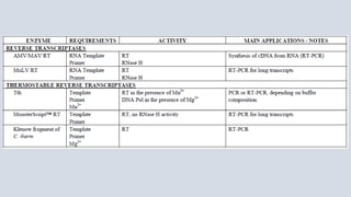
  • 7. • A reverse transcriptase (RT) is an enzyme used to generate complementary DNA (cDNA) from an RNA template, a process termed reverse transcription. REVERSE TRANSCRIPTASE FOR MOLECULAR BIOLOGY AMV/ MAV RT • The Avian Myeloblastosis Virus (AMV) Reverse Transcriptase is an RNA-directed DNA polymerase. This enzyme can synthesize a complementary DNA strand initiating from a primer using RNA (cDNA synthesis) or single- stranded DNA as a template. • AMV/MAV RT is widely used to copy total messenger RNAs using polydT or random primers. Applications: First- and second-strand synthesis of cDNA. Primer extensions and RNA sequencing.
  • 8. M-MuLV - Reverse Transcriptase • Moloney murine leukemia virus (M-MuLV) Reverse Transcriptase is a recombinant RNA-dependent DNA polymerase that uses single-stranded RNA as a template in the presence of a primer to synthesize a complementary DNA (cDNA) strand. • It can be used for the preparation of cDNA libraries, for first-strand synthesis in RT-PCR, or in dideoxy sequencing reactions in place of Klenow enzyme, for G+C–rich regions. • MuLV Reverse Transcriptase lacks endonuclease activity and has extremely low RNase H activity compared to AMV reverse transcriptase. Applications Reverse Transcriptase M-MuLV (from Moloney Murine Leukemia Virus) is used for: - In vitro synthesis of cDNA transcripts of specific RNA sequences - Preparation of cDNA libraries - Synthesis of first-strand cDNA for use in subsequent amplification reactions (RT-PCR) - Dideoxy-sequencing reactions in place of Klenow enzyme. Reverse Transcriptase M-MuLV will often synthesize through GC-rich regions where Klenow enzyme is hindered - Transcription of abundant, simple RNA target.
  • 10. • Phosphatase catalyses the cleavage of a phosphate (PO4-2) group from substrate by using a water molecule (hydrolytic cleavage). This reaction is not reversible. Phosphatase Schematic representation of hydrolytic cleavage of phosphate group (-PO4-2). • On the basis of their activity there are two types of phosphatase i.e acid phosphatase and alkaline phosphatase. In both forms the alkaline phosphatase are most common. Acid phosphatase: • It shows its optimal activity at pH between 3 and 6, e.g. a lysosomal enzyme that hydrolyze organic phosphates liberating one or more phosphate groups. They are found in prostatic epithelial cells, erythrocyte, prostatic tissue, spleen, kidney etc.
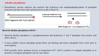
  • 11. • Homodimeric enzyme catalyzes the reactions like hydrolysis and transphosphophorylation of phosphate monoester and they show their optimal activity at pH of about 10. Alkaline phosphatase Bacterial alkaline phosphatase (BAP) – • Bacterial alkaline phosphate is a phosphomonoester that hydrolyzes 3’ and 5’ phosphate from nucleic acid (DNA/ RNA). • It more suitably removes phosphate group before end labeling and remove phosphate from vector prior to insert ligation. • BAP generally shows optimum activity at temperature 65°C. BAP is sensitive to inorganic phosphate so in presence of inorganic phosphates activity may reduce.
  • 12. Calf intestinal alkaline phosphatase (CIP) – It is isolated from calf intestine, which catalyzes the removal of phosphate group from 5’ end of DNA as well as RNA. This enzyme is highly used in gene cloning experiments, as to make a construct that could not undergo self- ligation. Hence after the treatment with CIP, without having a phosphate group at 5’ ends a vector cannot self ligate and recircularise. This step improves the efficiency of vector containing desired insert. Shrimp alkaline phosphatase (SAP) - Shrimp alkaline phosphatase is highly specific, heat labile phosphatase enzyme isolated from arctic shrimp (Pandalus borealis). It removes 5’ phosphate group from DNA, RNA, dNTPs and proteins. SAP has similar specificity as CIP but unlike CIP, it can be irreversibly inactivated by heat treatment at 65°C for 15mins. SAP is used for 5’ dephosphorylation during cloning experiments for various application as follows: Dephosphorylate 5’-phosphate group of DNA/RNA for subsequent labeling of the ends. To prevent self-ligation of the linearized plasmid. To prepare PCR product for sequencing. To inactivate remaining dNTPs from PCR product (for downstream sequencing appication).
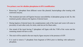
  • 13. Two primary uses for alkaline phosphatase in DNA modification: • Removing 5’ phosphate from different vector like plasmid, bacteriophage after treating with restriction enzyme. • This treatment prevents self ligation because unavailability of phosphate group at end. So, this treatment greatly enhances the ligation of desired insert. • During ligation of desired insert, the complementary ends of the insert and vector will come to proximity of each other (only for sticky ends but not for blunt ends). • One strand of the insert having 5’-phosphate will ligate with the 3’OH of the vector and the remaining strand will have a nick. • This nick will be sealed in the next step by ligase enzyme in the presence of ATP. • It is used to remove 5’ phosphate from fragment of DNA prior to labeling with radioactive phosphate.
  • 14. • The reversible phosphorylation of proteins, catalyzed by protein kinases and protein phosphatases, is a major mechanism for the regulation of many eukaryotic cellular processes, including metabolism, muscle contraction, pre-mRNA splicing and cell cycle control. • The eukaryotic protein phosphatases comprise several families of enzymes that catalyze the dephosphorylation of intracellular phosphoproteins. • The protein phosphatases responsible for dephosphorylation of serine and threonine residues in the cytoplasmic and nuclear compartments of eukaryotic cells. • Protein phosphatases originated from two ancestor genes, one serving as the prototype for the phosphotyrosine phosphatases family (the PTP family), and the other for phosphoserine/phosphothreonine phosphatases (the PPP and PPM family). Protein phosphatases
  • 15. T4 Polynucleotide kinase • T4 Polynucleotide Kinase catalyzes the transfer of the γ-phosphate from ATP to the 5´-hydroxyl terminus of double and single-stranded RNA and DNA, oligonucleotides or nucleoside 3´-monophosphates. • The enzyme is also capable of catalyzing the removal of 3´-phosphoryl groups from 3´-phosphoryl polynucleotides, deoxynucleoside 3´-monophosphates and deoxynucleoside 3´-diphosphates. • T4 PNK is widely used to end-label short oligonucleotide probes, DNA and RNA molecules.
  • 16. Protein Kinase and Phosphatase reactions
  • 17. PNK carries out two types of enzymatic activity: • Forward reaction: γ-phosphate is transferred from ATP to the 5' end of a polynucleotide (DNA or RNA). • Exchange reaction: target DNA or RNA having a 5' phosphate is incubated with an excess of ADP - where PNK transfers the phosphate from the nucleic acid to an ADP, forming ATP. • PNK then performs a forward reaction and transfer a phosphate from ATP to the target nucleic acid. Exchange reaction is used to label with radioactive phosphate group.
  • 18. • T4 Pnkp is a homotetramer of a 301-aa (33-kDa) polypeptide, which consists of an N-terminal kinase domain of the P-loop phosphotransferase superfamily and a C-terminal phosphatase domain of the DxD acylphosphatase superfamily. • The homotetramer is formed via pairs of phosphatase-phosphatase and kinase-kinase homodimer interfaces. • Phosphate transfer activity is optimum at 37°C, pH 7.6, Mg2+ and reducing reagents (DTT or β-mercaptoethanol), and 1 mM ATP and a 5:1 ratio of ATP over 5´-OH ends. T4 Polynucleotide kinase
  • 19. Applications • Labelling 5’-termini of DNA or RNA to be used as: - primers for DNA sequencing - primers for PCR - probes for hybridization - probes for transcript mapping - markers for gel electrophoresis • Addition of 5’-phosphates to oligonucleotides, PCR products, and DNA or RNA prior to ligation • Removal of 3´-phosphoryl groups • In addition to the kinase activity, T4 polynucleotide kinase also exhibits a 3´-phosphatase activity. • Optimum pH for 3’ phosphatase activity between 5 and 6, i.e. more acidic than for phosphate transfer activity. • Based on this property, protocols using T4 polynucleotide kinase as specific 3´-phosphatase have been developed (Cameron 1978). • The mutant polynucleotide kinase is very useful when the 3´-exonuclease activity must be avoided (e.g. 5´-32P terminal labeling of 3´-CMP in view of 3’ end-labeling of RNA species prior to fingerprinting or sequencing).
  • 21. Terminal transferase (TdT) is a template independent polymerase that catalyzes the addition of deoxynucleotides to the 3' hydroxyl terminus of DNA molecules. Protruding, recessed or blunt-ended double or single- stranded DNA molecules serve as a substrate for TdT. The 58.3 kDa enzyme does not have 5' or 3' exonuclease activity. The addition of Co2+ in the reacton makes tailing more efficient. Terminal nucleotidyl transferase (TdT) Applications Labeling of the 3′ ends of DNA with modified nucleotides (e.g., ddNTP, DIG-dUTP) Isolated from a recombinant source Supplied with 10X Reaction Buffer Tested for the absence of endonucleases and exonucleases
  • 22. Methyltransferases are a large group of enzymes that all methylate their substrates Class I Methyltransferases, all of which contain a Rossman fold for binding S-Adenosyl methionine (SAM). Class II methyltransferases contain a SET domain, which are exemplified by SET domain histone methyltransferases, Class III methyltransferases, which are membrane associated. Methyltransferases Methyltransferases can also used in protein methyltransferases, DNA/RNA methyltransferases, natural product methyltransferases, and non-SAM dependent methyltransferases.
  • 23. Methyltransferase can be classified in three groups: a) m6A-generates N6 methyladenosine, b) m4C-generates N4 methylcytosine, c) m5C-generatesN5 methylcytosine. m6A and m4C methyltransferase are primarily found in prokaryotes. Restriction enzyme EcoRI cleaves within the recognition sequence if the DNA is unmethylated. On methylation by methylases, the restriction enzyme EcoRI is inhibited from cleaving within the restriction site.