SlideShare a Scribd company logo
ERRORS OF
REFRACTIO
NMOHAMED ABDELZAHER MD, FRCS
Change in the direction of rays as they pass between media of different refractive
index.
Magnifying lens Minifying lens
• Diopter is the unit power of lenses
• Diopter = 1 / Focal length in meters
Real Image Virtual Image
• Diopter is the unit power of lenses
• Diopter = 1 / Focal length in meters
• A cylinder is specified by its
axis
• The axis of a cylindrical
lens is parallel to the
cylinder from which the lens
cut
• The power of a cylinder in
its axis meridian is zero.
• Maximum power is 90
degrees away from the
axis. This is known as the
power meridian.
• The image formed by the
power meridian is a focal
line parallel to the axis.
• There is no line focus
image formed by the axis
meridian, because the axis
• Carries both spherical & cylindrical correction
• Prism deviates light toward
its base, thus the image is
displaced toward its apex
• Prism Diopter =
Displacement of image for 1
cm at 1 m distance
Converging Diverging
Magnifying Minifying
Tow Prisms Base to
Base
Two Prisms Apex to
Apex
The instrument used for measurement of lenses & prisms power is the Lensmeter.
• Change in the refractive power of the eye to
maintain a clear focus on variable distances.
• For near vision, the ciliary muscle contracts,
reducing the zonular tension & allowing the
elastic capsule of the lens to contract,
causing a decrease in equatorial lens
diameter and an increase in the curvatures
of the anterior and posterior lens surfaces.
• Accommodation power
decreases with age (Presbyopia)
• Far Point (Punctum Remotum, PR): Position of an object when its image clearly
falls on the retina with no accommodation.
• Near Point (Punctum Proximum, PP): The nearest point clearly seen with maximum
accommodation
• Range of accommodation: The distance between far & near point
• Amplitude of Accommodation: The difference in Dioptric power between eye at rest
& eye at maximum accommodation
• Far Point (Punctum Remotum):
Emmetropic eye; at infinity
Myopic eye; anterior to the eye
Hyperopic eye; virtual point behind the
eye
• Near Point (Punctum Proximum):
Presbyopia is physiological recession of near point of accommodation
N’ Normal near point
N Near point in Presbyopia
• The amount of convergence measured in prism diopters per unit (diopter) change in
accommodation
• Normal Value: 3- 5 Prism Diopter / 1 D.
• Abnormalities in AC/A ratio causes strabismus
• Visual Axis: Line passing between Object of fixation & Fovea.
• Optic Axis: Line passing through the centre of refractive media of the eye.
• Angle Alpha : Angle between visual axis & optic axis (Normally +5 degrees)
• Nodal point of the eye: Point through which light passing undeviated
• Angle between visual axis & optic
axis (Normally +5 degrees)
• Normally, Visual axis cuts cornea
NASAL to the optic axis
• Parallel light rays
come into a focus
on the retina,
when
accommodation
is relaxed.
• No refractive
error is present.
• Parallel light rays come
into a focus anterior to the
retina, when
accommodation is
completely relaxed.
• AKA Near sightedness
• PR is in front of the eye,
(MR & M’ are conjugate
points)
A. Axial: Long axial length (Average AXL is 22 - 23 mm)
B. Refractive:
1. Index; Increased RI of lens nucleus e.g. nuclear cataract or decrease RI of lens
cortex e.g. DM
2. Curvature; increase curvature of cornea (KCN) or lens (Subluxation or
lenticonus)
1. Simple myopia
2. Degenerative (Progressive) myopia
3. Nocturnal myopia: Insufficient contrast, because of
dim illumination for an adequate accommodative
stimulus, the eye assumes the intermediate dark focus
accommodative position rather than focusing for infinity
4. Pseudo-myopia: Increase in ocular refractive power
due to overstimulation of the eye's accommodative
mechanism e.g. during exams or ciliary spasm
5. Induced (acquired) myopia: Result from exposure
to various pharmaceutical agents e.g. Phenothiazine,
active ocular inflammation (uveitis), variation in blood
sugar levels, nuclear sclerosis of the crystalline lens, or
other anomalous conditions. This myopia is often
temporary and reversible.
Nocturnal myopia
Causes of Night Myopia:
•Spherical aberration (The optical effect of the larger pupil decreases the depth
of focus)
•In dark adaptation the eye becomes more sensitive to
shorter wave lengths (Blue)
•Chromatic aberration: Shorter wavelengths come into focus in front of the
retina
•Ciliary spasm: the eye assumes the intermediate dark focus accommodative
position rather than focusing for infinity
1. Low Myopia: < 3D
2. Medium myopia: 3 -6
D
3. High myopia: > 6D
1. Congenital: Present since birth
2. Youth onset: < 20 years
3. Adult onset: > 20 years
Simple Myopia Progressive Myopia
Common Less Common
Starts at Puberty Starts at younger age
Usually less than 6 D Up to 25 D or more
Normal fundus Degenerative retinal changes
• Blurred far vision
• Good near vision
• Young age
• Frowning in trial to improve vision
• Defective night vision (Dilated pupil increases effect of aberrations)
• Retinal symptoms: musca, photopsia (In high myopia > 6 D)
• Peri-papillary myopic annulus or
temporal crescent
Of High Myopia
(Chorio-retinal degeneration)
• Tigroid (Tessellated) Fundus
• Caused by atrophy of the retinal
pigment epithelium
• Underlying streaks of normal
choroidal pigmentation are
visible
• Lacquer Cracks
Cracks in Bruchs
membrane
• Fuchs spots
Cracks in Bruchs
membrane allows
neovessels to proliferate
under the retina & causes
recurrent retinal
haemorrhage
• Posterior staphyloma
• Posterior protrusion of week
sclera lined with choroid
• Vitreous floaters
• Caused by vitreous
degeneration or posterior
vitreous detachment
• Peripheral Retinal Degeneration
• e.g. Lattice degeneration
• Predisopse to rhegmatogenous RD
The myopic retina
Retina
• Nuclear
cataract
Of High Myopia
• Open Angle
Glaucoma
• Rhegmatogenous Retinal Detachment,
caused by peripheral retinal degeneration
Of High Myopia
• Macular
Hole
• Squin
t
Of High Myopia
In high myopia, the fovea lies NASAL to the optic axis. So, the visual axis
crosses temporal to the optic axis at the cornea. (-ve angle alpha)
• Squin
t
Of High Myopia
Right exotropia when fixating at a 6 metres
objection (A and B). Eyes straight with
myopic
correction (C). Exotropia disappeared when
the eyes looked at a near object (D). Right
exotropia
worsened when the eyes looked at an
object further than 6 metres (E).
Concave
Lenses
Concave
Lenses
Indications:
A. Cosmetic
B. Anisometropia
Advantages:
A. Less minification of image, 10%
(nearer to the nodal point of the eye)
B. Less aberration of lens periphery
(wider field)
Prescribe
the
lowest
correctio
n that
gives the
best
vision
Indications:
A. Cosmetic
B. Anisometropia
• A corneal flap is fashioned by an
automated keratome
• Excimer laser is used to ablate the central
corneal stromal bed, thus reducing the
curvature of the central part of the cornea
• The flap is re-positioned
• Can correct up to - 12 D according to
corneal thickness & curvature
• A corneal flap is
fashioned by an Femto
laser
• Excimer laser is used to
ablate the corneal
stromal bed, thus
reducing the curvature
of the central part of the
cornea
• The flap is re-
positioned
LASIK
• A lenticule of corneal storm
is fashioned using the femto
laser
• The lenticule is extracted
through a curvilinear small
inferior corneal incision
SMILE
Indications:
A. Thin corneas
B. Low refractive errors
• Corneal epithelium is removed
either mechanically or with alcohol
• Excimer laser is used to ablate the
corneal stromal bed, thus reducing
the curvature of the central part of
the cornea
• No flap
• Can correct up to - 4 D according to
corneal thickness & curvature
Indications:
A. Thin corneas
B. High refractive errors
Indications:
A. Thin corneas
B. High refractive errors
C. Old age > 40 years
Disadvantages:
A. Loss of accommodation
B. High incidence of post-operative
complications e.g. RD
• Radial Partial thickness cuts in
corneal stroma
• Increases the curvature of corneal
periphery
• Obsolete
• Parallel light rays come
into a focus posterior to
the retina, when
accommodation is
completely relaxed.
• AKA Far sightedness
• PR is a virtual point
behind the eye, (MR & M’
are conjugate points)
A. Axial: Short axial length (Average AXL is 22 - 23 mm)
B. Refractive:
1. Index; Decreased RI of lens e.g. cortical cataract
2. Curvature; decrease curvature of cornea (Cornea Plana)
3. Aphakia
4. Posterior Dislocation of the lens
1. Total (Ht): Amount of hyperopia measured under the
effect of Atropine
2. Manifest (Hm): Amount of hyperopia measured
without Atropine
3. Latent (Hl): Amount of hyperopia caused by CB tone
(1D)
4. Facultative (Hf): Amount of hyperopia corrected by
Accommodation
5. Absolute (Ha): Amount of hyperopia NOT corrected
by accommodation (to be corrected optically)
• Ht = Hm + Hl
• Hm = Hf + Ha
• In young age with stronger accommodation, most, if not
all Hm is Hf
• Hf decreases with age as accommodation gets weaker
• None, in young age with small error because of strong accommodation
• Poor near vision
• Better far vision (later becomes poor, virtual PR)
• Ocular asthenia, because of continuous accommodation
• Early presbyopia
• Shallow
AC
Small eye
• Crowded Optic disc
(Pseudo swelling)
• with tortuous retinal
vessels
Small eye
• Narrow angle Glaucoma
• Squin
t
In high hyperopia, the fovea lies more Temporal to the optic axis than normal.
So, the visual axis crosses nasal to the optic axis at the cornea. (large +ve
angle alpha, > 5 degrees)
• Squin
t
Due to excessive accommodation
Convex
Lenses
Convex
Lenses
Indications:
A. Cosmetic
B. Anisometropia
Advantages:
A. Less magnification of image (nearer to
the nodal point of the eye)
B. Less aberration of lens periphery
(wider field)
Prescribe the
highest
correction
that gives
the best
vision
Indications:
A. Cosmetic
B. Anisometropia
• A corneal flap is fashioned by an
automated keratome
• Excimer laser is used to ablate the
periphery of corneal stromal bed, thus
increasing the curvature of the central
part of the cornea
• The flap is re-positioned
• Can correct up to + 6 D according to
corneal thickness & curvature
• A corneal flap is
fashioned by an Femto
laser
• Excimer laser is used to
ablate the corneal
stromal bed, thus
reducing the curvature
of the central part of the
cornea
• The flap is re-
positioned
LASIK
Indications:
A. Thin corneas
B. Low refractive errors
• Corneal epithelium is removed
either mechanically or with alcohol
• Excimer laser is used to ablate the
periphery of corneal stromal bed,
thus increasing the curvature of the
central part of the cornea
• No flap
Indications:
A. Thin corneas
B. High refractive errors
Might not be
applicable in some
cases with very
shallow AC & narrow
anterior segment
Indications:
A. Thin corneas
B. High refractive errors
C. Old age > 40 years
Disadvantages:
A. Loss of accommodation
Might not be a better
option than Phakic
IOL, especially in
cases with narrow AC
angle
Indications:
• Low refractive errors
Principle:
CTK delivers controlled-released
radio-frequency current (within
peripheral corneal stroma, raising the
temperature of the peripheral
collagen lamellae to the 65°C
resulting in controlled shrinkage of
the peripheral collagen lamellae &
subsequent central corneal
steepening.
• Parallel light rays do not
come into a focus but
rather a line.
A. Cornea:
1. Keratoconus
2. Corneal scars
3. Corneal stitches
4. Pressure over cornea
B. Lenticular:
Incipient cataract
Lenticonus
Lens subluxation
C. Retinal: e.g.
staphyloma in high
myopia, because of
posterior pole obliquity
• With (WTR)
• Against (ATR)
• Oblique
• Simple
• Compound
• Mixed
The two Principal Meridians (of highest & lowest power) are at right angle to
each others, and change of power from one meridian to the other is gradual &
regular
• Vertical meridian is more curved than
Horizontal meridian
• More common
• Vertical meridian is less curved than
Horizontal meridian
• Less common
• The two principal meridians at at right angle
to each other but they are not at 90 & 180
degrees
• Less common
• One principal meridian is emmetropic & the
other is ametropic, with accommodation
relaxed
• One principal meridian is Myopic & the
other is emmetropic
• One principal meridian is Hyperopic & the
other is emmetropic
• Both principal meridians are ametropic, with
same sign, with accommodation relaxed
• Both Principal meridians are myopic
• Both Principal meridians are hyperopic
• Both principal meridians are ametropic, with
opposite sign, with accommodation relaxed
• One Principal meridians myopic, the
other is hyperopic

More Related Content

PPTX
Introduction to binocular single vision (BSV)
PPTX
AC/A
PPTX
Subjective refraction
PPTX
Maddox rod n wing
PPTX
Synoptophore
PPTX
Maddox rod and double maddox rod
PPTX
Amblyopia
PPTX
Convergence insufficiency
Introduction to binocular single vision (BSV)
AC/A
Subjective refraction
Maddox rod n wing
Synoptophore
Maddox rod and double maddox rod
Amblyopia
Convergence insufficiency

What's hot (20)

PPTX
Synaptophore in ophthalmology
PPTX
Dissociated vertical deviation
PPTX
Synaptophore
PPTX
Dispensing glasses PPT for O.A. 2nd year
PPT
Acaratio
PPTX
Bandage Contact Lens
PPTX
Anisometropia
PPTX
IOL Master
PPT
Slit Lamp Illumination Techniques
PPT
Corneal topography
PPTX
Microtropia
PPTX
Dynamic retinoscopy
PPTX
Retinal correspondence
PPT
Frame adjustment and quality checking
PPTX
Amsler grid chart
PPTX
Prism in ophthalmology
PPT
Measuring interpupillary distance
PPTX
Accommodative Facility Testing.pptx
PPTX
divergent squint.pptx
PPTX
Active Vision Therapy in Management of Amblyopia (healthkura.com)
Synaptophore in ophthalmology
Dissociated vertical deviation
Synaptophore
Dispensing glasses PPT for O.A. 2nd year
Acaratio
Bandage Contact Lens
Anisometropia
IOL Master
Slit Lamp Illumination Techniques
Corneal topography
Microtropia
Dynamic retinoscopy
Retinal correspondence
Frame adjustment and quality checking
Amsler grid chart
Prism in ophthalmology
Measuring interpupillary distance
Accommodative Facility Testing.pptx
divergent squint.pptx
Active Vision Therapy in Management of Amblyopia (healthkura.com)
Ad

Similar to Errors 1 (20)

PPTX
High myopia and management
PPT
Refractive errors
PPT
Refractiveerrors 130309052054-phpapp01
PPT
Refractive errors (Dr. Ajinkya wankhede). presentation
PPTX
MYOPIA.pptx
PPTX
0HY5M94d7TShdydhnsuamalakga न्सकाकाfF8K5372.pptx
PPTX
Errors of refraction
PPTX
By pd aphakia & pseudophakia
PPTX
Accommodation by the eye.pptx
PPT
Retinoscopy and its principles
PPTX
Refractive errors of eye ophthalmology astigmatism hypermetropia myopia medic...
PPT
Reffraction myopia by Dr Abdul Basir safi eye surgeon from Afghanistan
PPTX
Accommodation and convergence
PPTX
Optics & Refraction-Dr.PrabhatDevkota.pptx
PPTX
Emmetropia and refractive errors of eyes
PPTX
Emmetropia and refractive errors of eyes
PPT
Error of Refraction
PPT
Accommodation reema
PPT
Accommodation
High myopia and management
Refractive errors
Refractiveerrors 130309052054-phpapp01
Refractive errors (Dr. Ajinkya wankhede). presentation
MYOPIA.pptx
0HY5M94d7TShdydhnsuamalakga न्सकाकाfF8K5372.pptx
Errors of refraction
By pd aphakia & pseudophakia
Accommodation by the eye.pptx
Retinoscopy and its principles
Refractive errors of eye ophthalmology astigmatism hypermetropia myopia medic...
Reffraction myopia by Dr Abdul Basir safi eye surgeon from Afghanistan
Accommodation and convergence
Optics & Refraction-Dr.PrabhatDevkota.pptx
Emmetropia and refractive errors of eyes
Error of Refraction
Accommodation reema
Accommodation
Ad

More from faculty of medicine -benha university (20)

Recently uploaded (20)

PDF
3rd Neelam Sanjeevareddy Memorial Lecture.pdf
PDF
Module 4: Burden of Disease Tutorial Slides S2 2025
PDF
2.FourierTransform-ShortQuestionswithAnswers.pdf
PPTX
Cell Types and Its function , kingdom of life
PPTX
Introduction-to-Literarature-and-Literary-Studies-week-Prelim-coverage.pptx
PPTX
202450812 BayCHI UCSC-SV 20250812 v17.pptx
PPTX
Cell Structure & Organelles in detailed.
PDF
Computing-Curriculum for Schools in Ghana
PDF
Anesthesia in Laparoscopic Surgery in India
PDF
Weekly quiz Compilation Jan -July 25.pdf
PDF
01-Introduction-to-Information-Management.pdf
PDF
The Lost Whites of Pakistan by Jahanzaib Mughal.pdf
PDF
RTP_AR_KS1_Tutor's Guide_English [FOR REPRODUCTION].pdf
PDF
A systematic review of self-coping strategies used by university students to ...
PDF
Chapter 2 Heredity, Prenatal Development, and Birth.pdf
PDF
Classroom Observation Tools for Teachers
PDF
Black Hat USA 2025 - Micro ICS Summit - ICS/OT Threat Landscape
PPTX
master seminar digital applications in india
PDF
RMMM.pdf make it easy to upload and study
PPTX
1st Inaugural Professorial Lecture held on 19th February 2020 (Governance and...
3rd Neelam Sanjeevareddy Memorial Lecture.pdf
Module 4: Burden of Disease Tutorial Slides S2 2025
2.FourierTransform-ShortQuestionswithAnswers.pdf
Cell Types and Its function , kingdom of life
Introduction-to-Literarature-and-Literary-Studies-week-Prelim-coverage.pptx
202450812 BayCHI UCSC-SV 20250812 v17.pptx
Cell Structure & Organelles in detailed.
Computing-Curriculum for Schools in Ghana
Anesthesia in Laparoscopic Surgery in India
Weekly quiz Compilation Jan -July 25.pdf
01-Introduction-to-Information-Management.pdf
The Lost Whites of Pakistan by Jahanzaib Mughal.pdf
RTP_AR_KS1_Tutor's Guide_English [FOR REPRODUCTION].pdf
A systematic review of self-coping strategies used by university students to ...
Chapter 2 Heredity, Prenatal Development, and Birth.pdf
Classroom Observation Tools for Teachers
Black Hat USA 2025 - Micro ICS Summit - ICS/OT Threat Landscape
master seminar digital applications in india
RMMM.pdf make it easy to upload and study
1st Inaugural Professorial Lecture held on 19th February 2020 (Governance and...

Errors 1

  • 2. Change in the direction of rays as they pass between media of different refractive index.
  • 3. Magnifying lens Minifying lens • Diopter is the unit power of lenses • Diopter = 1 / Focal length in meters Real Image Virtual Image
  • 4. • Diopter is the unit power of lenses • Diopter = 1 / Focal length in meters
  • 5. • A cylinder is specified by its axis • The axis of a cylindrical lens is parallel to the cylinder from which the lens cut • The power of a cylinder in its axis meridian is zero. • Maximum power is 90 degrees away from the axis. This is known as the power meridian. • The image formed by the power meridian is a focal line parallel to the axis. • There is no line focus image formed by the axis meridian, because the axis
  • 6. • Carries both spherical & cylindrical correction
  • 7. • Prism deviates light toward its base, thus the image is displaced toward its apex • Prism Diopter = Displacement of image for 1 cm at 1 m distance
  • 8. Converging Diverging Magnifying Minifying Tow Prisms Base to Base Two Prisms Apex to Apex
  • 9. The instrument used for measurement of lenses & prisms power is the Lensmeter.
  • 10. • Change in the refractive power of the eye to maintain a clear focus on variable distances. • For near vision, the ciliary muscle contracts, reducing the zonular tension & allowing the elastic capsule of the lens to contract, causing a decrease in equatorial lens diameter and an increase in the curvatures of the anterior and posterior lens surfaces.
  • 11. • Accommodation power decreases with age (Presbyopia)
  • 12. • Far Point (Punctum Remotum, PR): Position of an object when its image clearly falls on the retina with no accommodation. • Near Point (Punctum Proximum, PP): The nearest point clearly seen with maximum accommodation • Range of accommodation: The distance between far & near point • Amplitude of Accommodation: The difference in Dioptric power between eye at rest & eye at maximum accommodation
  • 13. • Far Point (Punctum Remotum): Emmetropic eye; at infinity Myopic eye; anterior to the eye Hyperopic eye; virtual point behind the eye
  • 14. • Near Point (Punctum Proximum): Presbyopia is physiological recession of near point of accommodation N’ Normal near point N Near point in Presbyopia
  • 15. • The amount of convergence measured in prism diopters per unit (diopter) change in accommodation • Normal Value: 3- 5 Prism Diopter / 1 D. • Abnormalities in AC/A ratio causes strabismus
  • 16. • Visual Axis: Line passing between Object of fixation & Fovea. • Optic Axis: Line passing through the centre of refractive media of the eye. • Angle Alpha : Angle between visual axis & optic axis (Normally +5 degrees) • Nodal point of the eye: Point through which light passing undeviated
  • 17. • Angle between visual axis & optic axis (Normally +5 degrees) • Normally, Visual axis cuts cornea NASAL to the optic axis
  • 18. • Parallel light rays come into a focus on the retina, when accommodation is relaxed. • No refractive error is present.
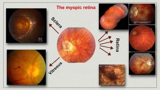
  • 19. • Parallel light rays come into a focus anterior to the retina, when accommodation is completely relaxed. • AKA Near sightedness • PR is in front of the eye, (MR & M’ are conjugate points)
  • 20. A. Axial: Long axial length (Average AXL is 22 - 23 mm) B. Refractive: 1. Index; Increased RI of lens nucleus e.g. nuclear cataract or decrease RI of lens cortex e.g. DM 2. Curvature; increase curvature of cornea (KCN) or lens (Subluxation or lenticonus)
  • 21. 1. Simple myopia 2. Degenerative (Progressive) myopia 3. Nocturnal myopia: Insufficient contrast, because of dim illumination for an adequate accommodative stimulus, the eye assumes the intermediate dark focus accommodative position rather than focusing for infinity 4. Pseudo-myopia: Increase in ocular refractive power due to overstimulation of the eye's accommodative mechanism e.g. during exams or ciliary spasm 5. Induced (acquired) myopia: Result from exposure to various pharmaceutical agents e.g. Phenothiazine, active ocular inflammation (uveitis), variation in blood sugar levels, nuclear sclerosis of the crystalline lens, or other anomalous conditions. This myopia is often temporary and reversible.
  • 22. Nocturnal myopia Causes of Night Myopia: •Spherical aberration (The optical effect of the larger pupil decreases the depth of focus) •In dark adaptation the eye becomes more sensitive to shorter wave lengths (Blue) •Chromatic aberration: Shorter wavelengths come into focus in front of the retina •Ciliary spasm: the eye assumes the intermediate dark focus accommodative position rather than focusing for infinity
  • 23. 1. Low Myopia: < 3D 2. Medium myopia: 3 -6 D 3. High myopia: > 6D 1. Congenital: Present since birth 2. Youth onset: < 20 years 3. Adult onset: > 20 years
  • 24. Simple Myopia Progressive Myopia Common Less Common Starts at Puberty Starts at younger age Usually less than 6 D Up to 25 D or more Normal fundus Degenerative retinal changes
  • 25. • Blurred far vision • Good near vision • Young age • Frowning in trial to improve vision • Defective night vision (Dilated pupil increases effect of aberrations) • Retinal symptoms: musca, photopsia (In high myopia > 6 D)
  • 26. • Peri-papillary myopic annulus or temporal crescent Of High Myopia (Chorio-retinal degeneration)
  • 27. • Tigroid (Tessellated) Fundus • Caused by atrophy of the retinal pigment epithelium • Underlying streaks of normal choroidal pigmentation are visible
  • 28. • Lacquer Cracks Cracks in Bruchs membrane
  • 29. • Fuchs spots Cracks in Bruchs membrane allows neovessels to proliferate under the retina & causes recurrent retinal haemorrhage
  • 30. • Posterior staphyloma • Posterior protrusion of week sclera lined with choroid
  • 31. • Vitreous floaters • Caused by vitreous degeneration or posterior vitreous detachment
  • 32. • Peripheral Retinal Degeneration • e.g. Lattice degeneration • Predisopse to rhegmatogenous RD
  • 34. • Nuclear cataract Of High Myopia • Open Angle Glaucoma
  • 35. • Rhegmatogenous Retinal Detachment, caused by peripheral retinal degeneration Of High Myopia • Macular Hole
  • 36. • Squin t Of High Myopia In high myopia, the fovea lies NASAL to the optic axis. So, the visual axis crosses temporal to the optic axis at the cornea. (-ve angle alpha)
  • 37. • Squin t Of High Myopia Right exotropia when fixating at a 6 metres objection (A and B). Eyes straight with myopic correction (C). Exotropia disappeared when the eyes looked at a near object (D). Right exotropia worsened when the eyes looked at an object further than 6 metres (E).
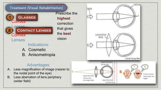
  • 38. Concave Lenses Concave Lenses Indications: A. Cosmetic B. Anisometropia Advantages: A. Less minification of image, 10% (nearer to the nodal point of the eye) B. Less aberration of lens periphery (wider field) Prescribe the lowest correctio n that gives the best vision
  • 39. Indications: A. Cosmetic B. Anisometropia • A corneal flap is fashioned by an automated keratome • Excimer laser is used to ablate the central corneal stromal bed, thus reducing the curvature of the central part of the cornea • The flap is re-positioned • Can correct up to - 12 D according to corneal thickness & curvature
  • 40. • A corneal flap is fashioned by an Femto laser • Excimer laser is used to ablate the corneal stromal bed, thus reducing the curvature of the central part of the cornea • The flap is re- positioned LASIK
  • 41. • A lenticule of corneal storm is fashioned using the femto laser • The lenticule is extracted through a curvilinear small inferior corneal incision SMILE
  • 42. Indications: A. Thin corneas B. Low refractive errors • Corneal epithelium is removed either mechanically or with alcohol • Excimer laser is used to ablate the corneal stromal bed, thus reducing the curvature of the central part of the cornea • No flap • Can correct up to - 4 D according to corneal thickness & curvature
  • 43. Indications: A. Thin corneas B. High refractive errors
  • 44. Indications: A. Thin corneas B. High refractive errors C. Old age > 40 years Disadvantages: A. Loss of accommodation B. High incidence of post-operative complications e.g. RD
  • 45. • Radial Partial thickness cuts in corneal stroma • Increases the curvature of corneal periphery • Obsolete
  • 46. • Parallel light rays come into a focus posterior to the retina, when accommodation is completely relaxed. • AKA Far sightedness • PR is a virtual point behind the eye, (MR & M’ are conjugate points)
  • 47. A. Axial: Short axial length (Average AXL is 22 - 23 mm) B. Refractive: 1. Index; Decreased RI of lens e.g. cortical cataract 2. Curvature; decrease curvature of cornea (Cornea Plana) 3. Aphakia 4. Posterior Dislocation of the lens
  • 48. 1. Total (Ht): Amount of hyperopia measured under the effect of Atropine 2. Manifest (Hm): Amount of hyperopia measured without Atropine 3. Latent (Hl): Amount of hyperopia caused by CB tone (1D) 4. Facultative (Hf): Amount of hyperopia corrected by Accommodation 5. Absolute (Ha): Amount of hyperopia NOT corrected by accommodation (to be corrected optically) • Ht = Hm + Hl • Hm = Hf + Ha • In young age with stronger accommodation, most, if not all Hm is Hf • Hf decreases with age as accommodation gets weaker
  • 49. • None, in young age with small error because of strong accommodation • Poor near vision • Better far vision (later becomes poor, virtual PR) • Ocular asthenia, because of continuous accommodation • Early presbyopia
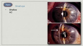
  • 51. • Crowded Optic disc (Pseudo swelling) • with tortuous retinal vessels Small eye
  • 52. • Narrow angle Glaucoma
  • 53. • Squin t In high hyperopia, the fovea lies more Temporal to the optic axis than normal. So, the visual axis crosses nasal to the optic axis at the cornea. (large +ve angle alpha, > 5 degrees)
  • 54. • Squin t Due to excessive accommodation
  • 55. Convex Lenses Convex Lenses Indications: A. Cosmetic B. Anisometropia Advantages: A. Less magnification of image (nearer to the nodal point of the eye) B. Less aberration of lens periphery (wider field) Prescribe the highest correction that gives the best vision
  • 56. Indications: A. Cosmetic B. Anisometropia • A corneal flap is fashioned by an automated keratome • Excimer laser is used to ablate the periphery of corneal stromal bed, thus increasing the curvature of the central part of the cornea • The flap is re-positioned • Can correct up to + 6 D according to corneal thickness & curvature
  • 57. • A corneal flap is fashioned by an Femto laser • Excimer laser is used to ablate the corneal stromal bed, thus reducing the curvature of the central part of the cornea • The flap is re- positioned LASIK
  • 58. Indications: A. Thin corneas B. Low refractive errors • Corneal epithelium is removed either mechanically or with alcohol • Excimer laser is used to ablate the periphery of corneal stromal bed, thus increasing the curvature of the central part of the cornea • No flap
  • 59. Indications: A. Thin corneas B. High refractive errors Might not be applicable in some cases with very shallow AC & narrow anterior segment
  • 60. Indications: A. Thin corneas B. High refractive errors C. Old age > 40 years Disadvantages: A. Loss of accommodation Might not be a better option than Phakic IOL, especially in cases with narrow AC angle
  • 61. Indications: • Low refractive errors Principle: CTK delivers controlled-released radio-frequency current (within peripheral corneal stroma, raising the temperature of the peripheral collagen lamellae to the 65°C resulting in controlled shrinkage of the peripheral collagen lamellae & subsequent central corneal steepening.
  • 62. • Parallel light rays do not come into a focus but rather a line.
  • 63. A. Cornea: 1. Keratoconus 2. Corneal scars 3. Corneal stitches 4. Pressure over cornea B. Lenticular: Incipient cataract Lenticonus Lens subluxation C. Retinal: e.g. staphyloma in high myopia, because of posterior pole obliquity
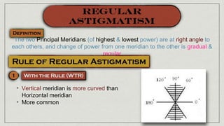
  • 64. • With (WTR) • Against (ATR) • Oblique • Simple • Compound • Mixed
  • 65. The two Principal Meridians (of highest & lowest power) are at right angle to each others, and change of power from one meridian to the other is gradual & regular • Vertical meridian is more curved than Horizontal meridian • More common
  • 66. • Vertical meridian is less curved than Horizontal meridian • Less common • The two principal meridians at at right angle to each other but they are not at 90 & 180 degrees • Less common
  • 67. • One principal meridian is emmetropic & the other is ametropic, with accommodation relaxed • One principal meridian is Myopic & the other is emmetropic • One principal meridian is Hyperopic & the other is emmetropic
  • 68. • Both principal meridians are ametropic, with same sign, with accommodation relaxed • Both Principal meridians are myopic • Both Principal meridians are hyperopic
  • 69. • Both principal meridians are ametropic, with opposite sign, with accommodation relaxed • One Principal meridians myopic, the other is hyperopic