SlideShare a Scribd company logo
Genus Streptococcus
Dr Ravi Kant Agrawal, MVSc, PhD
Senior Scientist (Veterinary Microbiology)
Food Microbiology Laboratory
Division of Livestock Products Technology
ICAR-Indian Veterinary Research Institute
Izatnagar 243122 (UP) India
Morphology
 Streptococcus is  a genus of Gram-positive cocci  or 
spherical  bacteria,  that  belongs  to  the 
family Streptococcaceae, within the order Lactobacillales 
(lactic acid bacteria), in the phylum Firmicutes.
 Gram positive spherical or oval cocci arranged in chains 
or pairs.
 Chain formation is due to the cocci dividing in one plane 
only  and  the  daughter  cells  failing  to  separate 
completely
 The  name  streptococci  was  given  in  1877  by  Viennese 
surgeon Albert Theodor Billroth (1829–1894).
 Streptos: easily twisted or coiled; coccus: berry)
 Individual coccus is 0.5-1.0 μm in diameter.
 They are non-motile and non-spore forming.
 Nutritionally fastidious
 Streptococci are catalase negative
 Facultative  anaerobes  but  don’t  use  oxygen 
metabolically (create lactic acid)
 Longest  chain  is  produced  by  Streptococcus salivarius
(Commensal)
 Chains are longer in liquid than in solid media 
 Some strains have capsule composed of  hyaluronic acid 
(non-immunogenic).
Habitat
Normal  flora  of  upper 
respiratory  tract  in  animals 
and human – Commensals and 
Saprophytes  of  decaying 
matter
Some  of  them  may  be 
pathogens  e.g.  Streptococcus
pyogenes causing  pyogenic 
infections with a tendency to 
spread  unlike  staphylococcal 
infections
It  produces  non-suppurative 
lesions, acute rheumatic fever 
and glomerulonephritis which 
occur as sequelae to infection 
Classification
 Initial  classification  based  on  hemolysis  on 
sheep blood agar plates: α (partial, reduction), β 
(complete), and γ (none).
 The cell wall structure of group A streptococci is 
among the most studied of any bacteria.
 The cell wall is composed of repeating units of  
N-acetylglucosamine and N-acetylmuramic acid, 
the standard peptidoglycan. 
 Rebecca Lancefield  (1895-1981) :     in 1922  while 
working  for  her  PhD  thesis,  Eighteen  group-
specific  antigens  were  established  on  basis  C 
Carbohydrate.
 1930’s:  Lancefield  defines  cell  wall  antigen 
groups
 Concentrated on virulent, β-hemolytic species
 Sherman: pyogenic, viridans, enterococci, lactic
 1980’s: Enterococci get own genus
 Lactic  acid  Streptococci  (Lancefield  N):  New 
genus Lactococcus
 Nutritionally  variant  Streptococci  (require 
pyridoxal): Abiotrophia and Granulicatella
Rebecca Craighill
Lancefield (1895-1981)
Classification of Streptococci
• Brown`s classification
• Lancefield grouping
• Griffith typing
Brown`s classification Lancefield grouping
V
Hemolysis
Hemolysis
Example –
Streptococcus viridans (Viridans = green)
Streptococcus pneumoniae
Example – enterococcus group
CLASSIFICATION:
StreptococciAerobes & facultative
anaerobes
Obligate anaerobes
Eg: PeptostreptococciBeta haemolytic
Gamma haemolytic
Eg: Enterococcus group
Alpha haemolytic
Eg: Viridans streptococci
20 Lancefield groups
(ABCDEFGHKLMNOPQRSTUV)80 Griffith types
(1,2,3,etc. up to 80)
02 requirement
Haemolysis
Serological Grouping (C carbohydrate antigen)
Group A- Streptococcus pyogens
Serological typing (M Protein)
Serology: Lancefield Classification
• Streptococci classified into many groups from A-K & H-V
• One or more species per group
• Classification based on C- carbohydrate antigen of cell wall
– Groupable streptococci
• A, B and D (more frequent)
• C, G and F (Less frequent)
– Non-groupable streptococci
• S. pneumoniae (pneumonia)
• viridans streptococci
– e.g. S. mutans
– Causing dental carries
Streptococci
Group A
S. pyogenes
Group B
S. agalactiae
Group C
S. equisimitis
Group D
Enterococcus
Lanciefield classification
Other groups
(E-U)
Group A Streptococci (GAS)
S. pyogenes: Cultural characteristics
• Aerobes and facultative anaerobes
• Optimum temperature: 37C
• Needs  enrichment  with  blood  or  serum  -  Growth 
occurs  only  in  media  containing  fermentable 
carbohydrates or enriched with blood or serum
i. Blood agar:
Small  (0.5-1.0  mm),  circular,  semi-transparent 
colonies 
Produce wide zone of β- hemolysis
Growth and hemolysis are promoted by 5-10% CO2
Virulent strains, on fresh isolation form lesions, 
produce a ‘matt’ (finely granular) colony while 
avirulent strains form ‘glossy’ colonies
Mucoid colonies are formed by strains that produce 
large capsules
ii.  Liquid media: 
Glucose or serum broth
Growth  occurs  as  a  granular  turbidity  with  a 
powdery deposit
No pellicle is formed
CULTURE:
Media used:
1. Non selective media:- Sheep blood agar
2. Selective media:-   Crystal violet blood agar
                              PNF medium
(Horse blood agar containing polymyxin B
sulphate, neomycin sulphate, and fusidic
acid inhibited the growth of Staph. aureus, Ps.
pyocyanea, Proteus mirabilis, E. coli, and
Klebsiella pneumoniae but allowed good
growth of, and haemolysis by, Str. pyogenes)
Biochemical reactions
 Catalase negative
 Bile insoluble
 Ferments  sugars  producing  acid  but  no  gas, 
faliure to ferment ribose
 PYR  test  positive:  Hydrolyse  pyrrolidonyl-beta-
napthylamide (PYR) due to presence of peptidase, 
the resulting napthylamide produces a red colour 
upon  the  addition  of  0.01%  cinnamaldehyde 
reagent
Bile insoluble
Positive Negative PYR test  Catalase
Resistance
 S. pyogenes is  a  delicate  organism,
easily destroyed by heat (54C for 30 min)
 Sensitive to bacitracin
 Has developed less resistance to drugs
 Dies in a few days in culture, unless stored at a 
low  temperature  (4C)  preferably  in 
Robertson’s cooked meat medium
 Rapidly  inactivated  by  antiseptics  and  many 
antibiotics
Fig. Zone of inhibition
shown by S. pyogenes
Antigenic structure
 Capsular hyaluronic acid: 
• Non  antigenic  as  hyaluronic  acid  is  identical  to  that  found  in 
human  connective  tissue  and  hence  bacteria  can  disguise 
themselves with an immunological self substance
• Has  weak  anti-phagocytic  activity  but  protects  streptococci 
against immunological attacks
Antigenic structure
A. Cell wall: 
1. Outer  layer:  Protein  and 
lipoteichoic acid
2. Middle  layer:  Group  specific 
carbohydrate
3. Inner  layer:  Peptidoglycan 
(mucoprotein)
Responsible  for  cell  wall 
rigidity
Enhances  non-specific 
resistance  (pyrogenic  and 
thrombolytic activity)
B. Group specific C-carbohydrates
Serological  grouping  of 
streptococci  is  done  on  its 
basis
Divided  into  20  Lancefield 
groups (A to V) except I and J 
on  the  basis  of  group  specific 
carbohydrates.
All streptococci except viridans 
(α-hemolytic)  group  have  a 
layer of carbohydrate
C. Proteins
Present  in  outermost 
layer
Produces  surface 
protein antigens (F, M, T 
and R) 
Useful  in  serological 
typing of S. pyogenes
M protein: Most virulent; 
heat and acid stable, but 
susceptible  to  tryptic 
digestion.  80  types 
identified.  Griffith 
typing.
T  protein:  Not  virulent, 
acid  labile,  trypsin 
resistant. 
R protein: Not virulent
Antigenic structure
Antigenic structure
F-protein
 Recognizes  host  fibronectin,  a  matrix  protein  that  is  present  in 
eukaryotic  cells.    Hence  helps  in  attachment  together  with 
lipoteichoic acid and M protein
M protein 
 Most antigenic
 Covered with lipoteichoic acid that enable the organism to attach to 
epithelial cell
 M protein is heat and acid stable but susceptible to tryptic digestion
 On the basis of antigenic difference in M protein, S. pyogenes can be 
divided into about 100 types
 The most distal part of M protein shows extensive variability among 
strains  hence  individual  may  suffer  from  recurrent  S. pyogenes
infections with strains expressing different versions of M protein.
 Acts as a virulence factor by inhibiting phagocytosis
T-Protein
• Common to many M- types 
• Not associated with virulence and is not a protective antigen
• It is strongly antigenic
R protein
• Non-type-specific and is associated with M- proteins of types 2,3 28 
and 48 known as M-associated protein (MAP)
• Not associated to virulence and not a protective antigen
• Strongly antigenic
Antigenic Structure
D. Pili (Fimbriae)
Hair like and project from capsule
Consist partly of M-protein 
Covered with lipoteichoic acid 
Important in the attachment of streptococci to epithelial cells
Virulence Factors: Toxins and enzymes
Toxins
1. Hemolysins 
(Streptolysin O and S)
2. Erythrogenic toxin 
(Pyrogenic Exotoxin)
Enzymes:
1. Streptokinase 
(Fibrinolysin)
2. Deoxyribonucleases 
(Streptodornase, 
DNAase)
3. Hyaluronidase 
4.NADase
5.Serum opacity factor
Cytolytic toxins and other exo-enzymes produced by
Streptococcus pyogenes
Hemolysins (Streptolysins)
 Produce complete disruption of RBC
Contribute to tissue invasion and destruction
There are two types of Streptolysins
Streptolysin O: sensitive to oxygensensitive to oxygen
Streptolysin S: insensitive to oxygeninsensitive to oxygen
Erythrogenic toxin
• Also known as pyrogenic exotoxin/ Dick/ 
Scarlatinal toxin
• Primary effect of the toxin is production 
of fever hence also called Streptococcal 
pyrogenic exotoxin (SPE)
• Known  as  erythrogenic  because  its 
intradermal  injection  into  susceptible 
individuals  produced  an  erythematous 
reaction (Dick test) 
• DICK  TEST-  Used  to  identify 
susceptibility to scarlet fever
• Three types of SPE (A,B,C)
• Mediate production of rash e.g. (scarlet 
fever)
• SUPERANTIGENS  hence massive release 
of cytokines occur that leads to variety 
of clinical signs including inflammation, 
shock and organ failure
Fig. Rash in scarlet fever
Streptokinase (Fibrinolysin)
Two types of streptokinase (A and B)
Antigenic protein
Fibrinolysin facilitates spread of  infection by breaking down the 
fibrin barrier around the lesions also known as spreading factor
Promotes  the  lysis  of  human  blood  clot  by  converting 
plasminogen to plasmin
It  is  given intravenously  for  the treatment  of early myocardial 
infarction and other thromboembolic disorders
Deoxyribonucleases (Streptodornase, DNAase)
• Degrades DNA
• Four antigenically distinct DNAases: A,B,C,D; B most antigenic
• Capable of liquefying DNA accumulated in thick pus derived
from nuclei of necrotic cells, hence the exudate is thin in
streptococcal infections
• Important therapeutically in liquefying localised collections of
thick exudates (Empyema)
• Demonstration of anti-DNAase B antibody in the diagnosis of S.
pyogenes infections when ASO titres is low
Hyaluronidase
• Breaks down hyaluronic acid of connective tissue and favors
spread of infection
• Antigenic and specific antibodies are formed
• Degrades capsule
• Others are proteinase, phosphatase, amylase, esterases,
NADase, C5a peptidase, lipase, Serum opacity factor (SOP) etc.
Pathogenicity
Produces pyrogenic
infection with a tendency
to spread locally, along
lymphatics and through
blood stream
Disease caused can be:
1. Suppurative diseases:
a. Respiratory infections
b.Skin and soft tissue
infections
c. Genital infections
2. Non suppurative sequelae:
a. Acute rheumatic fever
b.Acute glomerulonephritis
Pathogenicity
Structural components of S. pyogenes that cross react
with human tissues
Structural components of
S. pyogenes
Human tissue with which it
cross reacts
Capsular hyaluronic acid Synovial fluid
Cell wall protein Myocardium
Cell wall carbohydrtes Cardiac valves
Cytoplasmic membrane antigens Vascular intima
Peptidoglycans Skin antigens
Streptococcal Diseases
Suppurative Complications
1. Respiratory infections
Primary site of invasion is throat causing SORE THROAT
May be localized as tonsillitis or pharyngitis
Lipoteichoic acid covering surface pili binds to the glycoprotein
fibronectin on epithelial cells of pharynx
From the throat, spreads to surrounding tissues leading to suppurative
complications like
Otitis media
Mastoiditis
Quinsy
Suppurative adenitis
Meningitis (rare)
(it is an inflammation of the mucosal lining of the mastoid antrum and mastoid air cell system
inside[1]
the mastoid process. The mastoid process is the portion of the temporal bone of the skull
that is behind the ear which contains open, air-containing spaces.
(Peritonsillar abscess (PTA), also known as a quinsy, is pus due to an infection behind the
tonsil).
Adenitis is a general term for an inflammation of a gland
tonsillitis pharyngitispharyngitis
2. Skin and soft tissue infection
• S. pyogenes causes
subcutaneous infections
ranging cellutitis to
necrotising fascilitis
• Include infections of
wounds or burns, with a
predilection to produce
lymphangitis and cellulitis
• Infection of minor
abrasions may lead to
fatal septicemia
• S. pyogenes is also known
as ‘FLESH EATING
BACTERIA’ - extensive
necrosis of subcutaneous
and muscular tissue and
adjacent fascia – causes
Toxic shock like
syndrome
Quinsy
Mastoiditis
Pharyngitis
Otitis media
a) Impetigo (Pyoderma)
 Pyo-purulent and derma-skin
 Caused by higher numbered M
types S. pyrogen
 Superficial discrete crushed
spot of less than one inch in
diameter seen in children
 Lasts for 1-2 weeks and heals
spontaneously without any
scars
b) Erysipelas
 Erythros: red and pella: skin
 Hypersensitivity reaction to
Streptococcal antigen
 Causes acute spreading lesions
involving superficial lymphatics
 Affected skin is red, swollen,
indurated and sharply
demarcated from surrounding
healthy skin
 Rare and seen only in older
patients
Erysipelas
Impetigo
c) Cellulitis and
Necrotising fascilitis
Involves deeper
subcutaneous tissues
Local inflammation and
systemic
signs like erysipelas are
observed
Necrotising faciitis :
• M types 1 and 3 forming
pyrogenic exotoxin A
• High fatality
• Flesh eating bacteria
• Shock, DIC
• Treatment with
penicillin – not effective
• Vancomycin – DOC in life
threatening infections
Effect of flesh eating
bacteria 
Cellulitis
Necrotising fascitis
3. Genital infection
• Both aerobic and anaerobic Streptococci are normal habitat of
female genitalia
• Causes puerperal sepsis with exogenous infection
• Puerperal fever is caused due to endogenous infection with
anaerobic Streptococci
 Other suppurative infections: Abscesses in brain, lungs, kidney
and liver causing septicemia and pyemia
Non suppurative complications
• After a latent period of 1-4 weeks
• Followed by rheumatic fever and acute glomerulonephritis
a) Rheumatic fever
• Complication of S. pyogenes pharyngitis due to specific M
protein types
• Characterized by aschoff nodules (sub cutaneous nodule)
• Causes inflammatory myocardial lesion of connective tissue
degeneration of heart valves
• Results in chronic and progressive damage to heart valves,
arthralgias to frank arthritis
• Mimics epidemiologic character of streptococcal pharyngitis
Aschoff bodies are nodules found in the hearts of individuals with
rheumatic fever. They result from inflammation in the heart
muscle and are characteristic of rheumatic heart disease.
b) Glomerulonephritis
Caused by specific nephritogenic strains of group A
streptococcus
Characterized by acute inflammation of renal glomeruli with
edema, hypertension, hematuria and proteinuria
In contrast with rheumatic fever it is sequela of both pharyngeal
and pyodermal streptococcal infection differing in nephrogenic
M serotypes
Mimics epidemiologic character of streptococcal infection
Progressive, irreversible loss of renal function in young is
common
Epidemiology
 Group A Streptococci causes transient asymptomatic colonization of
oropharynx and skin
 Regulated by ability to mount specific immunity to M protein of
colonizing strain and presence of competitive organism in oropharynx
 Pharyngitis is primarily disease of children (5-15 yrs.), infants and
adults are also susceptible
 Pathogen spreads from respiratory droplets and direct contact
especially in winter season
 Soft tissue infections are proceeded by initial skin colonization to
superficial or deep tissue through a break in the skin
 Re-infection occur due to multiplicity of M protein serotypes
A) In acute suppurative infectionA) In acute suppurative infection
Specimens to be collected:
• Throat swab,
• Pus, Pus swab
• Tissue material,
• Blood,
• Swab from nose for detection of carriers.
Transport media: Pike’s medium
2. Direct Microscopy:
Gram-staining of pus can be examined
Presence of Gram-positive cocci in chains can be indication.
Direct microscopy with Gram stained smear is useful in case of
pus & CSF, where cocci in chains are seen.
This is of no value for specimen like sputum & genital swabs
where mixed flora are normally present.
LABORATORY DIAGNOSIS
Laboratory Diagnosis
a. Throat swab culture: Detection of group A antigen
b.Specific nucleic acid based test
c. Elevation of anti-hyaluronidase antibodies (strong evidence)
Culture:
Swab from the affected area is collected and are either plated
immediately or sent to laboratory in Pike’s medium.
The specimen should be plated on blood agar and incubated at
37˚C anaerobically or under 5-10% CO2, as hemolysis develops
better.
Bacitracin sensitivity
Bacitracin sensitivity:
Based on Maxted’s observation that they are
more sensitive to bacitracin than other
streptococci
A filter paper disc of 0.04U is applied on the
surface of an inoculated blood agar
After incubation, a wide zone of inhibition is seen
with S. pyrogenes but not with other streptococci
Principle:
– Bacitracin test is used for presumptive
identification of group A
– To distinguish between S. pyogenes (susceptible to
B) & non group A such as S. agalactiae (Resistant to
B)
– Bacitracin will inhibit the growth of group A Strep.
pyogenes giving zone of inhibition around the disk
Procedure:
– Inoculate BAP with heavy suspension of tested
organism
– Bacitracin disk (0.04 U) is applied to inoculated BAP
– After incubation, any zone of inhibition around the
disk is considered as susceptible
4. Identification:
Rapid diagnostic test kits are
available for the detection of
streptococcal group A antigen
from throat swab
f) Lancefield serogrouping: Based
on ‘C’ carbohydrate antigen
g) Serotyping: serotyping of S.
pyogens is required only for
epidemiological purposes.
III) Antigen detection:
ELISA & Agglutination tests are
used for detection of S.
pyogenes antigen from throat
swabs
• Serological tests are useful
• The tests are –
1. Anti Streptolysin O (ASO) test
2. Anti Deoxyribonuclease B (anti-DNAase B) test
3. Anti Hyaluronidase test
4. Streptozyme test
B) In Non-suppurative complicationsB) In Non-suppurative complications
5. Serology:
Antistreptolysin O titration
Standard test ASO titres higher than 200
are indicative of prior streptococcal
infection.
High levels are usually found in acute
rheumatic fever but in
glomerulonephritis, times are often low.
Strep throat is now most often
diagnosed quickly by a serological
diagnostic test. One of the most
common types is a latex particle
agglutination test, where the latex
particles are coated with specific Ab.
against the prevailing Ag types of Group
A Streptococcus. The swab from your
throat is tested for the presence of
these Ags by swirling in the tube of
latex particles with specific Ab clumping
being positive.
ASO TITER greater than 1 in 180 Todd
units is helpful in diagnosis of
Rheumatic fever.
5. Serology:
Antideoxyribonuclease B (anti-DNAase
B)
Commonly used
Titres higher than 300 are taken
Streptozyme test:
A passive slide hemagglutination test
using erthyrocytes sensitised with a
crude preparation of streptococci
It is a convenient, sensitive and
specific screening test.
Treatment, prevention and control
DRUGS USED:
• For streptococcal pharyngitis: Oral penicillin V or amoxicillin
• Oral cephalosporin or macrolides can be used for penicillin
sensitive patients
• For severe, systemic infection: Combined use of intravenous
penicillin with protein synthesis inhibiting antibiotics
(clindamycin) is recommended
• Streptococcal pyogenes have developed resistance over
tetracyclines and sulfonamides, newer macrolides
• Antimicrobial drugs has no effect on glomerulonephritis and
rheumatic fever
Prophylaxis
• Rheumatic fever requires long term antibiotic prophylaxis to
prevent recurrence of disease
• Penicillin is used in patients who have developed early signs of
rheumatic fever
• For acute glomerulonephritis no need of antibiotic therapy and
prophylactic therapy (no re-infection)
• For patients with serious soft tissue infection, drainage and
aggressive surgical debridement must be initiated
Group B beta hemolytic Streptococci
Streptococcus agalactiae
• Infect newborns - Neonatal infection
• Source – Infection acquired through maternal vagina during
birth
• Group B Streptococci (GBS) – puerperal sepsis, pneumonia
• Presents as meningitis, pneumonia or septicemia
• Most common cause of neonatal meningitis
• Diagnostic markers – Hippurate hydrolysis, CAMP test
CAMP test
• Christie, Atkins and Munch-Peterson
• When S. agalactiae is inoculated perpendicular to a streak of S.
aureus grown on blood agar  an accentuated zone of
hemolysis occurs
CAMP test
Principle:
Group B streptococci produce extracellular protein (CAMP
factor)
CAMP act synergistically with Staphylococcus aureus β-lysin to
cause lysis of RBCs
Procedure:
Single streak of Streptococcus to be tested and a Staph. aureus
are made perpendicular to each other
3-5 mm distance was left between two streaks
After incubation, a positive result appear as an arrowhead
shaped zone of complete hemolysis
S. agalactiae is CAMP test positive while non group B
streptococci are negative
CAMP TEST
S. aureus
(Spingomyelinase C)
Group B
Streptococcus
(CAMP Factor)
Group A
Streptococcus
Enhanced
Zone of
Hemolysis
Hippurate Test
Hippurase NEG
Streptococcus agalactiae not added
Hippurase POS
Grp B Streptococci added
Streptococcal Mastitis
 S. agalactiae (B), S, dysgalactiae (C) and S. uberis are main causes.
 S. pyogenes (A) and S. zooepidemicus (C) are less commonly isolated.
 S. agalactiae colonizes in milk ducts and produces persistent infection
with intermittent bouts of acute mastitis.
 S. dysgalactiae found in buccal cavity and genitalia and on the skin of
the mammary gland causes acute mastitis.
 S. uberis a normal inhabitant of skin, tonsils and vaginal mucosa is a
major cause of clinical mastitis, usually without systemic signs.
Hemolysis CAMP test Aesculin
hydrolysis
(Edwards
Medium)
Growth on
MacConkey
agar
Lancefield
Group
S. agalactiae β (α, γ) + - - B
S. dysgalactiae α - - - C
S. uberis α - + - Not
assigned
E. faecalis α - + + D
Group C beta hemolytic Streptococci
Streptococcus equi
• S. equi ssp equi causes Stangles in Horses.
• Highly contagious disease of horses.
• Febrile disease (>1010
C) involving the upper
respiratory tract with abscessation of regional
lymph nodes.
• Horses of all ages are susceptible however, most
common in young horses.
• Assembling horses at sales, shows and races
increases risk of infection.
• Transmission is via purulent exudates from the
upper respiratory tract or from discharging
abscesses.
• A chronic convalescent carrier state can develop
with bacteria present in the guttural pouch.
• An atypical mild form where S. equi are present
in small purulent foci has been described.
• Infected animals may shed S. equi for at least 04
weeks after development of clinical signs.
Strangles
• Incubation Period: 3-6 days
• There is high fever, depression and anorexia
followed by an occulo-nasal discharge that
becomes purulent.
• The lymph nodes of head and neck are
swollen and painful.
• Characteristically sub-mandibular lymph
nodes are affected and they eventually
rupture discharging purulent highly infectious
pus.
• Guttural pouch empyema is a common finding
• Morbidity up to 100% and mortality less than
5%
• Death occurs due to complications like
pneumonia, neurological involvement,
asphyxia due to pressure on pharynx from
enlarges lymph nodes or purpura
hemorrhagica (caused by bleeding
from capillaries which results in red spots on
the skin and mucous membranes together
with oedema (swelling) of the limbs and the
head).
• Bastard Strangles: in which abscessation
develops in many organs, is a serious
complication in about 1% of affected animals.
Clinical Signs
Diagnosis
Clinical signs and history of recent exposure to suspected
animals
Colonies are mucoid, up to 4.0 mm in diameter, surrounded by a
wide zone of beta-hemolysis
S. equisimillis and S. zooepidemicus, which causes mild upper
respiratory tract infections, must be differentiated from S. equi
by sugar fermentation tests in peptone water containing serum.
Trehalose Sorbitol Lactose Maltose
S. equi - - - +
S. equisimilis + - V +
S. zooepidemicus - + + +(-)
Treatment
Treatment
Penicillin to in-contact and infected horses is recommended
Antibiotics are of limited use when abscesses have developed.
Control:
Clinically suspected animals should be isolated.
Horses should be isolated for 10 days when first introduced or
returning to herd
Predisposing factors liking overcrowding and mixing of different
age groups should be avoided.
After outbreak, building and equipment should be disinfected.
Prophylaxis:
Inactivated vaccine is available in some countries
Group C beta hemolytic Streptococci
Streptococci equisimilis
• Upper respiratory infections
• Endocarditis, osteomyelitis, brain abscess
• Treat with penicillin and gentamicin
• Penicillin tolerance
• Source of streptokinase used for thrombolytic therapy in
patients
Classified into 2 groups:
1. Non enterococcal group: containing S. suis, S. bovis and S.
equinus.
2.The enterococcus group: which have been reclassified as a
separate genus called Enterococcus, containing- E. faecalis, E.
faecium and E. durans.
Group D Streptococci:
 S. suis is recognized as cause of significant losses in pig industry.
 Causes meningitis, arthritis, septicemia and bronchopneumonia in pigs of
all ages with sporadic cases of endocarditis, neonatal deaths and abortion.
 Belong to Lancefield group D.
 Symptomatic carrier pigs harbour S. suis in tonsillar tissue.
 Disease outbreaks most common in intensively reared pigs due to
overcrowding, poor ventilation and other stress factors.
 Sows carrying organisms may infect their litters leading to neonatal deaths
or carrier state animals
 Respiratory disease may occur in conjunction with Mycoplasma and
Pasteurella spp.
 S. suis may infect cattle, small ruminants, horses and cats.
 Infections occurs in human directly involved in pig husbandry or processing.
Control:
 These bacteria tend to become endemic in herd and eradication not
feasible.
 Improved husbandry practices
 Prophylactic Penicillins may be given to sows 01 week prior to farrowing
and to piglets during first 02 weeks of life- in herds experiencing neonatal
deaths and meningitis at weaning
S. suis
Formerly Streptococcus - GROUP D STREPTOCOCCI
Enterococcus faecalis
Enterococcus faecium
Enterococcus durans
Most commonMost common species in faecalisspecies in faecalis
Group D carbohydrate cell wall antigen
Present in intestine, genital tract and saliva - Normal inhabitant
Normal gut floraNormal gut flora
Enterococcus
Special features
• Grow in the presence of 40% bile
• 6.5% sodium chloride
• Resist pH of 9.6
• Growth at 45 deg C
• Heat resistant – survives 60 deg C for
30 minutes
• They grow in 0.1% methylene blue milk.
• Tiny magenta coloured colonies in Mac
Conkey agar
• Enterococci typically appear as pairs of
oval Gram positive cocci.
• The cells in a pair arranged at an angle
to each other.
• Non hemolytic
Source & mode of infection:
1. Endogenous: from colonized site.
2. Exogenous: through direct or indirect contact.
Common infections:
1. Urinary tract infection
2. Wound infection
3. Bacteremia
4. Intra-abdominal abscesses
5. Biliary tract infection
6. Sub acute bacterial endocarditis
Clinical significance
Laboratory diagnosis
Specimens collected: Urine, blood, pus &
exudates.
Methods of examination:
1. Direct microscopy- by doing Gram’s smear.
2. Culture- Blood agar and MacConkey’s agar is
used.
Identification:
• Gram’s stain
• Catalase Negative
• Mannitol, sucrose, sorbitol fermentation
• Bile Esculin hydrolysis
• Non-hemolytic on blood agar
• Intrinsically resistant to cephalosporins
Treatment:
• Strains resistant to penicillin & other
antibiotics occur frequently.
• Penicillin + Aminoglycosides
• Vancomycin is the alternative drug to
penicillin: Vancomycin resistant is also seen-
VRE - terminal D-ala replaced by D-lactateterminal D-ala replaced by D-lactate
Viridans group
• Streptococci normally resident in the mouth and upper
respiratory tract
• Alpha hemolysis on blood agar
• Cannot be categorized under Lancefield antigenic groups
Types:
1. S. mitis
2. S. mutans
3. S. salivarius
4.S. sanguis
• Causes dental caries
• Tooth extraction – seeding into blood stream - endocarditis –
hence give prophylactic antibiotics
Streptococcus pneumoniae
(pneumococcus)
Morphology:
• Lancet shaped (one end broad,
other pointed)
• Capsulated
• Diplococci
S. pneumoniae: lancet-shaped diplococcus
Growth characteristics
• Alpha hemolysis on blood
agar
• On further incubation, the
colonies become flat with
raised edges and central
elevation resembling
carrom coins
Example –
Streptococcus viridans (Viridans = green)
Streptococcus pneumoniae
Biochemical reactions
• Hiss serum water – fermentation of inulin
• Bile solubility
Resistance
• Pneumococcus is
sensitive to optochin –
used for typing
• Resistance to penicillin
• May be resistant to third
gen ceph also – third
generation cephalosporin
like ceftriaxone
Capsule
Pathogenicity and virulence
• Capsule
• Toxins – pneumolysin
• C reactive protein
Strep. pneumoniae capsule
Pneumolysin
• Membrane damaging toxin
• Cytotoxic activity
C reactive protein
• Abnormal immunoglobulin against C protein of S. pneumoniae
appears in serum of patients
• Also seen in acute phase of infections and inflammations
• CRP titre is tested in acute infections
Epidemiology
• Source – respiratory tract of humans
• Spread – droplets
• Carriers – pharynx
• Low immunity is responsible for fulminant infections
– RSV infection, pulmonary congestion, stress, malnutrition,
alcoholism
– Splenectomy
– Sickle cell disease
76
Diseases caused
• Middle ear – otitis media
• Para nasal sinuses – sinusitis
• Respiratory tract – pneumonia, bronchitis, empyema
• Meningitis is secondary to otitis media, penumonia, sinusitis and
conjunctivitis
Laboratory diagnosis
Sample
Pneumonia - Sputum
Septicemia - blood culture
Otitis media - fluid from middle ear
Meningitis - CSF
Method
– Gram stain, microscopy
– Culture
Latex agglutination for pneumococcal antigen in serum of patients
Differentiation between α-hemolytic streptococci
• The following definitive tests used to differentiate
between S. pneumoniae & viridans streptococci
Optochin Test
Bile Solubility Test
Inulin Fermentation
Optochin Susceptibility Test
Principle:
– Optochin (OP) test is presumptive
test that is used to identify S.
pneumoniae
– S. pneumoniae is inhibited by
Optochin reagent (<5 µg/ml) giving
a inhibition zone ≥14 mm in
diameter.
Procedure:
– BAP inoculated with organism to
be tested
– OP disk is placed on the center of
inoculated BAP
– After incubation at 37oC for 18 hrs,
accurately measure the diameter of
the inhibition zone by the ruler
– ≥14 mm zone of inhibition around
the disk is considered as positive
and ≤13 mm is considered negative
• S. pneumoniae is positive (S) while
S. viridans is negative (R)
Optochin
susceptible
S. pneumoniae
Optochin
resistant
S. viridans
Bile Solubility test
Principle:
– S. pneumoniae produce a self-
lysing enzyme to inhibit the
growth
– The presence of bile salt
accelerate this process
Procedure:
– Add ten parts (10 ml) of the
broth culture of the organism
to be tested to one part (1 ml)
of 2% Na deoxycholate (bile)
into the test tube
– Negative control is made by
adding saline instead of bile to
the culture
– Incubate at 37oC for 15 min
– Record the result after 15 min
Results:
– Positive test appears as
clearing in the presence of
bile while negative test
appears as turbid
– S. pneumoniae soluble in bile
whereas S. viridans insoluble
Differentiation between β-hemolytic streptococci
Hemolysis Bacitracin
sensitivity
CAMP test
S. pyogenes β Susceptible Negative
S. agalactiae β Resistant Positive
Hemolysis Optochin
sensitivit
y
Bile
solubility
Inulin
Fermentation
S. pneumoniae α Sensitive
(≥ 14 mm)
Soluble Not ferment
Viridans strep α Resistant
(≤13 mm)
Insoluble Ferment
Differentiation between α-hemolytic streptococci
Outline of differentiation between Gram-Positive cocci
e.g. S. epidermidis
Treatment
• Penicillin
• Amoxycillin
• ceftriaxone/ceftazidime
• Vancomycin
Prophylaxis
• Pneumococcal conjugate vaccine is available
• Given to splenectomy patients, sickle cell anemia patients and
susceptible individuals
Classification of Streptococci from Humans
Species Lancefield Hemolytic Comments
S. pyogenes A β Scarlet Fever, Septic Sore
throat, Rheumatic fever,
Glomerulonephritis
S. agalactiae B β (α, γ) Chronic Mastitis
S. dysgalactiae C α (β, γ) Acute mastitis
S. equisimilis C α (β, γ) Abscesses, Endometritis,
Mastitis
S. equi C β Strangles, suppurative
conditions, purpura
haemorrhagica
S. equi subsp
zooepidemicus
C β Bovine mastitis
S. suis D α, β septicaemia; meningitis,
arthritis, bronchopneumonia &
infrequent human meningitis
Zoonotic Streptococcus Species
Species Lancefield Hemol
ysis
Comments
S. bovis species
group
D α, γ Viridans; associated with colon
cancer; IE
S. mutans group not useful α, γ,
rarely β
Viridans; dental caries and IE
S. salivarius
group
not useful α, γ Viridans; opportunistic
S. mitis group not useful α Viridans; IE, opportunistic
S. anginosus
group
A, C, F, G, or
no
detectable
α, β, γ Viridans; formerly known as S.
milleri; 3 species S. anginosus, S.
constellatus, and S. intermedius;
purulent infections
S. canis G β Dogs; infrequent human pathogen
S. iniae None
detectable
β Fish; SSTI, sepsis
Group F beta hemolytic Streptococci
Streptococcus MG
• Grow poorly on blood agar
• Minute streptococci
• Streptococcus MG – alphalytic strain – isolated from Primary
atypical pneumonia
Minute colony streptococciMinute colony streptococci
Various groups/ hemolysis (e.g. group A)Various groups/ hemolysis (e.g. group A)
– genetically distinct from large colony (e.g. S. pyogenes)genetically distinct from large colony (e.g. S. pyogenes)
– no rheumatic feverno rheumatic fever
Large colonyLarge colony Minute colonyMinute colony
Thanks
Acknowledgement: All the material/presentations available online on the subject
are duly acknowledged.
Disclaimer: The author bear no responsibility with regard to the source and
authenticity of the content.
Questions???
Thanks
Acknowledgement: All the presentations available online on the
subject are duly acknowledged.
Disclaimer: The author bear no responsibility with regard to the
source and authenticity of the content.

More Related Content

PPT
Genus staphylococcus
PDF
Streptococcus
PPTX
Streptococcus
PPTX
Staphylococcus
PPTX
Corynebacterium
PPTX
AVIAN LEUCOSIS
PPTX
Conidial ontogeny akanksha
Genus staphylococcus
Streptococcus
Streptococcus
Staphylococcus
Corynebacterium
AVIAN LEUCOSIS
Conidial ontogeny akanksha

What's hot (20)

PPT
Salmonella
PDF
Medical Microbiology Laboratory (streptococcus spp.)
PPT
Bacillus anthracis
PPTX
ClOSTRIDIUM perfringens
PPTX
GIARDIA LAMBLIA
PPTX
Mycobacterium
PPTX
E coli lab types and laboratory diagnosis
PPTX
Camp test
PPTX
Treponema pallidum
PPT
Klebsiella Dr. Mahadi ppt
PPT
Enterobacteriaceae
PPTX
Streptococci
PPTX
Escherichia coli lecture
PPTX
Treponema pallidum
PPTX
Escherichia coli
PPTX
Mycobacterium tuberculosis
PPT
Enterobacteriaceae
Salmonella
Medical Microbiology Laboratory (streptococcus spp.)
Bacillus anthracis
ClOSTRIDIUM perfringens
GIARDIA LAMBLIA
Mycobacterium
E coli lab types and laboratory diagnosis
Camp test
Treponema pallidum
Klebsiella Dr. Mahadi ppt
Enterobacteriaceae
Streptococci
Escherichia coli lecture
Treponema pallidum
Escherichia coli
Mycobacterium tuberculosis
Enterobacteriaceae
Ad

Similar to Genus Streptococcus (20)

PDF
genusstreptococcus-.pdf
PPTX
Staphylococcus
PPTX
Genus Escherichia coli
PPTX
structure of bacteria PPT N.pptx
PPT
Vibrio by Dr. Rakesh Prasad Sah
PPTX
Gram positive and gram negitive bacteria
PPT
Cl Perf+ Cl Botu
PPT
Cl Perf+ Cl Botu
PPT
Vibrio by Dr. Rakesh Prasad Sah
PDF
Medical Microbiology Laboratory (Bacillus spp.)
PPT
Staphylococcus.ppt veterinary microbiology
PPTX
Staphylococcus Spp
PPT
Enterobacteriaceae & Brucella
PPTX
Bacterial toxoids
DOCX
BIOL 2420Identification of Streptococcus speciesLa
PPT
Bacterial Growth And Metabolism
PPTX
Food borne Diseases unit.pptx
PPTX
Shigella dysentriae | Medical Bacteriology.pptx.
PPTX
microbiology corynebacterium-11 fcm (1).pptx
genusstreptococcus-.pdf
Staphylococcus
Genus Escherichia coli
structure of bacteria PPT N.pptx
Vibrio by Dr. Rakesh Prasad Sah
Gram positive and gram negitive bacteria
Cl Perf+ Cl Botu
Cl Perf+ Cl Botu
Vibrio by Dr. Rakesh Prasad Sah
Medical Microbiology Laboratory (Bacillus spp.)
Staphylococcus.ppt veterinary microbiology
Staphylococcus Spp
Enterobacteriaceae & Brucella
Bacterial toxoids
BIOL 2420Identification of Streptococcus speciesLa
Bacterial Growth And Metabolism
Food borne Diseases unit.pptx
Shigella dysentriae | Medical Bacteriology.pptx.
microbiology corynebacterium-11 fcm (1).pptx
Ad

More from Ravi Kant Agrawal (20)

PPTX
Corynebacterium toxins
PPTX
Corynebacterial toxins
PPT
Biochemical tests for identification of bacteria
PPTX
Genus Yersinia
PPT
Genus Salmonella
PPT
Classification and nomenclature of bacterial toxins
PPT
Foodborne Infections and Intoxications
PPT
Microorganisms important in livestock products
PPT
Factors affecting microbial growth in Livestock products
PPT
Lect 01 introduction to antibiotics 19 08-142
PPT
Antibiotics acting on cell wall 2 cephalosporins 03-05-2018
PPT
Safety considerations and guidelines veterinary microbiology laboratory
PPT
Good lab practices (GLP)
PPT
Biosafety Levels, Biological Safety Cabinets and Biosafety Laboratory Constru...
PPT
Rapid pathogen detection methods
PPT
Novel non thermal preservation technologies
PPT
Microorganisms important in livestock products
PPT
Biopreservation
PPT
Genus listeria
PPT
Genus campylobacter
Corynebacterium toxins
Corynebacterial toxins
Biochemical tests for identification of bacteria
Genus Yersinia
Genus Salmonella
Classification and nomenclature of bacterial toxins
Foodborne Infections and Intoxications
Microorganisms important in livestock products
Factors affecting microbial growth in Livestock products
Lect 01 introduction to antibiotics 19 08-142
Antibiotics acting on cell wall 2 cephalosporins 03-05-2018
Safety considerations and guidelines veterinary microbiology laboratory
Good lab practices (GLP)
Biosafety Levels, Biological Safety Cabinets and Biosafety Laboratory Constru...
Rapid pathogen detection methods
Novel non thermal preservation technologies
Microorganisms important in livestock products
Biopreservation
Genus listeria
Genus campylobacter

Recently uploaded (20)

PPT
ASRH Presentation for students and teachers 2770633.ppt
PPTX
POLYCYSTIC OVARIAN SYNDROME.pptx by Dr( med) Charles Amoateng
PDF
Deadly Stampede at Yaounde’s Olembe Stadium Forensic.pdf
PPT
Management of Acute Kidney Injury at LAUTECH
PPT
CHAPTER FIVE. '' Association in epidemiological studies and potential errors
PPTX
History and examination of abdomen, & pelvis .pptx
PPTX
CEREBROVASCULAR DISORDER.POWERPOINT PRESENTATIONx
PPTX
Respiratory drugs, drugs acting on the respi system
PPT
genitourinary-cancers_1.ppt Nursing care of clients with GU cancer
PPTX
ca esophagus molecula biology detailaed molecular biology of tumors of esophagus
PPT
Copy-Histopathology Practical by CMDA ESUTH CHAPTER(0) - Copy.ppt
PPTX
Note on Abortion.pptx for the student note
PPTX
neonatal infection(7392992y282939y5.pptx
PPTX
CME 2 Acute Chest Pain preentation for education
PPTX
Acid Base Disorders educational power point.pptx
PPTX
JUVENILE NASOPHARYNGEAL ANGIOFIBROMA.pptx
DOCX
RUHS II MBBS Microbiology Paper-II with Answer Key | 6th August 2025 (New Sch...
DOCX
NEET PG 2025 | Pharmacology Recall: 20 High-Yield Questions Simplified
PPTX
Pathophysiology And Clinical Features Of Peripheral Nervous System .pptx
PDF
Handout_ NURS 220 Topic 10-Abnormal Pregnancy.pdf
ASRH Presentation for students and teachers 2770633.ppt
POLYCYSTIC OVARIAN SYNDROME.pptx by Dr( med) Charles Amoateng
Deadly Stampede at Yaounde’s Olembe Stadium Forensic.pdf
Management of Acute Kidney Injury at LAUTECH
CHAPTER FIVE. '' Association in epidemiological studies and potential errors
History and examination of abdomen, & pelvis .pptx
CEREBROVASCULAR DISORDER.POWERPOINT PRESENTATIONx
Respiratory drugs, drugs acting on the respi system
genitourinary-cancers_1.ppt Nursing care of clients with GU cancer
ca esophagus molecula biology detailaed molecular biology of tumors of esophagus
Copy-Histopathology Practical by CMDA ESUTH CHAPTER(0) - Copy.ppt
Note on Abortion.pptx for the student note
neonatal infection(7392992y282939y5.pptx
CME 2 Acute Chest Pain preentation for education
Acid Base Disorders educational power point.pptx
JUVENILE NASOPHARYNGEAL ANGIOFIBROMA.pptx
RUHS II MBBS Microbiology Paper-II with Answer Key | 6th August 2025 (New Sch...
NEET PG 2025 | Pharmacology Recall: 20 High-Yield Questions Simplified
Pathophysiology And Clinical Features Of Peripheral Nervous System .pptx
Handout_ NURS 220 Topic 10-Abnormal Pregnancy.pdf

Genus Streptococcus

  • 1. Genus Streptococcus Dr Ravi Kant Agrawal, MVSc, PhD Senior Scientist (Veterinary Microbiology) Food Microbiology Laboratory Division of Livestock Products Technology ICAR-Indian Veterinary Research Institute Izatnagar 243122 (UP) India
  • 2. Morphology  Streptococcus is  a genus of Gram-positive cocci  or  spherical  bacteria,  that  belongs  to  the  family Streptococcaceae, within the order Lactobacillales  (lactic acid bacteria), in the phylum Firmicutes.  Gram positive spherical or oval cocci arranged in chains  or pairs.  Chain formation is due to the cocci dividing in one plane  only  and  the  daughter  cells  failing  to  separate  completely  The  name  streptococci  was  given  in  1877  by  Viennese  surgeon Albert Theodor Billroth (1829–1894).  Streptos: easily twisted or coiled; coccus: berry)  Individual coccus is 0.5-1.0 μm in diameter.  They are non-motile and non-spore forming.  Nutritionally fastidious  Streptococci are catalase negative  Facultative  anaerobes  but  don’t  use  oxygen  metabolically (create lactic acid)  Longest  chain  is  produced  by  Streptococcus salivarius (Commensal)  Chains are longer in liquid than in solid media   Some strains have capsule composed of  hyaluronic acid  (non-immunogenic).
  • 3. Habitat Normal  flora  of  upper  respiratory  tract  in  animals  and human – Commensals and  Saprophytes  of  decaying  matter Some  of  them  may  be  pathogens  e.g.  Streptococcus pyogenes causing  pyogenic  infections with a tendency to  spread  unlike  staphylococcal  infections It  produces  non-suppurative  lesions, acute rheumatic fever  and glomerulonephritis which  occur as sequelae to infection 
  • 4. Classification  Initial  classification  based  on  hemolysis  on  sheep blood agar plates: α (partial, reduction), β  (complete), and γ (none).  The cell wall structure of group A streptococci is  among the most studied of any bacteria.  The cell wall is composed of repeating units of   N-acetylglucosamine and N-acetylmuramic acid,  the standard peptidoglycan.   Rebecca Lancefield  (1895-1981) :     in 1922  while  working  for  her  PhD  thesis,  Eighteen  group- specific  antigens  were  established  on  basis  C  Carbohydrate.  1930’s:  Lancefield  defines  cell  wall  antigen  groups  Concentrated on virulent, β-hemolytic species  Sherman: pyogenic, viridans, enterococci, lactic  1980’s: Enterococci get own genus  Lactic  acid  Streptococci  (Lancefield  N):  New  genus Lactococcus  Nutritionally  variant  Streptococci  (require  pyridoxal): Abiotrophia and Granulicatella Rebecca Craighill Lancefield (1895-1981)
  • 5. Classification of Streptococci • Brown`s classification • Lancefield grouping • Griffith typing Brown`s classification Lancefield grouping V
  • 7. Hemolysis Example – Streptococcus viridans (Viridans = green) Streptococcus pneumoniae Example – enterococcus group
  • 8. CLASSIFICATION: StreptococciAerobes & facultative anaerobes Obligate anaerobes Eg: PeptostreptococciBeta haemolytic Gamma haemolytic Eg: Enterococcus group Alpha haemolytic Eg: Viridans streptococci 20 Lancefield groups (ABCDEFGHKLMNOPQRSTUV)80 Griffith types (1,2,3,etc. up to 80) 02 requirement Haemolysis Serological Grouping (C carbohydrate antigen) Group A- Streptococcus pyogens Serological typing (M Protein)
  • 9. Serology: Lancefield Classification • Streptococci classified into many groups from A-K & H-V • One or more species per group • Classification based on C- carbohydrate antigen of cell wall – Groupable streptococci • A, B and D (more frequent) • C, G and F (Less frequent) – Non-groupable streptococci • S. pneumoniae (pneumonia) • viridans streptococci – e.g. S. mutans – Causing dental carries Streptococci Group A S. pyogenes Group B S. agalactiae Group C S. equisimitis Group D Enterococcus Lanciefield classification Other groups (E-U)
  • 10. Group A Streptococci (GAS) S. pyogenes: Cultural characteristics • Aerobes and facultative anaerobes • Optimum temperature: 37C • Needs  enrichment  with  blood  or  serum  -  Growth  occurs  only  in  media  containing  fermentable  carbohydrates or enriched with blood or serum i. Blood agar: Small  (0.5-1.0  mm),  circular,  semi-transparent  colonies  Produce wide zone of β- hemolysis Growth and hemolysis are promoted by 5-10% CO2 Virulent strains, on fresh isolation form lesions,  produce a ‘matt’ (finely granular) colony while  avirulent strains form ‘glossy’ colonies Mucoid colonies are formed by strains that produce  large capsules ii.  Liquid media:  Glucose or serum broth Growth  occurs  as  a  granular  turbidity  with  a  powdery deposit No pellicle is formed
  • 11. CULTURE: Media used: 1. Non selective media:- Sheep blood agar 2. Selective media:-   Crystal violet blood agar                               PNF medium (Horse blood agar containing polymyxin B sulphate, neomycin sulphate, and fusidic acid inhibited the growth of Staph. aureus, Ps. pyocyanea, Proteus mirabilis, E. coli, and Klebsiella pneumoniae but allowed good growth of, and haemolysis by, Str. pyogenes)
  • 12. Biochemical reactions  Catalase negative  Bile insoluble  Ferments  sugars  producing  acid  but  no  gas,  faliure to ferment ribose  PYR  test  positive:  Hydrolyse  pyrrolidonyl-beta- napthylamide (PYR) due to presence of peptidase,  the resulting napthylamide produces a red colour  upon  the  addition  of  0.01%  cinnamaldehyde  reagent Bile insoluble Positive Negative PYR test  Catalase
  • 13. Resistance  S. pyogenes is  a  delicate  organism, easily destroyed by heat (54C for 30 min)  Sensitive to bacitracin  Has developed less resistance to drugs  Dies in a few days in culture, unless stored at a  low  temperature  (4C)  preferably  in  Robertson’s cooked meat medium  Rapidly  inactivated  by  antiseptics  and  many  antibiotics Fig. Zone of inhibition shown by S. pyogenes
  • 14. Antigenic structure  Capsular hyaluronic acid:  • Non  antigenic  as  hyaluronic  acid  is  identical  to  that  found  in  human  connective  tissue  and  hence  bacteria  can  disguise  themselves with an immunological self substance • Has  weak  anti-phagocytic  activity  but  protects  streptococci  against immunological attacks
  • 15. Antigenic structure A. Cell wall:  1. Outer  layer:  Protein  and  lipoteichoic acid 2. Middle  layer:  Group  specific  carbohydrate 3. Inner  layer:  Peptidoglycan  (mucoprotein) Responsible  for  cell  wall  rigidity Enhances  non-specific  resistance  (pyrogenic  and  thrombolytic activity) B. Group specific C-carbohydrates Serological  grouping  of  streptococci  is  done  on  its  basis Divided  into  20  Lancefield  groups (A to V) except I and J  on  the  basis  of  group  specific  carbohydrates. All streptococci except viridans  (α-hemolytic)  group  have  a  layer of carbohydrate
  • 16. C. Proteins Present  in  outermost  layer Produces  surface  protein antigens (F, M, T  and R)  Useful  in  serological  typing of S. pyogenes M protein: Most virulent;  heat and acid stable, but  susceptible  to  tryptic  digestion.  80  types  identified.  Griffith  typing. T  protein:  Not  virulent,  acid  labile,  trypsin  resistant.  R protein: Not virulent Antigenic structure
  • 17. Antigenic structure F-protein  Recognizes  host  fibronectin,  a  matrix  protein  that  is  present  in  eukaryotic  cells.    Hence  helps  in  attachment  together  with  lipoteichoic acid and M protein M protein   Most antigenic  Covered with lipoteichoic acid that enable the organism to attach to  epithelial cell  M protein is heat and acid stable but susceptible to tryptic digestion  On the basis of antigenic difference in M protein, S. pyogenes can be  divided into about 100 types  The most distal part of M protein shows extensive variability among  strains  hence  individual  may  suffer  from  recurrent  S. pyogenes infections with strains expressing different versions of M protein.  Acts as a virulence factor by inhibiting phagocytosis T-Protein • Common to many M- types  • Not associated with virulence and is not a protective antigen • It is strongly antigenic R protein • Non-type-specific and is associated with M- proteins of types 2,3 28  and 48 known as M-associated protein (MAP) • Not associated to virulence and not a protective antigen • Strongly antigenic
  • 19. Virulence Factors: Toxins and enzymes Toxins 1. Hemolysins  (Streptolysin O and S) 2. Erythrogenic toxin  (Pyrogenic Exotoxin) Enzymes: 1. Streptokinase  (Fibrinolysin) 2. Deoxyribonucleases  (Streptodornase,  DNAase) 3. Hyaluronidase  4.NADase 5.Serum opacity factor
  • 20. Cytolytic toxins and other exo-enzymes produced by Streptococcus pyogenes
  • 22. Erythrogenic toxin • Also known as pyrogenic exotoxin/ Dick/  Scarlatinal toxin • Primary effect of the toxin is production  of fever hence also called Streptococcal  pyrogenic exotoxin (SPE) • Known  as  erythrogenic  because  its  intradermal  injection  into  susceptible  individuals  produced  an  erythematous  reaction (Dick test)  • DICK  TEST-  Used  to  identify  susceptibility to scarlet fever • Three types of SPE (A,B,C) • Mediate production of rash e.g. (scarlet  fever) • SUPERANTIGENS  hence massive release  of cytokines occur that leads to variety  of clinical signs including inflammation,  shock and organ failure Fig. Rash in scarlet fever
  • 23. Streptokinase (Fibrinolysin) Two types of streptokinase (A and B) Antigenic protein Fibrinolysin facilitates spread of  infection by breaking down the  fibrin barrier around the lesions also known as spreading factor Promotes  the  lysis  of  human  blood  clot  by  converting  plasminogen to plasmin It  is  given intravenously  for  the treatment  of early myocardial  infarction and other thromboembolic disorders
  • 24. Deoxyribonucleases (Streptodornase, DNAase) • Degrades DNA • Four antigenically distinct DNAases: A,B,C,D; B most antigenic • Capable of liquefying DNA accumulated in thick pus derived from nuclei of necrotic cells, hence the exudate is thin in streptococcal infections • Important therapeutically in liquefying localised collections of thick exudates (Empyema) • Demonstration of anti-DNAase B antibody in the diagnosis of S. pyogenes infections when ASO titres is low
  • 25. Hyaluronidase • Breaks down hyaluronic acid of connective tissue and favors spread of infection • Antigenic and specific antibodies are formed • Degrades capsule • Others are proteinase, phosphatase, amylase, esterases, NADase, C5a peptidase, lipase, Serum opacity factor (SOP) etc.
  • 26. Pathogenicity Produces pyrogenic infection with a tendency to spread locally, along lymphatics and through blood stream Disease caused can be: 1. Suppurative diseases: a. Respiratory infections b.Skin and soft tissue infections c. Genital infections 2. Non suppurative sequelae: a. Acute rheumatic fever b.Acute glomerulonephritis
  • 28. Structural components of S. pyogenes that cross react with human tissues Structural components of S. pyogenes Human tissue with which it cross reacts Capsular hyaluronic acid Synovial fluid Cell wall protein Myocardium Cell wall carbohydrtes Cardiac valves Cytoplasmic membrane antigens Vascular intima Peptidoglycans Skin antigens
  • 29. Streptococcal Diseases Suppurative Complications 1. Respiratory infections Primary site of invasion is throat causing SORE THROAT May be localized as tonsillitis or pharyngitis Lipoteichoic acid covering surface pili binds to the glycoprotein fibronectin on epithelial cells of pharynx From the throat, spreads to surrounding tissues leading to suppurative complications like Otitis media Mastoiditis Quinsy Suppurative adenitis Meningitis (rare) (it is an inflammation of the mucosal lining of the mastoid antrum and mastoid air cell system inside[1] the mastoid process. The mastoid process is the portion of the temporal bone of the skull that is behind the ear which contains open, air-containing spaces. (Peritonsillar abscess (PTA), also known as a quinsy, is pus due to an infection behind the tonsil). Adenitis is a general term for an inflammation of a gland tonsillitis pharyngitispharyngitis
  • 30. 2. Skin and soft tissue infection • S. pyogenes causes subcutaneous infections ranging cellutitis to necrotising fascilitis • Include infections of wounds or burns, with a predilection to produce lymphangitis and cellulitis • Infection of minor abrasions may lead to fatal septicemia • S. pyogenes is also known as ‘FLESH EATING BACTERIA’ - extensive necrosis of subcutaneous and muscular tissue and adjacent fascia – causes Toxic shock like syndrome Quinsy Mastoiditis Pharyngitis Otitis media
  • 31. a) Impetigo (Pyoderma)  Pyo-purulent and derma-skin  Caused by higher numbered M types S. pyrogen  Superficial discrete crushed spot of less than one inch in diameter seen in children  Lasts for 1-2 weeks and heals spontaneously without any scars b) Erysipelas  Erythros: red and pella: skin  Hypersensitivity reaction to Streptococcal antigen  Causes acute spreading lesions involving superficial lymphatics  Affected skin is red, swollen, indurated and sharply demarcated from surrounding healthy skin  Rare and seen only in older patients Erysipelas Impetigo
  • 32. c) Cellulitis and Necrotising fascilitis Involves deeper subcutaneous tissues Local inflammation and systemic signs like erysipelas are observed Necrotising faciitis : • M types 1 and 3 forming pyrogenic exotoxin A • High fatality • Flesh eating bacteria • Shock, DIC • Treatment with penicillin – not effective • Vancomycin – DOC in life threatening infections Effect of flesh eating bacteria  Cellulitis Necrotising fascitis
  • 33. 3. Genital infection • Both aerobic and anaerobic Streptococci are normal habitat of female genitalia • Causes puerperal sepsis with exogenous infection • Puerperal fever is caused due to endogenous infection with anaerobic Streptococci  Other suppurative infections: Abscesses in brain, lungs, kidney and liver causing septicemia and pyemia
  • 34. Non suppurative complications • After a latent period of 1-4 weeks • Followed by rheumatic fever and acute glomerulonephritis
  • 35. a) Rheumatic fever • Complication of S. pyogenes pharyngitis due to specific M protein types • Characterized by aschoff nodules (sub cutaneous nodule) • Causes inflammatory myocardial lesion of connective tissue degeneration of heart valves • Results in chronic and progressive damage to heart valves, arthralgias to frank arthritis • Mimics epidemiologic character of streptococcal pharyngitis Aschoff bodies are nodules found in the hearts of individuals with rheumatic fever. They result from inflammation in the heart muscle and are characteristic of rheumatic heart disease.
  • 36. b) Glomerulonephritis Caused by specific nephritogenic strains of group A streptococcus Characterized by acute inflammation of renal glomeruli with edema, hypertension, hematuria and proteinuria In contrast with rheumatic fever it is sequela of both pharyngeal and pyodermal streptococcal infection differing in nephrogenic M serotypes Mimics epidemiologic character of streptococcal infection Progressive, irreversible loss of renal function in young is common
  • 37. Epidemiology  Group A Streptococci causes transient asymptomatic colonization of oropharynx and skin  Regulated by ability to mount specific immunity to M protein of colonizing strain and presence of competitive organism in oropharynx  Pharyngitis is primarily disease of children (5-15 yrs.), infants and adults are also susceptible  Pathogen spreads from respiratory droplets and direct contact especially in winter season  Soft tissue infections are proceeded by initial skin colonization to superficial or deep tissue through a break in the skin  Re-infection occur due to multiplicity of M protein serotypes
  • 38. A) In acute suppurative infectionA) In acute suppurative infection Specimens to be collected: • Throat swab, • Pus, Pus swab • Tissue material, • Blood, • Swab from nose for detection of carriers. Transport media: Pike’s medium 2. Direct Microscopy: Gram-staining of pus can be examined Presence of Gram-positive cocci in chains can be indication. Direct microscopy with Gram stained smear is useful in case of pus & CSF, where cocci in chains are seen. This is of no value for specimen like sputum & genital swabs where mixed flora are normally present. LABORATORY DIAGNOSIS
  • 39. Laboratory Diagnosis a. Throat swab culture: Detection of group A antigen b.Specific nucleic acid based test c. Elevation of anti-hyaluronidase antibodies (strong evidence) Culture: Swab from the affected area is collected and are either plated immediately or sent to laboratory in Pike’s medium. The specimen should be plated on blood agar and incubated at 37˚C anaerobically or under 5-10% CO2, as hemolysis develops better.
  • 40. Bacitracin sensitivity Bacitracin sensitivity: Based on Maxted’s observation that they are more sensitive to bacitracin than other streptococci A filter paper disc of 0.04U is applied on the surface of an inoculated blood agar After incubation, a wide zone of inhibition is seen with S. pyrogenes but not with other streptococci Principle: – Bacitracin test is used for presumptive identification of group A – To distinguish between S. pyogenes (susceptible to B) & non group A such as S. agalactiae (Resistant to B) – Bacitracin will inhibit the growth of group A Strep. pyogenes giving zone of inhibition around the disk Procedure: – Inoculate BAP with heavy suspension of tested organism – Bacitracin disk (0.04 U) is applied to inoculated BAP – After incubation, any zone of inhibition around the disk is considered as susceptible
  • 41. 4. Identification: Rapid diagnostic test kits are available for the detection of streptococcal group A antigen from throat swab f) Lancefield serogrouping: Based on ‘C’ carbohydrate antigen g) Serotyping: serotyping of S. pyogens is required only for epidemiological purposes. III) Antigen detection: ELISA & Agglutination tests are used for detection of S. pyogenes antigen from throat swabs
  • 42. • Serological tests are useful • The tests are – 1. Anti Streptolysin O (ASO) test 2. Anti Deoxyribonuclease B (anti-DNAase B) test 3. Anti Hyaluronidase test 4. Streptozyme test B) In Non-suppurative complicationsB) In Non-suppurative complications
  • 43. 5. Serology: Antistreptolysin O titration Standard test ASO titres higher than 200 are indicative of prior streptococcal infection. High levels are usually found in acute rheumatic fever but in glomerulonephritis, times are often low. Strep throat is now most often diagnosed quickly by a serological diagnostic test. One of the most common types is a latex particle agglutination test, where the latex particles are coated with specific Ab. against the prevailing Ag types of Group A Streptococcus. The swab from your throat is tested for the presence of these Ags by swirling in the tube of latex particles with specific Ab clumping being positive. ASO TITER greater than 1 in 180 Todd units is helpful in diagnosis of Rheumatic fever.
  • 44. 5. Serology: Antideoxyribonuclease B (anti-DNAase B) Commonly used Titres higher than 300 are taken Streptozyme test: A passive slide hemagglutination test using erthyrocytes sensitised with a crude preparation of streptococci It is a convenient, sensitive and specific screening test.
  • 45. Treatment, prevention and control DRUGS USED: • For streptococcal pharyngitis: Oral penicillin V or amoxicillin • Oral cephalosporin or macrolides can be used for penicillin sensitive patients • For severe, systemic infection: Combined use of intravenous penicillin with protein synthesis inhibiting antibiotics (clindamycin) is recommended • Streptococcal pyogenes have developed resistance over tetracyclines and sulfonamides, newer macrolides • Antimicrobial drugs has no effect on glomerulonephritis and rheumatic fever
  • 46. Prophylaxis • Rheumatic fever requires long term antibiotic prophylaxis to prevent recurrence of disease • Penicillin is used in patients who have developed early signs of rheumatic fever • For acute glomerulonephritis no need of antibiotic therapy and prophylactic therapy (no re-infection) • For patients with serious soft tissue infection, drainage and aggressive surgical debridement must be initiated
  • 47. Group B beta hemolytic Streptococci Streptococcus agalactiae • Infect newborns - Neonatal infection • Source – Infection acquired through maternal vagina during birth • Group B Streptococci (GBS) – puerperal sepsis, pneumonia • Presents as meningitis, pneumonia or septicemia • Most common cause of neonatal meningitis • Diagnostic markers – Hippurate hydrolysis, CAMP test
  • 48. CAMP test • Christie, Atkins and Munch-Peterson • When S. agalactiae is inoculated perpendicular to a streak of S. aureus grown on blood agar  an accentuated zone of hemolysis occurs
  • 49. CAMP test Principle: Group B streptococci produce extracellular protein (CAMP factor) CAMP act synergistically with Staphylococcus aureus β-lysin to cause lysis of RBCs Procedure: Single streak of Streptococcus to be tested and a Staph. aureus are made perpendicular to each other 3-5 mm distance was left between two streaks After incubation, a positive result appear as an arrowhead shaped zone of complete hemolysis S. agalactiae is CAMP test positive while non group B streptococci are negative
  • 50. CAMP TEST S. aureus (Spingomyelinase C) Group B Streptococcus (CAMP Factor) Group A Streptococcus Enhanced Zone of Hemolysis
  • 51. Hippurate Test Hippurase NEG Streptococcus agalactiae not added Hippurase POS Grp B Streptococci added
  • 52. Streptococcal Mastitis  S. agalactiae (B), S, dysgalactiae (C) and S. uberis are main causes.  S. pyogenes (A) and S. zooepidemicus (C) are less commonly isolated.  S. agalactiae colonizes in milk ducts and produces persistent infection with intermittent bouts of acute mastitis.  S. dysgalactiae found in buccal cavity and genitalia and on the skin of the mammary gland causes acute mastitis.  S. uberis a normal inhabitant of skin, tonsils and vaginal mucosa is a major cause of clinical mastitis, usually without systemic signs. Hemolysis CAMP test Aesculin hydrolysis (Edwards Medium) Growth on MacConkey agar Lancefield Group S. agalactiae β (α, γ) + - - B S. dysgalactiae α - - - C S. uberis α - + - Not assigned E. faecalis α - + + D
  • 53. Group C beta hemolytic Streptococci Streptococcus equi • S. equi ssp equi causes Stangles in Horses. • Highly contagious disease of horses. • Febrile disease (>1010 C) involving the upper respiratory tract with abscessation of regional lymph nodes. • Horses of all ages are susceptible however, most common in young horses. • Assembling horses at sales, shows and races increases risk of infection. • Transmission is via purulent exudates from the upper respiratory tract or from discharging abscesses. • A chronic convalescent carrier state can develop with bacteria present in the guttural pouch. • An atypical mild form where S. equi are present in small purulent foci has been described. • Infected animals may shed S. equi for at least 04 weeks after development of clinical signs.
  • 54. Strangles • Incubation Period: 3-6 days • There is high fever, depression and anorexia followed by an occulo-nasal discharge that becomes purulent. • The lymph nodes of head and neck are swollen and painful. • Characteristically sub-mandibular lymph nodes are affected and they eventually rupture discharging purulent highly infectious pus. • Guttural pouch empyema is a common finding • Morbidity up to 100% and mortality less than 5% • Death occurs due to complications like pneumonia, neurological involvement, asphyxia due to pressure on pharynx from enlarges lymph nodes or purpura hemorrhagica (caused by bleeding from capillaries which results in red spots on the skin and mucous membranes together with oedema (swelling) of the limbs and the head). • Bastard Strangles: in which abscessation develops in many organs, is a serious complication in about 1% of affected animals.
  • 56. Diagnosis Clinical signs and history of recent exposure to suspected animals Colonies are mucoid, up to 4.0 mm in diameter, surrounded by a wide zone of beta-hemolysis S. equisimillis and S. zooepidemicus, which causes mild upper respiratory tract infections, must be differentiated from S. equi by sugar fermentation tests in peptone water containing serum. Trehalose Sorbitol Lactose Maltose S. equi - - - + S. equisimilis + - V + S. zooepidemicus - + + +(-)
  • 57. Treatment Treatment Penicillin to in-contact and infected horses is recommended Antibiotics are of limited use when abscesses have developed. Control: Clinically suspected animals should be isolated. Horses should be isolated for 10 days when first introduced or returning to herd Predisposing factors liking overcrowding and mixing of different age groups should be avoided. After outbreak, building and equipment should be disinfected. Prophylaxis: Inactivated vaccine is available in some countries
  • 58. Group C beta hemolytic Streptococci Streptococci equisimilis • Upper respiratory infections • Endocarditis, osteomyelitis, brain abscess • Treat with penicillin and gentamicin • Penicillin tolerance • Source of streptokinase used for thrombolytic therapy in patients
  • 59. Classified into 2 groups: 1. Non enterococcal group: containing S. suis, S. bovis and S. equinus. 2.The enterococcus group: which have been reclassified as a separate genus called Enterococcus, containing- E. faecalis, E. faecium and E. durans. Group D Streptococci:
  • 60.  S. suis is recognized as cause of significant losses in pig industry.  Causes meningitis, arthritis, septicemia and bronchopneumonia in pigs of all ages with sporadic cases of endocarditis, neonatal deaths and abortion.  Belong to Lancefield group D.  Symptomatic carrier pigs harbour S. suis in tonsillar tissue.  Disease outbreaks most common in intensively reared pigs due to overcrowding, poor ventilation and other stress factors.  Sows carrying organisms may infect their litters leading to neonatal deaths or carrier state animals  Respiratory disease may occur in conjunction with Mycoplasma and Pasteurella spp.  S. suis may infect cattle, small ruminants, horses and cats.  Infections occurs in human directly involved in pig husbandry or processing. Control:  These bacteria tend to become endemic in herd and eradication not feasible.  Improved husbandry practices  Prophylactic Penicillins may be given to sows 01 week prior to farrowing and to piglets during first 02 weeks of life- in herds experiencing neonatal deaths and meningitis at weaning S. suis
  • 61. Formerly Streptococcus - GROUP D STREPTOCOCCI Enterococcus faecalis Enterococcus faecium Enterococcus durans Most commonMost common species in faecalisspecies in faecalis Group D carbohydrate cell wall antigen Present in intestine, genital tract and saliva - Normal inhabitant Normal gut floraNormal gut flora Enterococcus
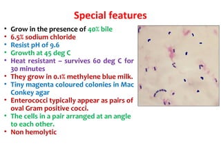
  • 62. Special features • Grow in the presence of 40% bile • 6.5% sodium chloride • Resist pH of 9.6 • Growth at 45 deg C • Heat resistant – survives 60 deg C for 30 minutes • They grow in 0.1% methylene blue milk. • Tiny magenta coloured colonies in Mac Conkey agar • Enterococci typically appear as pairs of oval Gram positive cocci. • The cells in a pair arranged at an angle to each other. • Non hemolytic
  • 63. Source & mode of infection: 1. Endogenous: from colonized site. 2. Exogenous: through direct or indirect contact. Common infections: 1. Urinary tract infection 2. Wound infection 3. Bacteremia 4. Intra-abdominal abscesses 5. Biliary tract infection 6. Sub acute bacterial endocarditis Clinical significance
  • 64. Laboratory diagnosis Specimens collected: Urine, blood, pus & exudates. Methods of examination: 1. Direct microscopy- by doing Gram’s smear. 2. Culture- Blood agar and MacConkey’s agar is used. Identification: • Gram’s stain • Catalase Negative • Mannitol, sucrose, sorbitol fermentation • Bile Esculin hydrolysis • Non-hemolytic on blood agar • Intrinsically resistant to cephalosporins Treatment: • Strains resistant to penicillin & other antibiotics occur frequently. • Penicillin + Aminoglycosides • Vancomycin is the alternative drug to penicillin: Vancomycin resistant is also seen- VRE - terminal D-ala replaced by D-lactateterminal D-ala replaced by D-lactate
  • 65. Viridans group • Streptococci normally resident in the mouth and upper respiratory tract • Alpha hemolysis on blood agar • Cannot be categorized under Lancefield antigenic groups Types: 1. S. mitis 2. S. mutans 3. S. salivarius 4.S. sanguis • Causes dental caries • Tooth extraction – seeding into blood stream - endocarditis – hence give prophylactic antibiotics
  • 66. Streptococcus pneumoniae (pneumococcus) Morphology: • Lancet shaped (one end broad, other pointed) • Capsulated • Diplococci S. pneumoniae: lancet-shaped diplococcus
  • 67. Growth characteristics • Alpha hemolysis on blood agar • On further incubation, the colonies become flat with raised edges and central elevation resembling carrom coins Example – Streptococcus viridans (Viridans = green) Streptococcus pneumoniae
  • 68. Biochemical reactions • Hiss serum water – fermentation of inulin • Bile solubility
  • 69. Resistance • Pneumococcus is sensitive to optochin – used for typing • Resistance to penicillin • May be resistant to third gen ceph also – third generation cephalosporin like ceftriaxone
  • 71. Pathogenicity and virulence • Capsule • Toxins – pneumolysin • C reactive protein
  • 73. Pneumolysin • Membrane damaging toxin • Cytotoxic activity
  • 74. C reactive protein • Abnormal immunoglobulin against C protein of S. pneumoniae appears in serum of patients • Also seen in acute phase of infections and inflammations • CRP titre is tested in acute infections
  • 75. Epidemiology • Source – respiratory tract of humans • Spread – droplets • Carriers – pharynx • Low immunity is responsible for fulminant infections – RSV infection, pulmonary congestion, stress, malnutrition, alcoholism – Splenectomy – Sickle cell disease
  • 76. 76 Diseases caused • Middle ear – otitis media • Para nasal sinuses – sinusitis • Respiratory tract – pneumonia, bronchitis, empyema • Meningitis is secondary to otitis media, penumonia, sinusitis and conjunctivitis
  • 77. Laboratory diagnosis Sample Pneumonia - Sputum Septicemia - blood culture Otitis media - fluid from middle ear Meningitis - CSF Method – Gram stain, microscopy – Culture Latex agglutination for pneumococcal antigen in serum of patients
  • 78. Differentiation between α-hemolytic streptococci • The following definitive tests used to differentiate between S. pneumoniae & viridans streptococci Optochin Test Bile Solubility Test Inulin Fermentation
  • 79. Optochin Susceptibility Test Principle: – Optochin (OP) test is presumptive test that is used to identify S. pneumoniae – S. pneumoniae is inhibited by Optochin reagent (<5 µg/ml) giving a inhibition zone ≥14 mm in diameter. Procedure: – BAP inoculated with organism to be tested – OP disk is placed on the center of inoculated BAP – After incubation at 37oC for 18 hrs, accurately measure the diameter of the inhibition zone by the ruler – ≥14 mm zone of inhibition around the disk is considered as positive and ≤13 mm is considered negative • S. pneumoniae is positive (S) while S. viridans is negative (R) Optochin susceptible S. pneumoniae Optochin resistant S. viridans
  • 80. Bile Solubility test Principle: – S. pneumoniae produce a self- lysing enzyme to inhibit the growth – The presence of bile salt accelerate this process Procedure: – Add ten parts (10 ml) of the broth culture of the organism to be tested to one part (1 ml) of 2% Na deoxycholate (bile) into the test tube – Negative control is made by adding saline instead of bile to the culture – Incubate at 37oC for 15 min – Record the result after 15 min Results: – Positive test appears as clearing in the presence of bile while negative test appears as turbid – S. pneumoniae soluble in bile whereas S. viridans insoluble
  • 81. Differentiation between β-hemolytic streptococci Hemolysis Bacitracin sensitivity CAMP test S. pyogenes β Susceptible Negative S. agalactiae β Resistant Positive Hemolysis Optochin sensitivit y Bile solubility Inulin Fermentation S. pneumoniae α Sensitive (≥ 14 mm) Soluble Not ferment Viridans strep α Resistant (≤13 mm) Insoluble Ferment Differentiation between α-hemolytic streptococci
  • 82. Outline of differentiation between Gram-Positive cocci e.g. S. epidermidis
  • 83. Treatment • Penicillin • Amoxycillin • ceftriaxone/ceftazidime • Vancomycin
  • 84. Prophylaxis • Pneumococcal conjugate vaccine is available • Given to splenectomy patients, sickle cell anemia patients and susceptible individuals
  • 85. Classification of Streptococci from Humans Species Lancefield Hemolytic Comments S. pyogenes A β Scarlet Fever, Septic Sore throat, Rheumatic fever, Glomerulonephritis S. agalactiae B β (α, γ) Chronic Mastitis S. dysgalactiae C α (β, γ) Acute mastitis S. equisimilis C α (β, γ) Abscesses, Endometritis, Mastitis S. equi C β Strangles, suppurative conditions, purpura haemorrhagica S. equi subsp zooepidemicus C β Bovine mastitis S. suis D α, β septicaemia; meningitis, arthritis, bronchopneumonia & infrequent human meningitis
  • 86. Zoonotic Streptococcus Species Species Lancefield Hemol ysis Comments S. bovis species group D α, γ Viridans; associated with colon cancer; IE S. mutans group not useful α, γ, rarely β Viridans; dental caries and IE S. salivarius group not useful α, γ Viridans; opportunistic S. mitis group not useful α Viridans; IE, opportunistic S. anginosus group A, C, F, G, or no detectable α, β, γ Viridans; formerly known as S. milleri; 3 species S. anginosus, S. constellatus, and S. intermedius; purulent infections S. canis G β Dogs; infrequent human pathogen S. iniae None detectable β Fish; SSTI, sepsis
  • 87. Group F beta hemolytic Streptococci Streptococcus MG • Grow poorly on blood agar • Minute streptococci • Streptococcus MG – alphalytic strain – isolated from Primary atypical pneumonia
  • 88. Minute colony streptococciMinute colony streptococci Various groups/ hemolysis (e.g. group A)Various groups/ hemolysis (e.g. group A) – genetically distinct from large colony (e.g. S. pyogenes)genetically distinct from large colony (e.g. S. pyogenes) – no rheumatic feverno rheumatic fever Large colonyLarge colony Minute colonyMinute colony
  • 89. Thanks Acknowledgement: All the material/presentations available online on the subject are duly acknowledged. Disclaimer: The author bear no responsibility with regard to the source and authenticity of the content. Questions???
  • 90. Thanks Acknowledgement: All the presentations available online on the subject are duly acknowledged. Disclaimer: The author bear no responsibility with regard to the source and authenticity of the content.