DEVELOPMENT OF
HEART TUBE
SHREEYA NAIK
ROLL NO. -141
• Development of blood cells and circulatory system – later part of 3rd
week of gestation
• Heart and blood vessels are mesodermal in origin
• Progenitor heart cells lie in the epiblast, adjacent to the cranial end
of the primitive streak. From there, they migrate through the streak
and into the splanchnic layer of lateral plate mesoderm where they
form a horseshoe-shaped cluster of cells called the primary heart
field (PHF) .
PHF induced by the underlying endoderm to
form cardiac myoblasts and blood islands that
will form blood cells and vessels by the
process of vasculogenesis. With time, the
islands unite and form a horseshoe-shaped
endothelial-lined heart tube surrounded by
myoblasts. This region is known as the
cardiogenic region.
• CRANIOCAUDAL FOLDING • LATERAL FOLDING
Initially the cardiogenic area was anterior to
oropharyngeal membrane ,as a result of growth CNS
system and CRANIOCAUDAL FOLDING of the embryo the
oropharyngeal membrane is pulled forward ,while the
heart and pericardial cavity move to the cervical region
and gradually to the thorax
The lateral folding cause the two heart tubes to fuse
except at the caudal most ends .
• Thus the heart becomes a continuous expanded tube having inner endothelial
lining and outer myocardial layer.
• Heart tube attached dorsal side of pericardial cavity by fold of mesocardium
tissue –DORSAL MESOCARDIUM.
• With futher development the dorsal mesocardium disappears ,creating the
transverse pericardial sinus that connects both side of pericardial cavity
• HEART TUBE consist of 3 layers
1. Inner ENDOTHELIUM
2. Myocardium,muscular wall
3. Epicardium/visceral pericardium
ATRIAL END
VENOUS END
cordis
The cephalic portion of the tube bends ventrally,
caudally, and to the right and the atrial (caudal)
portion shifts dorsocranially and to the left. This
bending, creates the cardiac loop.
LOOPING OF HEART
Development Of Right
Atrium
Roll No - 143
•The main part of
Right Atrium is
derived from Right
half of Primitive
Atrium
•The Sinus Venosus
is absorbed into the
Right Atrium
•The left horn of the
Sinus Venosus
becomes Coronary
Sinus
•The Right common
Cardinal vein becomes
part of Superior Vena
Cava
•The right Vitelline vein
forms the Inferior Vena
Cava
Development Of The Conducting System
Of The Heart
•After fusion of the
two tubes the
pacemaker lies in
the Sinus Venosus
•Afterwards it comes
to lie near the
opening of SVC
•Similarly the AV Node and the AVBundle
form in the left wall of Sinus Venosus and
afterwards it comes to lie near the Interatrial
Septum
FORMATION OF
SEPTUM
INTERMEDIUM
Septum
Intermedium
.
Septum intermedium
Heart development
Development Of Left Atrium
Roll no - 144
The left atrium is derived from :-
Left half of primitive atrial chamber
Left half of AV canal
Absorbed proximal parts of pulmonary
veins
Absorption of Pulmonary
Veins into Left Atrium
At first only one pulmonary
vein enters the atria
The proximal part of vein is
gradually absorbed and is
incorporated into the wall of
the atrium
As a result , four vein (two
on each side) finally open
into atrium
6th Week
8th Week
Development of the ventricles
The part of the heart tube lying within the pericardial cavity grows rapidly
and, therefore, becomes folded on itself to form a “U”-shaped
bulboventricular loop.
Bulboventricular sulcus gradually becomes shallower so that the conus,
the proximal part of the bulbus cordis, and the ventricle, come to form one
chamber which communicates with the truncus arteriosus.
DEVELOPMENT OF INTER-ATRIAL SEPTUM
Beginning at the end of the fourth week, the primordial atrium is divided into
right and left atria by the formation and subsequent modification and fusion of
two septa
Septum primum
Begins at the end of the fourth week
Septum primum, a thin crescent shaped membrane
Grows toward the fusing endocardial cushions from the roof of the
primordial atrium, partially dividing the common atrium into right and
left halves.
Fused endocardial cushion
Before the foramen primum disappears, perforations, produced by
apoptosis, appear in the central part of the septum primum.
During the fifth and sixth weeks septum secundum, a thick
crescentic muscular fold, grows from the ventrocranial wall of the
right atrium, immediately adjacent to the septum primum
How does this septum formation helps to prevent backflow
of oxygenated blood?
Septum Primum - thin and mobile
Septum Secundum - thick and rigid
Heart development
INTERVENTRICULAR SEPTUM
Roll No. 150
Interventricular Septum Divided into :-
• Muscular Part :-
• The Medial Walls of the expanding ventricles become apposed
and gradually merge forming the muscular interventricular septum
• Membranous Part :-
• Derived from Endocardial Cushions.
• Derived from Conus Septum.
Conus Septum :-
Heart Tube is divided into Bulbar Cordis , Primitive Ventricle , Primitive
Atrium , Sinus Venosus.
• Bulbus Cordis gets further divided into 3 parts :- Truncus Arteriosus ,
Conus Cordis and proximal part of Bulbus Cordis
Conus Cordis further develop Conus Swellings which gets further
developed into Conus Septum.
*Trunkus Arteriosus of Bulbus Cordis give rise to
aorta and Pulmonary Trunk seperated by
Spiral Septum.
*Proximal part of Bulbus Cordis give rise to
smooth parts of ventricle :-
Vestibule and Infundibulum.
Heart development
Applied :- Tetralogy of Fallot
Anterior Displacement of Spiral Septum
Ventricular Septal Defect
Congenital
anomalies of
the heart
Roll no.146
(I) DEXTROCARDIA - (i) with situs inversus (ii) with laterality
1. CARDIAC LOOPING ANOMALIES
Normal Dextrocardia
2. ATRIAL SEPTAL DEFECT (asd)
Normal Interatrial Septum Formation
Foramen Ovale
SP
SS
S
V
C
I
V
C
1. Excess Resorption of Septum Primum
90% ASDs- Ostium Secundum Defects
Foramen
Ovale Ostium
Secundum
2. Incomplete Development of Septum Secundum
ASDs- Ostium Secundum Defects
Foramen
Ovale Ostium
Secundum
Courtesy: MSD Manuals
Cor Triloculare Biventriculare
3. Ventricular septal defect (VSd)
Normal Interventricular Septum Development
Muscular Region
Membranous Region
(I) Muscular region of IV Septum (II) Membranous region of IV Septum
Muscular Region
Membranous Region
5. Tricuspid atresia
Normal Development of Atrioventricular Valves
(1) Underdevelopment of
Right Ventricle
(3) Ventricular Septal Defect
(4) Patent Foramen Ovale
Absence/Fusion of Tricuspid Valves
Normal Abnormal
(2) Left Ventricular Hypertrophy
THANK YOU !

More Related Content

PPTX
Embryology of the heart
PDF
Development of the heart tube and fetal circulation
PPTX
cardiovascular system embryology ppt pptx
PPT
Cardiovascularsystem 110514061056-phpapp02
PPTX
Cardiac embryology
PPT
Development of heart
PPTX
Development of heart.pptx
PPT
EMBRYOLOGY OF CVS
Embryology of the heart
Development of the heart tube and fetal circulation
cardiovascular system embryology ppt pptx
Cardiovascularsystem 110514061056-phpapp02
Cardiac embryology
Development of heart
Development of heart.pptx
EMBRYOLOGY OF CVS

Similar to Heart development (20)

PDF
Development of the heart tube
PPT
CVS Embryology (Amoud University)
PPTX
Cardiac development final
PDF
heart yuktawankhede006-220103133857 (1).pdf
PPTX
Development of Heart (Embryology)
PPTX
Cardiovascular System.pptx
PPTX
Cardiovascular System Embryology 2024 .pptx
PPTX
Development of heart
PPTX
Cardiac Embryology basics.pptx
PPT
11 development of the heart
PPTX
Development of the heart
PDF
development of heart and it’s clinical abnormal
PPT
Embryology [heart.].ppt
PPTX
CVS-EMBRYOLOGYyyyyyyyyyyyyyyyyyyyyy-1.pptx
PPTX
1st ppt developmental anatomy of heart.pptx
PPTX
Embryology of heart
PPTX
Final heart development.pptx
PPT
Cvs emb
PPTX
Heart and lung development
Development of the heart tube
CVS Embryology (Amoud University)
Cardiac development final
heart yuktawankhede006-220103133857 (1).pdf
Development of Heart (Embryology)
Cardiovascular System.pptx
Cardiovascular System Embryology 2024 .pptx
Development of heart
Cardiac Embryology basics.pptx
11 development of the heart
Development of the heart
development of heart and it’s clinical abnormal
Embryology [heart.].ppt
CVS-EMBRYOLOGYyyyyyyyyyyyyyyyyyyyyy-1.pptx
1st ppt developmental anatomy of heart.pptx
Embryology of heart
Final heart development.pptx
Cvs emb
Heart and lung development
Ad

Recently uploaded (20)

PDF
Transcultural that can help you someday.
DOCX
PEADIATRICS NOTES.docx lecture notes for medical students
PDF
AGE(Acute Gastroenteritis)pdf. Specific.
PPT
Infections Member of Royal College of Physicians.ppt
PDF
OSCE Series Set 1 ( Questions & Answers ).pdf
PPTX
Post Op complications in general surgery
PDF
focused on the development and application of glycoHILIC, pepHILIC, and comm...
PPTX
Human Reproduction: Anatomy, Physiology & Clinical Insights.pptx
PPTX
Reading between the Rings: Imaging in Brain Infections
PDF
B C German Homoeopathy Medicineby Dr Brij Mohan Prasad
PPTX
09. Diabetes in Pregnancy/ gestational.pptx
PDF
Lecture 8- Cornea and Sclera .pdf 5tg year
PPTX
Electrolyte Disturbance in Paediatric - Nitthi.pptx
PPTX
NUCLEAR-MEDICINE-Copy.pptxbabaabahahahaahha
PPTX
Introduction to Medical Microbiology for 400L Medical Students
PDF
OSCE SERIES ( Questions & Answers ) - Set 5.pdf
PDF
OSCE Series ( Questions & Answers ) - Set 6.pdf
PPTX
IMAGING EQUIPMENiiiiìiiiiiTpptxeiuueueur
PDF
Copy of OB - Exam #2 Study Guide. pdf
PPTX
Vaccines and immunization including cold chain , Open vial policy.pptx
Transcultural that can help you someday.
PEADIATRICS NOTES.docx lecture notes for medical students
AGE(Acute Gastroenteritis)pdf. Specific.
Infections Member of Royal College of Physicians.ppt
OSCE Series Set 1 ( Questions & Answers ).pdf
Post Op complications in general surgery
focused on the development and application of glycoHILIC, pepHILIC, and comm...
Human Reproduction: Anatomy, Physiology & Clinical Insights.pptx
Reading between the Rings: Imaging in Brain Infections
B C German Homoeopathy Medicineby Dr Brij Mohan Prasad
09. Diabetes in Pregnancy/ gestational.pptx
Lecture 8- Cornea and Sclera .pdf 5tg year
Electrolyte Disturbance in Paediatric - Nitthi.pptx
NUCLEAR-MEDICINE-Copy.pptxbabaabahahahaahha
Introduction to Medical Microbiology for 400L Medical Students
OSCE SERIES ( Questions & Answers ) - Set 5.pdf
OSCE Series ( Questions & Answers ) - Set 6.pdf
IMAGING EQUIPMENiiiiìiiiiiTpptxeiuueueur
Copy of OB - Exam #2 Study Guide. pdf
Vaccines and immunization including cold chain , Open vial policy.pptx
Ad

Heart development

  • 2. • Development of blood cells and circulatory system – later part of 3rd week of gestation • Heart and blood vessels are mesodermal in origin • Progenitor heart cells lie in the epiblast, adjacent to the cranial end of the primitive streak. From there, they migrate through the streak and into the splanchnic layer of lateral plate mesoderm where they form a horseshoe-shaped cluster of cells called the primary heart field (PHF) .
  • 3. PHF induced by the underlying endoderm to form cardiac myoblasts and blood islands that will form blood cells and vessels by the process of vasculogenesis. With time, the islands unite and form a horseshoe-shaped endothelial-lined heart tube surrounded by myoblasts. This region is known as the cardiogenic region.
  • 4. • CRANIOCAUDAL FOLDING • LATERAL FOLDING Initially the cardiogenic area was anterior to oropharyngeal membrane ,as a result of growth CNS system and CRANIOCAUDAL FOLDING of the embryo the oropharyngeal membrane is pulled forward ,while the heart and pericardial cavity move to the cervical region and gradually to the thorax The lateral folding cause the two heart tubes to fuse except at the caudal most ends .
  • 5. • Thus the heart becomes a continuous expanded tube having inner endothelial lining and outer myocardial layer. • Heart tube attached dorsal side of pericardial cavity by fold of mesocardium tissue –DORSAL MESOCARDIUM. • With futher development the dorsal mesocardium disappears ,creating the transverse pericardial sinus that connects both side of pericardial cavity • HEART TUBE consist of 3 layers 1. Inner ENDOTHELIUM 2. Myocardium,muscular wall 3. Epicardium/visceral pericardium
  • 7. The cephalic portion of the tube bends ventrally, caudally, and to the right and the atrial (caudal) portion shifts dorsocranially and to the left. This bending, creates the cardiac loop. LOOPING OF HEART
  • 9. •The main part of Right Atrium is derived from Right half of Primitive Atrium •The Sinus Venosus is absorbed into the Right Atrium
  • 10. •The left horn of the Sinus Venosus becomes Coronary Sinus •The Right common Cardinal vein becomes part of Superior Vena Cava •The right Vitelline vein forms the Inferior Vena Cava
  • 11. Development Of The Conducting System Of The Heart
  • 12. •After fusion of the two tubes the pacemaker lies in the Sinus Venosus •Afterwards it comes to lie near the opening of SVC
  • 13. •Similarly the AV Node and the AVBundle form in the left wall of Sinus Venosus and afterwards it comes to lie near the Interatrial Septum
  • 17. Development Of Left Atrium Roll no - 144 The left atrium is derived from :- Left half of primitive atrial chamber Left half of AV canal Absorbed proximal parts of pulmonary veins
  • 18. Absorption of Pulmonary Veins into Left Atrium At first only one pulmonary vein enters the atria The proximal part of vein is gradually absorbed and is incorporated into the wall of the atrium As a result , four vein (two on each side) finally open into atrium
  • 20. Development of the ventricles The part of the heart tube lying within the pericardial cavity grows rapidly and, therefore, becomes folded on itself to form a “U”-shaped bulboventricular loop.
  • 21. Bulboventricular sulcus gradually becomes shallower so that the conus, the proximal part of the bulbus cordis, and the ventricle, come to form one chamber which communicates with the truncus arteriosus.
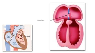
  • 22. DEVELOPMENT OF INTER-ATRIAL SEPTUM Beginning at the end of the fourth week, the primordial atrium is divided into right and left atria by the formation and subsequent modification and fusion of two septa
  • 23. Septum primum Begins at the end of the fourth week Septum primum, a thin crescent shaped membrane
  • 24. Grows toward the fusing endocardial cushions from the roof of the primordial atrium, partially dividing the common atrium into right and left halves. Fused endocardial cushion
  • 25. Before the foramen primum disappears, perforations, produced by apoptosis, appear in the central part of the septum primum.
  • 26. During the fifth and sixth weeks septum secundum, a thick crescentic muscular fold, grows from the ventrocranial wall of the right atrium, immediately adjacent to the septum primum
  • 27. How does this septum formation helps to prevent backflow of oxygenated blood? Septum Primum - thin and mobile Septum Secundum - thick and rigid
  • 31. • Muscular Part :- • The Medial Walls of the expanding ventricles become apposed and gradually merge forming the muscular interventricular septum • Membranous Part :- • Derived from Endocardial Cushions. • Derived from Conus Septum.
  • 32. Conus Septum :- Heart Tube is divided into Bulbar Cordis , Primitive Ventricle , Primitive Atrium , Sinus Venosus. • Bulbus Cordis gets further divided into 3 parts :- Truncus Arteriosus , Conus Cordis and proximal part of Bulbus Cordis Conus Cordis further develop Conus Swellings which gets further developed into Conus Septum. *Trunkus Arteriosus of Bulbus Cordis give rise to aorta and Pulmonary Trunk seperated by Spiral Septum. *Proximal part of Bulbus Cordis give rise to smooth parts of ventricle :- Vestibule and Infundibulum.
  • 34. Applied :- Tetralogy of Fallot Anterior Displacement of Spiral Septum
  • 37. (I) DEXTROCARDIA - (i) with situs inversus (ii) with laterality 1. CARDIAC LOOPING ANOMALIES
  • 39. 2. ATRIAL SEPTAL DEFECT (asd)
  • 42. 1. Excess Resorption of Septum Primum 90% ASDs- Ostium Secundum Defects Foramen Ovale Ostium Secundum
  • 43. 2. Incomplete Development of Septum Secundum ASDs- Ostium Secundum Defects Foramen Ovale Ostium Secundum
  • 46. 3. Ventricular septal defect (VSd) Normal Interventricular Septum Development Muscular Region Membranous Region
  • 47. (I) Muscular region of IV Septum (II) Membranous region of IV Septum Muscular Region Membranous Region
  • 48. 5. Tricuspid atresia Normal Development of Atrioventricular Valves
  • 49. (1) Underdevelopment of Right Ventricle (3) Ventricular Septal Defect (4) Patent Foramen Ovale Absence/Fusion of Tricuspid Valves Normal Abnormal (2) Left Ventricular Hypertrophy