Hla typing and its role in tissue transplantation
Download as PPTX, PDF172 likes58,352 views

This document discusses HLA typing and its role in tissue transplantation. It begins by introducing the major histocompatibility complex (MHC) and its role in transplant rejection. It then describes MHC polymorphism, HLA nomenclature, and various methods for HLA typing including serology and molecular techniques. The document concludes by discussing the applications of HLA typing in organ transplantation, including the mechanisms of allograft recognition and rejection.


Hla typing and its role in tissue transplantation
- 1. HLA system- HLA typing and its role in tissue transplantation Presenter: Dr. Animesh Debbarma Moderator: Dr. Sharmila
- 2. Objectives Major histocompatibility complex MHC polymorphism Regulation of MHC expression HLA nomenclature, typing, methods, application Organ transplantation
- 3. Major histocompatibility complex Group of genes coding for a set of host surface molecules that bind to a peptide fragments derived from pathogens and foreign antigens, and display them on host cell surface for recognition by the appropriate T cells. Also called human leukocyte antigens (HLA). Serves as a unique identification marker for every individual. Following transplantation of a graft the recipient mount an immune response against the graft’s MHC molecules and vice versa. Also called histocompatibility antigens.
- 4. HLA complex (MHC genes) and their products In humans, HLA complex coding for MHC proteins are located on short arm of chromosome 6. Around 4000 kbp in length covering >100 genes. Genes are clustered in three regions namely MHC region-I, II and III.
- 5. MHC region I About 2000 kbp in length. Comprises of three class I genes called HLA-A, HLA-B and HLA-C genes encoding HLA-A, HLA-B and HLA-C proteins respectively. Each protein is capable of forming the α-chain of MHC class I molecules. MHC class I molecules are present on the surface of all nucleated cells (except sperm cells) and platelets. Present the peptide antigen to CD8+ T cells.
- 6. MHC region II About 1000 kbp length. Comprises of three genes namely DP, DQ and DR genes encoding DP, DQ and DR proteins respectively. Each protein is capable of forming α and β-chain of MHC class II molecules. In addition MHC region also contain non classical genes such as DM, DO, LMP and TAP that help in antigen processing and presentation. MHC-II proteins are located on the surface of APC. Present the peptide to CD4+ T cells.
- 7. MHC region III About 1000 kbp in length. Not involved in Ag presentation. Comprises of genes that code for complement factors, heat shock proteins (HSP), tumor necrosis factor (TNF α and β), steroid 21 hydroxylases etc.
- 8. STRUCTURE OF THE MHC MOLECULES MHC class I molecules They are heterodimer composed of polymorphic α-chain (glycoprotein, 45 KDa, coded by HLA class I genes) linked non-covalently to smaller non polymorphic β2 microglobulin (non glycosylated 12 KDa protein, encoded by non MHC gene from chromosome 15). α-chain is organized into 3 extracellular globular domain (N terminal) α1, α2 and α3 (each containing 90 AA); A hydrophobic transmembrane domain of about 25 AA followed by a short stretch of charged (hydrophilic) amino acids; Cytoplasmic anchor segment of 30 amino acids. Antigen peptide groove is formed by the cleft between α1 and α2 domains. Polymorphic amino acid residues lines the side and base of the peptide binding groove. The α3 domain bind to CD8 molecules of cytotoxic T cell during Ag presentation.
- 9. MHC class II molecules They are heterodimer consisting of noncovalently associated α chain (33 KDa) and β chain (28 KDa), both of them are polymorphic. Like class I molecule they have external domain, a transmembrane segment and a cytoplasmic anchoring segment. Each chain contain 2 extracellular domain: α1, α2 and β1, β2 respectively. The Ag peptide groove is formed by the α1 and β1 domains. β2 domain interacts with CD4 molecules of Helper T cell during Ag presentation
- 12. Difference between MHC I and MHC II molecules MHC class I MHC class II Present on All nucleated cells (except sperm cells) and platelets Ag presenting cells Peptide Ag is presented to CD8+ T cells CD4+ T cells Nature of peptide Ag Endogenous or intracellular (viral/tumor Ag) Exogenous General size of bound antigens 8-10 amino acids 13-18 amino acids Peptide binding site α1 and α2 groove α1 and β1 groove CD4 or CD8 binding site α3 binds to CD8 molecules on Tc cells β2 binds to CD4 molecules on TH cells. Ag presentation pathways Cytosolic pathway Endocytic pathway
- 13. MHC polymorphism Three mechanism: 1. Multiple gene loci: Ex- MHC I molecules (α chain) are encoded by any of the three loci of MHC I region, i.e. HLA-A, HLA-B or HLA-C loci. MHC II molecules (α and β chain) are encoded by any of the three loci of MHC II region, i.e. DP, DQ or DR loci.
- 14. 2. Multiple allele for each locus: Ex- For Class I MHC region in humans, there are 240 alleles for HLA-A, 470 alleles for HLA-B and 110 alleles for HLA-C. So MHC Class I region of any of the individual would have one of the allele from each HLA-A, HLA-B and HLA-C allele bank. So there are total 240X470X110 theoretical combinations possible for MHC Class I region. These allele encode for products that differ from each other by 5-10 % of their DNA sequence. Similar polymorphism also exists for alleles of Class II DP, DQ and DR loci. 3. Codominant expression: MHC genes are expressed in codominant fashion, i.e. the alleles inherited from parents (one from father and one from mother) are simultaneously and equally expressed.
- 15. Regulation of MHC expression Transcription factors: MHC genes have promoter sequence at their 5’ end which are regulated by certain transcription factors such as CIITA and RFX (both bind to MHC II molecules and increase their transcription). Defect in CIITA and RFX- Bare lymphocyte syndrome. Cytokines: IFN-γ activates both MHC-I and II promoter genes. IL-4 increases the expression of MHC II molecules in resting B cells.
- 16. Corticosteroids and prostaglandins decrease the expression of MHC II molecules. Viral infection: Some viral antigens inhibit various components of MHC I (e.g. adenovirus protein inhibit TAP, cytomegalovirus protein inhibit β2 microglobulin). As a result, MHC-I expression is suppressed.
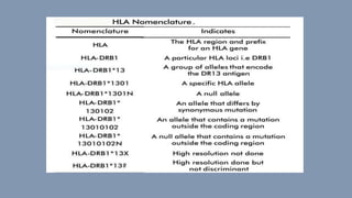
- 17. HLA Nomenclature There are a number of ways of writing an HLA antigen. For example, it may be expressed as HLA-DR3, HLA-DRI7, HLA-DRB* 03 or HLA- DRBI*0301. These could all refer to the same antigen. Firstly, “HLA” is the name for the gene cluster which tends to be inherited en-bloc. Second part -e.g. DR- is the name of the specific locus. There are 6 loci normally referred to. These are A, B, C, DR, DQ and DP.
- 18. Third part, the number, e.g. 3, 17, 03, 0301, refers to the actual antigen at the locus. For example, the DNA in the HLA-DR locus tends to be different from person to person. This difference will result in a different type of HLA-DR molecule. These different types of HLA-DR molecules are given names, such as DR17. When we look at the antigens above: -HLADR3 is the broadest description of the antigen. It is the name for a specific group of antigens. The DR3 group can be divided into HLA-DR17 and HLA- DR18 by using antibodies (serology).
- 19. When we look at this antigen at the DNA level we call the DR locus DRB 1 and the antigen 03 and 01 for the specific variant of the 03. So, HLA-DR17 is now called HLA-DRB1*0301. This is similar for other antigens in the system, at either HLA Class II or Class I e.g. HLA-B60 (HLA-B*4001 molecularly). Fig: HLA nomenclature
- 21. HLA TYPING In this test, donor’s antigens expressed on the surface of leukocytes or their genes are matched with that of the recipient. The closer the HLA antigens on the transplanted organ match the recipient, the more likely that the recipient’s body will not reject the transplant. Value of HLA matching between donor and recipient varies in different solid organ transplantation. In kidney transplants, there is substantial benefit if all the polymorphic HLA alleles are matched.
- 22. Every person inherits each of the following antigens from each parent: HLA-A antigen HLA-B antigen HLA-C antigen HLA-DR antigen HLA-DQ antigen and HLA-DP antigen Fig: Major histocompatibility complex at chromosome 6
- 23. When performing an HLA typing test for a kidney transplant, the following HLA antigens are looked at: HLA-A HLA-B HLA-DR Six HLA antigens are looked at for each person.
- 24. Each person has two of each of the antigens (one inherited from the mother and one inherited from the father).
- 25. By analyzing which six of these HLA-antigens both the donor and recipient have, scientists are able to determine the closeness of tissue matching. A six-antigen match is the best compatibility between a donor and recipient. This match occurs 25% of the time between siblings who have the same mother and father.
- 26. METHODS OF HLA TYPING A. Phenotypic method: Serology: Microcytotoxicity Tissue typing: Mixed lymphocyte reaction B. Genotypic methods: PCR detecting HLA genes PCR-RFLP (restriction fragment length polymorphism) Variable number tandem repeat (VNTR) typing Short tandem repeat (STR) typing DNA sequence based typing Karyosome analysis
- 27. Serology: Microlymphocytotoxic test: Viable WBC’s of the individual to be typed are incubated with HLA (class I and II) specific antibodies. If the specific Ag is present on the cell, the antibody is bound. Complement is added and incubated. If the antibody is bound, it will activate the complement which damages the cell membrane making it permeable to vital stains.
- 28. Pros Cons Easily performed, does not require expensive equipments Require large volume of blood 3 hrs Require viable WBC’s With good antisera results are reliable Difficult to find good antisera for rarer antigens.
- 29. Cellular: Mixed lymphocyte culture (MLC) Used to quantify the degree of class II MHC compatibility between potential donors and recipients. It is based on the principle that if immunocompetent T cells from one individual is incubated with APC’s of a genetically different individual, the T-cell of the first individual will proliferate. Proliferation of the recipient T-cells is measured by the uptake of thymidine into the cell DNA.
- 30. Molecular methods: Commonly used molecular techniques of the HLA typing utilizes DNA extraction and direct DNA typing The phenotypic methods were used widely in the past. But with the advent of molecular methods, they are not preferred now. VNTR and DNA sequence based typing are the most reliable method of HLA typing.
- 31. 1. Organ and tissue transplantation In organ and tissue transplantation, HLA antigens of the donor identified as invaders by the recipient causing rejection. Careful selection of the matched donor and recipient critically affect the outcome of transplantation. 2. Diagnosing some disease : In autoimmunity: Many HLA combination are potentially indicative of autoimmune disorders, e.g. Application of HLA typing
- 32. HLA allele Associated disease HLA B27 Ankylosing spondylitis, Reactive arthritis, Reiter’s syndrome DR2 Multiple sclerosis, Good pasture’s syndrome DR3 Myasthenia gravis, SLE DR3/DR4 Insulin dependent DM DR4 Rheumatoid arthritis A3/B14 Hereditary hemochromatosis
- 33. Susceptibility to viral infections: There is a link between certain HLA antigens and susceptibility to some viral infections such as AIDS (HIV virus), Hepatitis B (Hep B), Hepatitis C (Hep C), Infectious mononucleosis (EVB), Rubella (Rubella virus) etc. 3. Paternal testing HLA typing can be used alongside other test for paternity testing.
- 34. 4. Infertility (recurrent pregnancy loss): Infertility due to recurrent pregnancy loss can be attributed to immune factors (40%) one of which is presence of certain common HLA antigens between the parents. 5. Phylogenetic studies: Some HLA haplotypes have distinctive geographical distribution and are found only in some population. These haplotypes can be used to trace human migration.
- 35. ORGAN TRANSPLANTATION Based on the genetic relationship between the donor and the recipient: Autograft: Self tissue transferred from one part of the body site to another in the same individual (e.g. transfering healthy skin to a burned area in burned patients). Isograft of syngeneic graft: Tissue transferred between genetically identical individuals (e.g. monozygotic twins).
- 36. Allograft: Tissue transferred between genetically non-identical members of the same species (e.g. kidney or heart transplant). Xenograft: Tissue transferred between different species (e.g. the graft of a baboon heart into a man).
- 37. In humans allograft are the most commonly used graft in transplant centers. Histocompatibility between the graft and the recipient would decide whether the graft is going to be accepted or rejected. Transplantation antigens are the antigens of allograft against which the recipient would mount an immune response. MHC molecules are the most important transplantation antigens. Apart from that, ABO and Rh group system also play an important role in determining the histocompatibility.
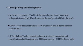
- 38. Mechanism of recognition and rejection of allograft The major antigenic differences between a donor and the recipient that result in rejection of transplants are differences in HLA alleles. Following transplantation, the recipient’s T cells recognize donor antigens from the graft (the allogeneic antigens, or alloantigens) by two pathways, called direct and indirect.
- 39. Direct pathway of allorecognition. In the direct pathway, T cells of the transplant recipient recognize allogeneic (donor) MHC molecules on the surface of APCs in the graft. CD8+ T cells recognize class I MHC molecules and differentiate into active CTLs. CD4+ helper T cells recognize allogeneic class II molecules and proliferate and differentiate into TH1 (and possibly TH17) effector cells
- 40. Indirect pathway of allorecognition: In the indirect pathway, recipient T lymphocytes recognize MHC antigens of the graft donor after they are presented by the recipient’s own APCs. This process involves the uptake and processing of MHC molecules from the grafted organ by host APCs. The peptides derived from the donor tissue are presented by the host’s own MHC molecules, like any other foreign peptide.
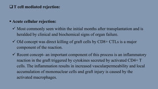
- 42. T cell mediated rejection: Acute cellular rejection: Most commonly seen within the initial months after transplantation and is heralded by clinical and biochemical signs of organ failure. Old concept was direct killing of graft cells by CD8+ CTLs is a major component of the reaction. Recent concept- an important component of this process is an inflammatory reaction in the graft triggered by cytokines secreted by activated CD4+ T cells. The inflammation results in increased vascularpermeability and local accumulation of mononuclear cells and graft injury is caused by the activated macrophages.
- 43. Chronic rejection: Lymphocytes reacting against alloantigens in the vessel wall secrete cytokines that induce local inflammation and may stimulate the proliferation of vascular endothelial and smooth muscle cells.
- 44. Antibody mediated reaction: Hyperacute rejection Occurs when preformed antidonor antibodies are present in the circulation of the recipient. Such antibodies may be present in a recipient who has previously rejected a transplant. Multiparous women who develop antibodies against paternal HLA antigens shed from the fetus may have preformed antibodies that will react with grafts taken from their husbands or children, or even from unrelated individuals who share HLA alleles with the husbands. Prior blood transfusions can also lead to presensitization, because platelets and white blood cells are rich in HLA antigens and donors and recipients are usually not HLA-identical.
- 45. Acute antibody-mediated rejection: caused by antidonor antibodies produced after transplantation. In recipients not previously sensitized to transplantation antigens, exposure to the class I and class II HLA antigens of the donor graft, as well as other antigens that differ between donor and recipient, may evoke antibodies. The antibodies formed by the recipient may cause injury by several mechanisms:- a. complementdependent cytotoxicity, b. inflammation, and c. antibodydependent cell-mediated cytotoxicity.
- 46. Chronic antibody-mediated rejection: usually develops insidiously, without preceding acute rejection, and primarily affects vascular components.
- 47. References: 1. Robbins and Clotran Pathologic Basis of Disease 2. Essential of medical microbiology by Dr. Apurba Sankar 3. Owen Kuby’s Immunology 4. Internet source 5. Slide of Dr. Anupama’s
- 48. THANK YOU For patience hearing