SlideShare a Scribd company logo
2
Most read
3
Most read
13
Most read
Imaging Geometry
Mithun kumar kar
Department of Electrical Engineering
BALASORE COLLEGE OF ENGINEERING AND TECHNOLOGY, BALASORE
August 12, 2020
Mithun kumar kar (BCET) Imaging Geometry August 12, 2020 1 / 17
Human vision system
The human visual system consist of an eye that transforms light in to
the neural signals and create neural activity in the brain and extract
necessary information for human vision.
In human visual perception, the eyes act as the sensor or camera, neu-
rons act as the connecting cable and the brain acts as the processor.
The basic elements of visual perceptions are
Structure of Eye
Image Formation in the Eye
Brightnes Adaptation and Discrimination
Mithun kumar kar (BCET) Imaging Geometry August 12, 2020 2 / 17
Structure of Eye
The human visual system consist of an eye that transforms light in to
the neural signals and create neural activity in the brain and extract
necessary information for human vision.
Mithun kumar kar (BCET) Imaging Geometry August 12, 2020 3 / 17
Structure of Eye
Mithun kumar kar (BCET) Imaging Geometry August 12, 2020 4 / 17
Structure of Eye
The pupil allows light to enter the eye to fall on the retina. It contracts
when exposed to bright light and controls the amount of light entering
to eye.
A colored circular pigmented muscle called irish gives us our eye colour
and controls the size of the pupil so that appropriate amount of light
is allowed to enter the eye.
A transparent external surface called cornea covers both the pupil and
the irish. This is the first and most powerful lens of the optical system
of the eye and together with crystalline lens, it allows the production
of a sharpe image at the retinal photo receptor level.
The upper white part of the eye is called sclera which form a supporting
wall of the eyeball. The sclera is a nearly spherical shell with a radius
of 11mm and 1mm thick.‘
Mithun kumar kar (BCET) Imaging Geometry August 12, 2020 5 / 17
Image formation in the eye
When the lens of the eye focus an image of the outside world onto a
light-sensitive membrane in the back of the eye, called retina the image
is formed. The lens of the eye focuses light on the photoreceptive
cells of the retina which detects the photons of light and responds by
producing neural impulses.
In an photographic camera, the lens has a fixed focal length and fo-
cusing at various distances is achieved by varying the distance between
the lens and the imaging plane.
In human eye the distance between the lens and the imaging region(retina)
is fixed, and the focal length needed to achieve proper focus is obtained
by varying the shape of the lens. The fibers in the ciliary body accom-
plish this by flattening or thickening the lens for distant or near objects.
Mithun kumar kar (BCET) Imaging Geometry August 12, 2020 6 / 17
Image formation in the eye
The distance between the center of the lens and the retina along the
visual axis is approximately 17mm and the range of the focal lengths
is approximately 14mm to 17mm.
Suppose that a person is looking at a tree 15m high at a distance of
100m. Let h denote the height of that object in the retinal image, then
from geometry we get the relation 15/100 = h/17 or h = 2.55mm
Mithun kumar kar (BCET) Imaging Geometry August 12, 2020 7 / 17
Brightness Adaptation and Discrimination:
Digital images are displayed as a discrete set of intensities. The eyes
ability to discriminate black and white at different intensity levels is an
important consideration in presenting image processing result.
Mithun kumar kar (BCET) Imaging Geometry August 12, 2020 8 / 17
Electromagnetic spectrum:
Mithun kumar kar (BCET) Imaging Geometry August 12, 2020 9 / 17
Brightness Adaptation and Discrimination:
The range of light intensity levels to which the human visual system
can adapt is of the order of 1010 that is from scotopic threshold to
glare limit.
The intensity perceived by the human visual system(subjective bright-
ness) is a logarithmic function of the light intensity incedent on the
eye.
The long solid curve represents the range of intensities to which the
visual system can adapt.
For any given set of conditions the current sensitivity level of the visual
system is called the brightness adaption level which may correspond to
brightness Ba.
The short intersecting curve represents the range of subjective bright-
ness that the eye can perceive when adapted to level Ba. This range is
restricted up to level Bb and below which all stimuli are percieved as
black.
Mithun kumar kar (BCET) Imaging Geometry August 12, 2020 10 / 17
Photopic and Scotopic Vision
The two types of receptors responsible for human vision are rods and
cones. The rods are sensitive to very low illumination and are respon-
sible for night vision (scotopic vision).
The cones which are tightly packed in the fovea, lie inline with the
visual axis and are mainly responsible for color vision (photopic vision).
The minimum sensitivity of cones is of the order of a microlambert(mL)
The cones in the human eye can be devided in to three principal sensing
categories corresponding to red, green, blue.
Mithun kumar kar (BCET) Imaging Geometry August 12, 2020 11 / 17
Photopic and Scotopic Vision
The cones in each eye numbers between 6 to 7 million and are located
in the central portion of retina called fovea which are highly sensitive
to color.
The number of rods is much larger, between 75 to 150 million. Rods
serve to give a general overall picture of the field of view and are
sensitive to low levels of illumination.
Objects appear coloured in daylight appears colourless in moon light
because only rods are stimulated. This phenomenon is known as sco-
topic vision.
Mithun kumar kar (BCET) Imaging Geometry August 12, 2020 12 / 17
Weber ratio
Weber ratio gives an measure of brightness discrimination that is the
ability of the eye to discriminate between changes in light intensity at
any specific adaptation level.
Weber ratio is defined as the ratio of the increment of illumination(∆Ic)
discriminable 50 percent of the time to the background illumination I.
Hence
w =
∆Ic
I
A small value of w means that a small percentage change in intensity
is discriminable which represents good brightness discrimination. Simi-
larly large value of w means that a large percentage change in intensity
is required and represents poor brightness discrimination.
Mithun kumar kar (BCET) Imaging Geometry August 12, 2020 13 / 17
Machband effect
The spatial interaction of luminance from an object and its surroundings
creates the Machband effect, which shows that brightness is not a
monotonic function of luminance.
The intensity is uniform over the width of the bar. However the visual
appearance is that each strip is darker at its left side than its right.
Machbanding is caused by lateral inhibition of the receptors in the eye.
As the receptors recieve light, they draw light sensitive chemical com-
pounds from adjacent regions, thus inhibiting the response of receptors
in those regions.
Perceived intensity is not a simple function of actual intensity)
Mithun kumar kar (BCET) Imaging Geometry August 12, 2020 14 / 17
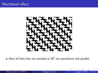
Machband effect
Here all lines that are oriented at 450 are equidistant and parallel.
Mithun kumar kar (BCET) Imaging Geometry August 12, 2020 15 / 17
Machband effect
The two horizontal line segments are of the same length.
Mithun kumar kar (BCET) Imaging Geometry August 12, 2020 16 / 17
Machband effect
The outline of a square is seen clearly, despite the fact that no lines
defining such a figure are part of the image.
Mithun kumar kar (BCET) Imaging Geometry August 12, 2020 17 / 17

More Related Content

PPTX
Image restoration and degradation model
PPTX
Object Recognition
PPT
pattern classification
PPT
Chapter 4 Image Processing: Image Transformation
PDF
Gaussian filtering 1up
PPT
Thresholding.ppt
PPT
introduction to Digital Image Processing
PPTX
Intensity Transformation and Spatial filtering
Image restoration and degradation model
Object Recognition
pattern classification
Chapter 4 Image Processing: Image Transformation
Gaussian filtering 1up
Thresholding.ppt
introduction to Digital Image Processing
Intensity Transformation and Spatial filtering

What's hot (20)

PPTX
Digital image processing
PPTX
IMAGE SEGMENTATION.
PPT
morphological image processing
PPTX
Region filling
PPTX
Image transforms
PPTX
Image compression
PPTX
Erosion and dilation
PPSX
Color Image Processing: Basics
PPTX
Image compression .
PPTX
Smoothing Filters in Spatial Domain
PPTX
Smoothing in Digital Image Processing
PPTX
Ip elements of image processing
PPTX
Chapter 8 image compression
PDF
Lecture 4 Relationship between pixels
PDF
Statistical Pattern recognition(1)
PPT
Dip Image Segmentation
PPTX
Chapter 9 morphological image processing
PPTX
Texture,pattern and pattern classes
PPT
Ajay ppt region segmentation new copy
PPT
Computer Vision: Shape from Specularities and Motion
Digital image processing
IMAGE SEGMENTATION.
morphological image processing
Region filling
Image transforms
Image compression
Erosion and dilation
Color Image Processing: Basics
Image compression .
Smoothing Filters in Spatial Domain
Smoothing in Digital Image Processing
Ip elements of image processing
Chapter 8 image compression
Lecture 4 Relationship between pixels
Statistical Pattern recognition(1)
Dip Image Segmentation
Chapter 9 morphological image processing
Texture,pattern and pattern classes
Ajay ppt region segmentation new copy
Computer Vision: Shape from Specularities and Motion
Ad

Similar to Imaging geometry (20)

PPTX
Elements of Visual Perceptions Structure
PPT
Lecture_4_Human_eye_Digital_Image_Processing.ppt
PPTX
Lect 02 first portion
PPTX
Lect 02 first portion
PPTX
Elements of visual perception Eye vision .pptx
PPTX
What is Image Processing. Image Process
PPTX
Dip 1.2 12
PDF
Digital_Image_Processing with examples.pdf
PDF
PPT s02-machine vision-s2
PDF
ECE-Digital-Image-Processing Introduction to Digital Image Processing.pdf
PPT
Visionbf 1h-fin
PPTX
Introduction to Elements of Digital image processing
KEY
IB Sl Physics Sight and Waves Flippingphysics by Nothingnerdy
PPT
Optical system of eye gauri s
PPTX
Digital-Image-ProcessinghJFDfhofhjHFbbbcccccccccc.pptx
PPT
Physiology.ppt
PPT
digitalimagefundamentals.ppt
PDF
human eye_merged (1) (1).pdf. TOTAL HUMAN EYE AND COLORFUL WORLD CHAPTER.
PDF
Chapter 7 physics
PPT
digital image processing cresent ppt slides
Elements of Visual Perceptions Structure
Lecture_4_Human_eye_Digital_Image_Processing.ppt
Lect 02 first portion
Elements of visual perception Eye vision .pptx
What is Image Processing. Image Process
Dip 1.2 12
Digital_Image_Processing with examples.pdf
PPT s02-machine vision-s2
ECE-Digital-Image-Processing Introduction to Digital Image Processing.pdf
Visionbf 1h-fin
Introduction to Elements of Digital image processing
IB Sl Physics Sight and Waves Flippingphysics by Nothingnerdy
Optical system of eye gauri s
Digital-Image-ProcessinghJFDfhofhjHFbbbcccccccccc.pptx
Physiology.ppt
digitalimagefundamentals.ppt
human eye_merged (1) (1).pdf. TOTAL HUMAN EYE AND COLORFUL WORLD CHAPTER.
Chapter 7 physics
digital image processing cresent ppt slides
Ad

Recently uploaded (20)

PDF
BIO-INSPIRED HORMONAL MODULATION AND ADAPTIVE ORCHESTRATION IN S-AI-GPT
PDF
The CXO Playbook 2025 – Future-Ready Strategies for C-Suite Leaders Cerebrai...
PDF
keyrequirementskkkkkkkkkkkkkkkkkkkkkkkkkkkkkkkkkkkkk
PPTX
UNIT - 3 Total quality Management .pptx
PDF
SMART SIGNAL TIMING FOR URBAN INTERSECTIONS USING REAL-TIME VEHICLE DETECTI...
PPTX
Fundamentals of Mechanical Engineering.pptx
PDF
R24 SURVEYING LAB MANUAL for civil enggi
PDF
Artificial Superintelligence (ASI) Alliance Vision Paper.pdf
PDF
COURSE DESCRIPTOR OF SURVEYING R24 SYLLABUS
PPTX
Fundamentals of safety and accident prevention -final (1).pptx
PPTX
MET 305 2019 SCHEME MODULE 2 COMPLETE.pptx
PPTX
UNIT 4 Total Quality Management .pptx
PDF
Human-AI Collaboration: Balancing Agentic AI and Autonomy in Hybrid Systems
PPTX
Artificial Intelligence
PDF
Abrasive, erosive and cavitation wear.pdf
PDF
Enhancing Cyber Defense Against Zero-Day Attacks using Ensemble Neural Networks
PDF
Integrating Fractal Dimension and Time Series Analysis for Optimized Hyperspe...
PPT
INTRODUCTION -Data Warehousing and Mining-M.Tech- VTU.ppt
PPT
Total quality management ppt for engineering students
PPT
Occupational Health and Safety Management System
BIO-INSPIRED HORMONAL MODULATION AND ADAPTIVE ORCHESTRATION IN S-AI-GPT
The CXO Playbook 2025 – Future-Ready Strategies for C-Suite Leaders Cerebrai...
keyrequirementskkkkkkkkkkkkkkkkkkkkkkkkkkkkkkkkkkkkk
UNIT - 3 Total quality Management .pptx
SMART SIGNAL TIMING FOR URBAN INTERSECTIONS USING REAL-TIME VEHICLE DETECTI...
Fundamentals of Mechanical Engineering.pptx
R24 SURVEYING LAB MANUAL for civil enggi
Artificial Superintelligence (ASI) Alliance Vision Paper.pdf
COURSE DESCRIPTOR OF SURVEYING R24 SYLLABUS
Fundamentals of safety and accident prevention -final (1).pptx
MET 305 2019 SCHEME MODULE 2 COMPLETE.pptx
UNIT 4 Total Quality Management .pptx
Human-AI Collaboration: Balancing Agentic AI and Autonomy in Hybrid Systems
Artificial Intelligence
Abrasive, erosive and cavitation wear.pdf
Enhancing Cyber Defense Against Zero-Day Attacks using Ensemble Neural Networks
Integrating Fractal Dimension and Time Series Analysis for Optimized Hyperspe...
INTRODUCTION -Data Warehousing and Mining-M.Tech- VTU.ppt
Total quality management ppt for engineering students
Occupational Health and Safety Management System

Imaging geometry

  • 1. Imaging Geometry Mithun kumar kar Department of Electrical Engineering BALASORE COLLEGE OF ENGINEERING AND TECHNOLOGY, BALASORE August 12, 2020 Mithun kumar kar (BCET) Imaging Geometry August 12, 2020 1 / 17
  • 2. Human vision system The human visual system consist of an eye that transforms light in to the neural signals and create neural activity in the brain and extract necessary information for human vision. In human visual perception, the eyes act as the sensor or camera, neu- rons act as the connecting cable and the brain acts as the processor. The basic elements of visual perceptions are Structure of Eye Image Formation in the Eye Brightnes Adaptation and Discrimination Mithun kumar kar (BCET) Imaging Geometry August 12, 2020 2 / 17
  • 3. Structure of Eye The human visual system consist of an eye that transforms light in to the neural signals and create neural activity in the brain and extract necessary information for human vision. Mithun kumar kar (BCET) Imaging Geometry August 12, 2020 3 / 17
  • 4. Structure of Eye Mithun kumar kar (BCET) Imaging Geometry August 12, 2020 4 / 17
  • 5. Structure of Eye The pupil allows light to enter the eye to fall on the retina. It contracts when exposed to bright light and controls the amount of light entering to eye. A colored circular pigmented muscle called irish gives us our eye colour and controls the size of the pupil so that appropriate amount of light is allowed to enter the eye. A transparent external surface called cornea covers both the pupil and the irish. This is the first and most powerful lens of the optical system of the eye and together with crystalline lens, it allows the production of a sharpe image at the retinal photo receptor level. The upper white part of the eye is called sclera which form a supporting wall of the eyeball. The sclera is a nearly spherical shell with a radius of 11mm and 1mm thick.‘ Mithun kumar kar (BCET) Imaging Geometry August 12, 2020 5 / 17
  • 6. Image formation in the eye When the lens of the eye focus an image of the outside world onto a light-sensitive membrane in the back of the eye, called retina the image is formed. The lens of the eye focuses light on the photoreceptive cells of the retina which detects the photons of light and responds by producing neural impulses. In an photographic camera, the lens has a fixed focal length and fo- cusing at various distances is achieved by varying the distance between the lens and the imaging plane. In human eye the distance between the lens and the imaging region(retina) is fixed, and the focal length needed to achieve proper focus is obtained by varying the shape of the lens. The fibers in the ciliary body accom- plish this by flattening or thickening the lens for distant or near objects. Mithun kumar kar (BCET) Imaging Geometry August 12, 2020 6 / 17
  • 7. Image formation in the eye The distance between the center of the lens and the retina along the visual axis is approximately 17mm and the range of the focal lengths is approximately 14mm to 17mm. Suppose that a person is looking at a tree 15m high at a distance of 100m. Let h denote the height of that object in the retinal image, then from geometry we get the relation 15/100 = h/17 or h = 2.55mm Mithun kumar kar (BCET) Imaging Geometry August 12, 2020 7 / 17
  • 8. Brightness Adaptation and Discrimination: Digital images are displayed as a discrete set of intensities. The eyes ability to discriminate black and white at different intensity levels is an important consideration in presenting image processing result. Mithun kumar kar (BCET) Imaging Geometry August 12, 2020 8 / 17
  • 9. Electromagnetic spectrum: Mithun kumar kar (BCET) Imaging Geometry August 12, 2020 9 / 17
  • 10. Brightness Adaptation and Discrimination: The range of light intensity levels to which the human visual system can adapt is of the order of 1010 that is from scotopic threshold to glare limit. The intensity perceived by the human visual system(subjective bright- ness) is a logarithmic function of the light intensity incedent on the eye. The long solid curve represents the range of intensities to which the visual system can adapt. For any given set of conditions the current sensitivity level of the visual system is called the brightness adaption level which may correspond to brightness Ba. The short intersecting curve represents the range of subjective bright- ness that the eye can perceive when adapted to level Ba. This range is restricted up to level Bb and below which all stimuli are percieved as black. Mithun kumar kar (BCET) Imaging Geometry August 12, 2020 10 / 17
  • 11. Photopic and Scotopic Vision The two types of receptors responsible for human vision are rods and cones. The rods are sensitive to very low illumination and are respon- sible for night vision (scotopic vision). The cones which are tightly packed in the fovea, lie inline with the visual axis and are mainly responsible for color vision (photopic vision). The minimum sensitivity of cones is of the order of a microlambert(mL) The cones in the human eye can be devided in to three principal sensing categories corresponding to red, green, blue. Mithun kumar kar (BCET) Imaging Geometry August 12, 2020 11 / 17
  • 12. Photopic and Scotopic Vision The cones in each eye numbers between 6 to 7 million and are located in the central portion of retina called fovea which are highly sensitive to color. The number of rods is much larger, between 75 to 150 million. Rods serve to give a general overall picture of the field of view and are sensitive to low levels of illumination. Objects appear coloured in daylight appears colourless in moon light because only rods are stimulated. This phenomenon is known as sco- topic vision. Mithun kumar kar (BCET) Imaging Geometry August 12, 2020 12 / 17
  • 13. Weber ratio Weber ratio gives an measure of brightness discrimination that is the ability of the eye to discriminate between changes in light intensity at any specific adaptation level. Weber ratio is defined as the ratio of the increment of illumination(∆Ic) discriminable 50 percent of the time to the background illumination I. Hence w = ∆Ic I A small value of w means that a small percentage change in intensity is discriminable which represents good brightness discrimination. Simi- larly large value of w means that a large percentage change in intensity is required and represents poor brightness discrimination. Mithun kumar kar (BCET) Imaging Geometry August 12, 2020 13 / 17
  • 14. Machband effect The spatial interaction of luminance from an object and its surroundings creates the Machband effect, which shows that brightness is not a monotonic function of luminance. The intensity is uniform over the width of the bar. However the visual appearance is that each strip is darker at its left side than its right. Machbanding is caused by lateral inhibition of the receptors in the eye. As the receptors recieve light, they draw light sensitive chemical com- pounds from adjacent regions, thus inhibiting the response of receptors in those regions. Perceived intensity is not a simple function of actual intensity) Mithun kumar kar (BCET) Imaging Geometry August 12, 2020 14 / 17
  • 15. Machband effect Here all lines that are oriented at 450 are equidistant and parallel. Mithun kumar kar (BCET) Imaging Geometry August 12, 2020 15 / 17
  • 16. Machband effect The two horizontal line segments are of the same length. Mithun kumar kar (BCET) Imaging Geometry August 12, 2020 16 / 17
  • 17. Machband effect The outline of a square is seen clearly, despite the fact that no lines defining such a figure are part of the image. Mithun kumar kar (BCET) Imaging Geometry August 12, 2020 17 / 17