ODONTOGENIC TUMORS & TUMOR
LIKE LESIONS OF THE JAW
Dr. Haydar Munir Salih Alnamer
BDS, PhD (Board Certified)
Definition
• neoplasm is a type of abnormal and excessive growth,
called neoplasia, of tissue. The growth of a neoplasm is
uncoordinated with that of the normal surrounding
tissue, and it persists growing abnormally, even if the
original trigger is removed his abnormal growth usually
(but not always) forms a mass. When it forms a mass, it
may be called a tumor.
Neoplasm
• The word is from Ancient Greek neo ("new")
and plasma ("formation", "creation").
Important causes of tumors (swellings) of the jaws
• Cysts, predominantly odontogenic cysts
• Odontogenic tumors
• Giant cell lesions
• Fibro-osseous lesions
• Primary (non-odontogenic) neoplasms of bone
Metastatic neoplasms
Odontogenic tumour
Odontogenic tumors comprise a complex group of lesions of
diverse histopathologic types and clinical behavior. Some of
these lesions are true neoplasms and may rarely exhibit
malignant behavior. Others may represent tumor-like
malformations (hamartomas)
Classification of odontogenic tumors
I. Tumors of odontogenic epithelium
A. Ameloblastoma
1. Malignant ameloblastoma
2. Ameloblastic carcinoma
B. Clear cell odontogenic carcinoma
C. Adenomatoid odontogenic tumor
D. Calcifying epithelial odontogenic tumor
E. Squamous odontogenic tumor
Classification of Odontogenic Tumors
II. Mixed odontogenic tumors
A. Ameloblastic fibroma
B. Ameloblasticfibro-odontoma
C. Ameloblastic fibrosarcoma
D. Odontoameloblastoma
E. Compound odontoma
F. Complex odontoma
Classification of Odontogenic Tumors
III. Tumors of odontogenic ectomesenchyme
A. Odontogenic fibroma
B. Granular cell odontogenic tumor
C. Odontogenic myxoma
D. Cementoblastoma
I. TUMOURS OF ODONTGENIC EPITHELIUM
AMELOBLASTOMA
The ameloblastoma is the most common clinically significant
odontogenic tumor.
Ameloblastomas are slow-growing, locally invasive tumors
They occur in 4 different clinic-radiographic situations:
1. Conventional solid or multicystic (about 86% of all cases)
2. Unicystic (about 13% of all cases)
3. Peripheral (extraosseous) (about 1% of all cases)
4. Desmoplastic type
AMELOBLASTOMA
• is the second most common odontogenic tumor
• 80% to 85% of conventional ameloblastomas occur in the
mandible
• The most typical radiographic feature is a “soap bubble”
appearance (when the radiolucent loculations are large) or
as being “honeycombed” (when the loculations are small)
• Buccal and lingual cortical expansion is frequently present.
Resorption of the roots of teeth adjacent to the tumor is
common
AMELOBLASTOMA
Radiographic features
Soap bubbles appearance Honeycombed appearance
Treatment
• The conventional ameloblastoma tends to infiltrate between
intact cancellous bone trabeculae at the periphery of the
lesion before bone resorption becomes radiographically
evident. Therefore, the actual margin of the tumor often
extends beyond its apparent radiographic or clinical margin
• Solid/multicystic ameloblastomas should be treated radically
i.e., by resection with a margin of normal tissue around
the tumor. Long-term, even lifelong follow-up is indicated.
AMELOBLASTOMA
AMELOBLASTOMA
• Ameloblastomas of the posterior maxilla are
particularly dangerous because of the difficulty of
obtaining an adequate surgical margin around the
tumor. Orbital invasion by maxillary ameloblastomas
occasionally has been described
AMELOBLASTOMA
AMELOBLASTOMA
UNICYSTIC AMELOBLASTOMA
DESMOPLASTIC AMELOBLASTOMA
PRIPHERAL AMELOBLASTOMA
CARCINOMA OR CANCER ?
• Cancer has always been with us: dinosaur fossils from
over 60 million years ago show evidence of
malignancy; Egyptian mummies had cancer.
CANCER = CRAB
AMELOBLASTIC CARCINOMA/ MALIGNANT AMELOBLASTOMA
ADENOMATOID ODNTOGENIC TUMORS (AOT)
• largely limited to younger patients
• Females are affected about twice as often as males
• It has a striking tendency to occur in the anterior portions of
the jaws and is found twice as often in the maxilla as in the
mandible
• In about 75% of cases, the tumor appears as a
circumscribed, unilocular radiolucency that involves the
crown of an unerupted tooth, most often a canine
• tumor sometimes extends apically along the root and it it
may contains fine (snowflake) calcifications
• Treatment: enculeation
ADENOMATOID ODNTOGENIC TUMORS (AOT)
CALCIFYING EPITHELIAL ODNTOGENIC TUMOR (PINDBORG TUMOR)
• uncommon lesion that accounts for less than 1% of all
odontogenic
• found in the mandible, most often in the posterior areas
• unilocular or a multilocular radiolucent The margins of the
lytic defect are often scalloped and usually relatively well
defined
• The lesion may be entirely radiolucent, but the defect usually
contains calcified structures of varying size and density.
• The tumor is frequently associated with an impacted tooth,
most often a mandibular molar. Calcifications are usually
scattered within the tumor.
CALCIFYING EPITHELIAL ODNTOGENIC TUMOR (PINDBORG TUMOR)
SQUAMOUS ODNTOGENIC TUMOR (SOT)
• Squamous odontogenic tumor is locally aggressive
neoplasm
• Develop in the periodontal ligament between the
roots of vital, erupted permanent teeth. Mobility of
teeth, local pain, swelling of the gums, osseous
expansion, or mild gingival erythema may be observed
• Conservative surgical treatment is considered to be
sufficient. Recurrences are rare.
SQUAMOUS ODNTOGENIC TUMOR (SOT)
II. MIXED ODNTOGENIC TUMOURS
AMELOBLASTIC FIBROMA & AMELOBLASTIC FIBRO-ODNTOMA
• The ameloblastic fibroma is considered to be a true mixed
tumor in which the epithelial and mesenchymal tissues are both
neoplastic
• The posterior mandible is the most common site an unerupted
tooth is associated with the lesion in about 75% of cases
• The ameloblastic fibro-odontoma is defined as a tumor with the
general features of an ameloblastic fibroma but that also
contains enamel and dentin
• the tumor shows a well-circumscribed unilocular or, rarely,
multilocular radiolucent defect that contains a variable amount
of calcified material with the radiodensity of tooth structure.
AMELOBLASTIC FIBROMA
AMELOBLASTIC FIBRO-ODONTOMA
AMELOBLASTIC FIBROSARCOMA
ODONTOMA
• Odontomas are the most common types of
odontogenic tumors. Their prevalence exceeds that of
all other odontogenic tumors combined
• Odontomas are considered to be developmental
anomalies (hamartomas), rather than true neoplasms.
When fully developed odontomas consist chiefly of
enamel and dentin, with variable amounts of pulp and
cementum
ODONTOMA
• Odontomas are further subdivided into compound and
complex types.
• The compound odontoma is composed of multiple, small
tooth-like structures.
• The complex odontoma consists of a conglomerate mass of
enamel and dentin, which bears no anatomic resemblance
to a tooth. In most series, compound odontomas are more
frequently diagnosed than complex
Complex odontoma Compound odontoma
ODONTOMA
• Most are detected during the first two decades of life
• the compound type is more often seen in the anterior
maxilla; complex odontomas occur more often in the molar
regions of either jaw
• the compound odontoma appears as a collection of tooth-
like structures of varying size and shape surrounded by a
narrow radiolucent zone. The complex odontoma appears
as a calcified mass with the radiodensity of tooth structure,
which is also surrounded by a narrow radiolucent rim
COMPOUND ODNTOMA COMPLEX ODONTOMA
ODONTOMA
• An un-erupted tooth is frequently associated with the
odontoma, and the odontoma prevents eruption of the
tooth
• Odontomas may erupt!, displace teeth or block their
eruption or become involved in cyst formation
• Treatment: Odontomas are treated by simple local
excision, and the prognosis is excellent.
ERUPTED ODONTOMA
ODONTOMA
III. TUMORS OF ODONTOGENIC ECTOMESENCHYME
MYXOMA
• mandible is involved more commonly than the maxilla
• Larger lesions are often associated with a painless
expansion of the involved bone
• may show a “soap bubble” radiolucent pattern, which
is indistinguishable from that seen in ameloblastoma
• For larger lesions, more extensive resection may be
required because myxomas are not encapsulated and
tend to infiltrate the surrounding bone
MYXOMA
MYXOMA
MYXOMA
CEMENTOBLASTOMA
• Cementoblastoma is an odontogenic neoplasm of
cementoblasts
• Almost 50% involve the first permanent molar.
Cementoblastomas rarely affect deciduous teeth
• two thirds of reported patients signs of locally aggressive
behavior may be observed, including bony expansion,
cortical erosion, displacement of adjacent teeth,
envelopment of multiple adjacent teeth, maxillary sinus
involvement, and infiltration into the pulp chamber and root
canals
CEMENTOBLASTOMA
• Radiographic features: the tumor appears as a
radiopaque mass that is fused to one or more tooth
roots and is surrounded by a thin radiolucent rim
• Treatment: usually consists of surgical extraction of the
tooth together with the attached calcified mass.
CEMENTOBLASTOMA
CEMENTOBLASTOMA
OTHER TUMOURS AND DYSPLASIAS OF CEMENTUM
• CEMENTO-OSSEOUS DYPLASIAS
These are non-neoplastic proliferations. They are of
periodontal ligament origin and all the variants involve
the same pathological processes and differ mainly in their
extent and radiographic appearances.
Treatment is not indicated except rarely for cosmetic
reasons
PERIAPICAL CEMENTAL DYSPLASIA
FLORID CEMENTO-OSSOUS DYSPLASIA
FOCAL CEMENTO-OSSOUS DYSPLASIA
LEC 2: ODONTOGENIC TUMORS AND TUMOR LIKE LESIONS OF THE JAW

More Related Content

PPTX
Orthognathic surgery
PPTX
Lec 3 non odntogenic tumors and tumor like lesions of orofacial region
PPTX
Salivary gland.pptx
PPTX
Lec 7 endodontic surgery
PDF
PPTX
Cyst Of Jaw
Orthognathic surgery
Lec 3 non odntogenic tumors and tumor like lesions of orofacial region
Salivary gland.pptx
Lec 7 endodontic surgery
Cyst Of Jaw

What's hot (20)

PPTX
Lec 4 implant
PPTX
Pre prosthetic surgery
PPTX
Premalignant condition
PPTX
Biopsy in Oral Surgery
PPTX
Classifications of salivary glands diseases
PPTX
odontogenic tumor 2022.pptx
PPSX
Management of jaw tumors
PPTX
Odontogeniccysts OKC
PPTX
Lec 6 implant [autosaved]
PPTX
Odontoma (Doctor Faris Alabeedi MSc, MMedSc, PgDip, BDS.)
PPTX
Giant cell lesions of the jaws
PPTX
mixed radiolucent and radiopaque lesions / oral surgery courses
PPTX
Odontogenic tumors
DOCX
mixed radiolucent radiopaque lesions of oral cavity
PPTX
Jaw bone lesions
PPTX
CARNOY’S SOLUTION AS A SURGICAL MEDICAMENT IN THE TREATMENT OF KERATOCYSTIC O...
PPTX
Ridge Split Techniques.pptx
Lec 4 implant
Pre prosthetic surgery
Premalignant condition
Biopsy in Oral Surgery
Classifications of salivary glands diseases
odontogenic tumor 2022.pptx
Management of jaw tumors
Odontogeniccysts OKC
Lec 6 implant [autosaved]
Odontoma (Doctor Faris Alabeedi MSc, MMedSc, PgDip, BDS.)
Giant cell lesions of the jaws
mixed radiolucent and radiopaque lesions / oral surgery courses
Odontogenic tumors
mixed radiolucent radiopaque lesions of oral cavity
Jaw bone lesions
CARNOY’S SOLUTION AS A SURGICAL MEDICAMENT IN THE TREATMENT OF KERATOCYSTIC O...
Ridge Split Techniques.pptx

Similar to LEC 2: ODONTOGENIC TUMORS AND TUMOR LIKE LESIONS OF THE JAW (20)

PDF
Odontogenic tumours
PPTX
Odontogenic tumors v / dental implant courses by Indian dental academy 
PPT
Cysts and Odontogenic tumors
PPTX
Jaw tumors beningn and malignant
PPTX
Jelaskan apa itu ameloblastoma, insidensi, lokasi.pptx
PPTX
Odontogenic Tumors presintation for dental students
PPTX
Odontomes & Odontogenic Tumors management.pptx
PPTX
ODONTOGENIC TUMORS.pptx
PPTX
oral hamartomas detailed second part.pptx
PPTX
Presentation
PPT
Odontogenic tumors- By Romeo Papa
PDF
Odontogenic tumors iii
PPTX
differential diagnosis
PPTX
Radiographic Interpretation of Benign Tumors of the Jaws
PPTX
Odontogenic tumors ppt
PPTX
Radiopaque Lesions
PPTX
Adenomatoid odontogenic tumour and others
PPTX
Benign Odontogenic Tumors of the Jaws
PPTX
Practical oral radiology 2 2016
PPTX
Benign Bone. Tumor of the Jaw.pptx
Odontogenic tumours
Odontogenic tumors v / dental implant courses by Indian dental academy 
Cysts and Odontogenic tumors
Jaw tumors beningn and malignant
Jelaskan apa itu ameloblastoma, insidensi, lokasi.pptx
Odontogenic Tumors presintation for dental students
Odontomes & Odontogenic Tumors management.pptx
ODONTOGENIC TUMORS.pptx
oral hamartomas detailed second part.pptx
Presentation
Odontogenic tumors- By Romeo Papa
Odontogenic tumors iii
differential diagnosis
Radiographic Interpretation of Benign Tumors of the Jaws
Odontogenic tumors ppt
Radiopaque Lesions
Adenomatoid odontogenic tumour and others
Benign Odontogenic Tumors of the Jaws
Practical oral radiology 2 2016
Benign Bone. Tumor of the Jaw.pptx

More from Dr. Haydar Muneer Salih (20)

PPTX
lec: vascular lesion in maxillofacial region .pptx
PPTX
PPTX
lec 19&20.pptx
PPTX
PPTX
PPTX
lec 14 [Autosaved].pptx
PPTX
PPTX
Armamentarium Used in Oral and Maxillofacial Surgery
PPTX
Complications of tooth extraction 2
PPTX
complications of tooth extraction
PPTX
Mechanical Principles Involved in Tooth Extraction 2
PPTX
Mechanical Principles Involved in Tooth Extraction 1
PPTX
Armamentarium Used in Oral and Maxillofacial Surgery
PPTX
PPTX
PPTX
control of infection in surgical practice part 2.pptx
PPTX
infection control in surgical practice .pptx
PPTX
Art of diagnosis part 2 .pptx
PPTX
Art of Diagnosis part 1.pptx
lec: vascular lesion in maxillofacial region .pptx
lec 19&20.pptx
lec 14 [Autosaved].pptx
Armamentarium Used in Oral and Maxillofacial Surgery
Complications of tooth extraction 2
complications of tooth extraction
Mechanical Principles Involved in Tooth Extraction 2
Mechanical Principles Involved in Tooth Extraction 1
Armamentarium Used in Oral and Maxillofacial Surgery
control of infection in surgical practice part 2.pptx
infection control in surgical practice .pptx
Art of diagnosis part 2 .pptx
Art of Diagnosis part 1.pptx

Recently uploaded (20)

PPTX
Neurology of Systemic disease all systems
PPTX
Macbeth play - analysis .pptx english lit
PPTX
4. Diagnosis and treatment planning in RPD.pptx
PPTX
2025 High Blood Pressure Guideline Slide Set.pptx
PDF
Laparoscopic Dissection Techniques at WLH
PDF
Solved Past paper of Pediatric Health Nursing PHN BS Nursing 5th Semester
PPT
Acidosis in Dairy Herds: Causes, Signs, Management, Prevention and Treatment
PDF
Fun with Grammar (Communicative Activities for the Azar Grammar Series)
PDF
Physical education and sports and CWSN notes
PPTX
Neurological complocations of systemic disease
PDF
Hospital Case Study .architecture design
PDF
Nurlina - Urban Planner Portfolio (english ver)
PPTX
Why I Am A Baptist, History of the Baptist, The Baptist Distinctives, 1st Bap...
PPTX
Designing Adaptive Learning Paths in Virtual Learning Environments
PPTX
Integrated Management of Neonatal and Childhood Illnesses (IMNCI) – Unit IV |...
PPTX
pharmaceutics-1unit-1-221214121936-550b56aa.pptx
PDF
African Communication Research: A review
DOCX
EDUCATIONAL ASSESSMENT ASSIGNMENT SEMESTER MAY 2025.docx
PPTX
principlesofmanagementsem1slides-131211060335-phpapp01 (1).ppt
PDF
fundamentals-of-heat-and-mass-transfer-6th-edition_incropera.pdf
Neurology of Systemic disease all systems
Macbeth play - analysis .pptx english lit
4. Diagnosis and treatment planning in RPD.pptx
2025 High Blood Pressure Guideline Slide Set.pptx
Laparoscopic Dissection Techniques at WLH
Solved Past paper of Pediatric Health Nursing PHN BS Nursing 5th Semester
Acidosis in Dairy Herds: Causes, Signs, Management, Prevention and Treatment
Fun with Grammar (Communicative Activities for the Azar Grammar Series)
Physical education and sports and CWSN notes
Neurological complocations of systemic disease
Hospital Case Study .architecture design
Nurlina - Urban Planner Portfolio (english ver)
Why I Am A Baptist, History of the Baptist, The Baptist Distinctives, 1st Bap...
Designing Adaptive Learning Paths in Virtual Learning Environments
Integrated Management of Neonatal and Childhood Illnesses (IMNCI) – Unit IV |...
pharmaceutics-1unit-1-221214121936-550b56aa.pptx
African Communication Research: A review
EDUCATIONAL ASSESSMENT ASSIGNMENT SEMESTER MAY 2025.docx
principlesofmanagementsem1slides-131211060335-phpapp01 (1).ppt
fundamentals-of-heat-and-mass-transfer-6th-edition_incropera.pdf

LEC 2: ODONTOGENIC TUMORS AND TUMOR LIKE LESIONS OF THE JAW

  • 1. ODONTOGENIC TUMORS & TUMOR LIKE LESIONS OF THE JAW Dr. Haydar Munir Salih Alnamer BDS, PhD (Board Certified)
  • 2. Definition • neoplasm is a type of abnormal and excessive growth, called neoplasia, of tissue. The growth of a neoplasm is uncoordinated with that of the normal surrounding tissue, and it persists growing abnormally, even if the original trigger is removed his abnormal growth usually (but not always) forms a mass. When it forms a mass, it may be called a tumor.
  • 3. Neoplasm • The word is from Ancient Greek neo ("new") and plasma ("formation", "creation").
  • 4. Important causes of tumors (swellings) of the jaws • Cysts, predominantly odontogenic cysts • Odontogenic tumors • Giant cell lesions • Fibro-osseous lesions • Primary (non-odontogenic) neoplasms of bone Metastatic neoplasms
  • 5. Odontogenic tumour Odontogenic tumors comprise a complex group of lesions of diverse histopathologic types and clinical behavior. Some of these lesions are true neoplasms and may rarely exhibit malignant behavior. Others may represent tumor-like malformations (hamartomas)
  • 6. Classification of odontogenic tumors I. Tumors of odontogenic epithelium A. Ameloblastoma 1. Malignant ameloblastoma 2. Ameloblastic carcinoma B. Clear cell odontogenic carcinoma C. Adenomatoid odontogenic tumor D. Calcifying epithelial odontogenic tumor E. Squamous odontogenic tumor
  • 7. Classification of Odontogenic Tumors II. Mixed odontogenic tumors A. Ameloblastic fibroma B. Ameloblasticfibro-odontoma C. Ameloblastic fibrosarcoma D. Odontoameloblastoma E. Compound odontoma F. Complex odontoma
  • 8. Classification of Odontogenic Tumors III. Tumors of odontogenic ectomesenchyme A. Odontogenic fibroma B. Granular cell odontogenic tumor C. Odontogenic myxoma D. Cementoblastoma
  • 9. I. TUMOURS OF ODONTGENIC EPITHELIUM
  • 10. AMELOBLASTOMA The ameloblastoma is the most common clinically significant odontogenic tumor. Ameloblastomas are slow-growing, locally invasive tumors They occur in 4 different clinic-radiographic situations: 1. Conventional solid or multicystic (about 86% of all cases) 2. Unicystic (about 13% of all cases) 3. Peripheral (extraosseous) (about 1% of all cases) 4. Desmoplastic type
  • 11. AMELOBLASTOMA • is the second most common odontogenic tumor • 80% to 85% of conventional ameloblastomas occur in the mandible • The most typical radiographic feature is a “soap bubble” appearance (when the radiolucent loculations are large) or as being “honeycombed” (when the loculations are small) • Buccal and lingual cortical expansion is frequently present. Resorption of the roots of teeth adjacent to the tumor is common
  • 13. Radiographic features Soap bubbles appearance Honeycombed appearance
  • 14. Treatment • The conventional ameloblastoma tends to infiltrate between intact cancellous bone trabeculae at the periphery of the lesion before bone resorption becomes radiographically evident. Therefore, the actual margin of the tumor often extends beyond its apparent radiographic or clinical margin • Solid/multicystic ameloblastomas should be treated radically i.e., by resection with a margin of normal tissue around the tumor. Long-term, even lifelong follow-up is indicated.
  • 16. AMELOBLASTOMA • Ameloblastomas of the posterior maxilla are particularly dangerous because of the difficulty of obtaining an adequate surgical margin around the tumor. Orbital invasion by maxillary ameloblastomas occasionally has been described
  • 22. CARCINOMA OR CANCER ? • Cancer has always been with us: dinosaur fossils from over 60 million years ago show evidence of malignancy; Egyptian mummies had cancer.
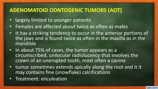
  • 25. ADENOMATOID ODNTOGENIC TUMORS (AOT) • largely limited to younger patients • Females are affected about twice as often as males • It has a striking tendency to occur in the anterior portions of the jaws and is found twice as often in the maxilla as in the mandible • In about 75% of cases, the tumor appears as a circumscribed, unilocular radiolucency that involves the crown of an unerupted tooth, most often a canine • tumor sometimes extends apically along the root and it it may contains fine (snowflake) calcifications • Treatment: enculeation
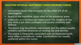
  • 27. CALCIFYING EPITHELIAL ODNTOGENIC TUMOR (PINDBORG TUMOR) • uncommon lesion that accounts for less than 1% of all odontogenic • found in the mandible, most often in the posterior areas • unilocular or a multilocular radiolucent The margins of the lytic defect are often scalloped and usually relatively well defined • The lesion may be entirely radiolucent, but the defect usually contains calcified structures of varying size and density. • The tumor is frequently associated with an impacted tooth, most often a mandibular molar. Calcifications are usually scattered within the tumor.
  • 28. CALCIFYING EPITHELIAL ODNTOGENIC TUMOR (PINDBORG TUMOR)
  • 29. SQUAMOUS ODNTOGENIC TUMOR (SOT) • Squamous odontogenic tumor is locally aggressive neoplasm • Develop in the periodontal ligament between the roots of vital, erupted permanent teeth. Mobility of teeth, local pain, swelling of the gums, osseous expansion, or mild gingival erythema may be observed • Conservative surgical treatment is considered to be sufficient. Recurrences are rare.
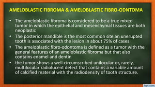
  • 32. AMELOBLASTIC FIBROMA & AMELOBLASTIC FIBRO-ODNTOMA • The ameloblastic fibroma is considered to be a true mixed tumor in which the epithelial and mesenchymal tissues are both neoplastic • The posterior mandible is the most common site an unerupted tooth is associated with the lesion in about 75% of cases • The ameloblastic fibro-odontoma is defined as a tumor with the general features of an ameloblastic fibroma but that also contains enamel and dentin • the tumor shows a well-circumscribed unilocular or, rarely, multilocular radiolucent defect that contains a variable amount of calcified material with the radiodensity of tooth structure.
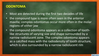
  • 36. ODONTOMA • Odontomas are the most common types of odontogenic tumors. Their prevalence exceeds that of all other odontogenic tumors combined • Odontomas are considered to be developmental anomalies (hamartomas), rather than true neoplasms. When fully developed odontomas consist chiefly of enamel and dentin, with variable amounts of pulp and cementum
  • 37. ODONTOMA • Odontomas are further subdivided into compound and complex types. • The compound odontoma is composed of multiple, small tooth-like structures. • The complex odontoma consists of a conglomerate mass of enamel and dentin, which bears no anatomic resemblance to a tooth. In most series, compound odontomas are more frequently diagnosed than complex
  • 39. ODONTOMA • Most are detected during the first two decades of life • the compound type is more often seen in the anterior maxilla; complex odontomas occur more often in the molar regions of either jaw • the compound odontoma appears as a collection of tooth- like structures of varying size and shape surrounded by a narrow radiolucent zone. The complex odontoma appears as a calcified mass with the radiodensity of tooth structure, which is also surrounded by a narrow radiolucent rim
  • 41. ODONTOMA • An un-erupted tooth is frequently associated with the odontoma, and the odontoma prevents eruption of the tooth • Odontomas may erupt!, displace teeth or block their eruption or become involved in cyst formation • Treatment: Odontomas are treated by simple local excision, and the prognosis is excellent.
  • 44. III. TUMORS OF ODONTOGENIC ECTOMESENCHYME
  • 45. MYXOMA • mandible is involved more commonly than the maxilla • Larger lesions are often associated with a painless expansion of the involved bone • may show a “soap bubble” radiolucent pattern, which is indistinguishable from that seen in ameloblastoma • For larger lesions, more extensive resection may be required because myxomas are not encapsulated and tend to infiltrate the surrounding bone
  • 49. CEMENTOBLASTOMA • Cementoblastoma is an odontogenic neoplasm of cementoblasts • Almost 50% involve the first permanent molar. Cementoblastomas rarely affect deciduous teeth • two thirds of reported patients signs of locally aggressive behavior may be observed, including bony expansion, cortical erosion, displacement of adjacent teeth, envelopment of multiple adjacent teeth, maxillary sinus involvement, and infiltration into the pulp chamber and root canals
  • 50. CEMENTOBLASTOMA • Radiographic features: the tumor appears as a radiopaque mass that is fused to one or more tooth roots and is surrounded by a thin radiolucent rim • Treatment: usually consists of surgical extraction of the tooth together with the attached calcified mass.
  • 53. OTHER TUMOURS AND DYSPLASIAS OF CEMENTUM • CEMENTO-OSSEOUS DYPLASIAS These are non-neoplastic proliferations. They are of periodontal ligament origin and all the variants involve the same pathological processes and differ mainly in their extent and radiographic appearances. Treatment is not indicated except rarely for cosmetic reasons