Lec 3 non odntogenic tumors and tumor like lesions of orofacial region
Download as PPTX, PDF17 likes1,212 views

This document discusses various non-odontogenic tumors and tumor-like lesions of the orofacial region. It begins by covering non-odontogenic bone tumors including both benign lesions like ossifying fibroma, osteoma, central giant cell granuloma, and malignant lesions such as osteosarcoma. It then discusses other non-neoplastic bone diseases and soft tissue tumors, providing details on specific lesions like cherubism, fibrous dysplasia, hemangioma and fibroma. For each lesion, it describes characteristics such as location, appearance, symptoms and treatment.


More Related Content


Similar to Lec 3 non odntogenic tumors and tumor like lesions of orofacial region (20)


More from Dr. Haydar Muneer Salih (20)


![lec 14 [Autosaved].pptx](https://cdn.slidesharecdn.com/ss_thumbnails/lec14autosaved-230315142106-831cdef1-thumbnail.jpg?width=560&fit=bounds)


Lec 3 non odntogenic tumors and tumor like lesions of orofacial region
- 1. NON-ODONTOGENIC TUMORS & TUMOR LIKE LESIONS OF THE OROFACIAL REGION DR. HAYDAR MUNIR SALIH BDS, PHD (BOARD CERTIFIED)
- 2. The most important aspect of clinical examination is for the clinician to know what you look for .. Leonardo da vinci call this Saper vedere or.. ‘Knowing how to see’
- 3. NONODNTOGENIC TUMORS I. NON- ODONTOGENIC BONE TUMORS A. Benign: Ossifying Fibroma Juvenile Ossifying Fibroma Osteoma Osteoblastoma and Osteoid Osteoma Central Giant Cell Granuloma Chondroma Hemangioma B. Malignant: Osteosarcoma Chondrosarcoma Ewing Sarcoma Langerhans cell histocytosis Metastatic Tumors to the Jaws
- 4. NONODNTOGENIC TUMORS II. OTHER NON-NEOPLASTIC BONE DISEASES Osteogenesis Imperfecta Osteopetrosis Cleidocranial Dysplasia Fibrous Dysplasia Paget’s Disease of Bone Cherubism Simple Bone Cyst Aneurysmal Bone Cyst
- 5. NONODNTOGENIC TUMORS III.SOFT TISSUE TUMORS A. Benign: Fibroma Giant Cell Fibroma Pyogenic Granuloma Peripheral Giant Cell Granuloma Peripheral Ossifying Fibroma Lipoma Neurofibroma Melanotic Neuroectodermal Tumor of Infancy Hemangioma and Vascular Malformations Lymphangioma
- 6. NONODNTOGENIC TUMORS B. Malignant (sarcomas): Fibrosarcoma Angiosarcoma Liposarcoma Rhabdomyosarcoma Metastases to the Oral Soft Tissues
- 7. OSSIFYING FIBROMA The mandibular premolar and molar area is the most common site. Larger tumors result in a painless swelling of the involved bone The lesion most often is well defined and unilocular varying degrees of radiopacity are noted. Root divergence or resorption of roots of teeth associated with the tumor may be seen. Large lesions demonstrate a characteristic downward bowing of the inferior cortex of the mandible. Treatment: enucleation
- 11. OSTEOMA Osteomas are benign tumors composed of mature compact or cancellous bone. essentially restricted to the craniofacial skeleton Osteomas of the jaws may arise on the surface of the bone, as a polypoid or sessile mass (periosteal, peripheral, or exophytic osteoma), or they may be located in the medullary bone (endosteal or central osteoma) osteomas appear as circumscribed sclerotic masses Larger osteomas of the mandibular body causing symptoms or cosmetic deformity are treated by conservative surgical excision
- 18. OSTEOBLASTOMA & OSTOID OSTEOMA They are closely related benign bone tumors that arise from osteoblasts. Classically, the distinction depends on the size of the lesion, with osteoid osteoma being smaller than 2 cm and osteoblastoma being larger than 2 cm the osteoid osteoma appears as a well-circumscribed radiolucent defect the osteoblastoma may appear as a well-defined or ill-defined radiolucent lesion often with patchy areas of mineralization Most cases of osteoid osteoma and osteoblastoma are treated by local excision or curettage
- 20. OSTOID OSTEOMA
- 21. CENTRAL GIANT CELL GRANULOMA Giant-cell granulomas are hyperplastic rather than neoplastic. More common in females and approximately 70% arise in the mandible. Lesions are more common in the anterior portions of the jaws central giant cell lesions appear as radiolucent defects, which may be unilocular or multilocular.
- 22. CENTRAL GIANT CELL GRANULOMA
- 23. CENTRAL GIANT CELL GRANULOMA central giant cell lesions of the jaws may be divided into two categories 1. Nonaggressive lesions make up most cases, exhibit few or no symptoms, demonstrate slow growth, and do not show cortical perforation or root resorption of teeth involved in the lesion. 2. Aggressive lesions are characterized by pain, rapid growth, cortical perforation, and root resorption. They show a marked tendency to recur after treatment, compared with the non- aggressive types
- 24. NON AGGRESSIVE CENTRAL GIANT CELL GRANULOMA
- 25. AGGRESSIVE GIANT CELL GRANULOMA
- 26. Differential diagnosis of giant cell lesions of the jaws Hyperparathyroidism. Histologically indistinguishable from giant cell granuloma but serum calcium levels are raised) Cherubism. lesions are symmetrical, near the angles of the mandible Aneurysmal bone cysts. may contain many giant cells but consist predominantly of multiple blood-filled spaces Fibrous dysplasia. Only limited foci of giant cells. No defined margins radiographically. Growth ceases with skeletal maturity
- 28. CHONDROMA Chondromas are benign tumors composed of mature hyaline cartilage Most gnathic examples have been found in the condyle or anterior maxilla of adult patient chondromas typically appear as radiolucencies with central areas of radiopacity. It is wise to consider any lesion diagnosed as chondroma of the jaws to represent a potential chondrosarcoma
- 29. CHONDROMA
- 30. HEMANGIOMA OF BONE hemangiomas cause progressive painless swellings which, when the overlying bone is resorbed, may become pulsatile. Teeth may be loosened and there may be bleeding, particularly from the gingival margins involved by the tumor there is a rounded or pseudoloculated radiolucent area with ill-defined margins or a soap-bubble appearance wide en bloc resection is the only practical treatment
- 33. Osteosarcoma Excluding hematopoietic neoplasms, osteosarcoma is the most common type of malignancy to originate within bone The maxilla and mandible are involved with about equal frequency Swelling and pain are the most common symptoms. Loosening of teeth, paresthesia, and nasal obstruction (in the case of maxillary tumors) also may be noted
- 34. OSTEOSARCOMA
- 37. LANGERHANS CELL HISTOCYTOSIS It is a disorder in which excess immune system cells called Langerhans cells build up in the body excess immature Langerhans cells usually form tumors called granulomas. Three forms are recognized; 1. Solitary eosinophilic granuloma 2. Multifocal eosinophilic granuloma (including Hand-Schuller-Christian disease) 3. Letterer-Siwe syndrome.
- 38. HISTCYTOSIS X (Solitary eosinophilic granuloma)
- 40. METASTATIC TUMORS TO THE JAW Although metastasis to a jaw bone may arise from primary carcinomas of any anatomic site, carcinomas of the breast, lung, thyroid, prostate, and kidney give rise to the majority of gnathic metastases pain or swelling of the jaw, and there may be paraesthesia or anaesthesia of the lip. There is typically an area of radiolucency with a hazy outline.
- 41. METASTATIC TUMORS TO THE JAW
- 42. “ ” II. OTHER NON-NEOPLASTIC BONE DISEASES
- 43. OSTEOGENESIS IMPERFECTA (BRITLE BONE SYNDROME) Osteogenesis imperfecta characterized by impairment of collagen maturation It is a rare disorder in addition to bone fragility, some affected individuals also have blue sclera, altered teeth, hypoacusis (hearing loss), long bone and spine deformities, and joint hyper- extensibility. Care must be taken during dental extractions, but fractures of the jaws are uncommon in this disease
- 45. OSTEOPETROSIS (MARBEL BONE DISEASE) characterized by a marked increase in bone density resulting from a defect in remodeling caused by failure of normal osteoclast function anemia is common. Osteomyelitis is a recognized complication and prevention of dental infections is important.
- 46. OSTEOPETROSIS (MARBEL BONE DISEASE)
- 47. CLEIDOCRANIAL DYSPLASIA Partial or complete absence of clavicles allows the patient to bring the shoulders together in front of the chest. This disorder is one of the few recognizable causes of delayed eruption of the permanent dentition. Many permanent teeth may remain embedded in the jaw and frequently become enveloped in dentigerous cysts
- 49. CHERUBISM
- 50. CHERUBISM the disease usually occurs between the ages of 2 and 5 years. In mild cases the diagnosis may not be made until the patient reaches 10 to 12 years of age. The clinical alterations typically progress until puberty, then stabilize and slowly regress. The cherub like facies arises from bilateral involvement of the posterior mandible that produces angelic chubby cheeks. In addition, there is an “eyes upturned to heaven” appearance Radiographically, the lesions are typically multilocular, expansile radiolucencies The appearance is diagnostic as a result of their bilateral location.
- 51. CHERUBISM
- 52. CHERUBISM “eyes upturned to heaven”
- 53. CHERUBISM
- 54. FIBRO-OSSOUS LESIONS Fibro-osseous lesions are a diverse group of processes that are characterized by replacement of normal bone by fibrous tissue Fibrous dysplasia Cemento-osseous dysplasia Periapical cemental dysplasia Focal cemento-osseous dysplasia Florid cemento-osseous dysplasia Fibro-osseous neoplasms Cemento-ossifying fibroma
- 55. MONOSTOTIC FIBROUS DYSPLASIA monostotic fibrous dysplasia are diagnosed during the second decade of life The maxilla is involved more often than the mandible. Teeth involved in the lesion usually remain firm but may be displaced by the bony mass. The chief radiographic feature is a fine “ground- glass” opacification
- 59. CAFÉ AU LAIT
- 61. PAGETS DISEASE OF THE BONE Paget’s disease of bone is a condition characterized by abnormal and anarchic resorption and deposition of bone, resulting in distortion and weakening of the affected bones. The cause of Paget’s disease is unknown Bone pain, which may be quite severe, is a common complaint. The patchy sclerotic areas often are described as having a “cotton wool” appearance On radiographic examination, the teeth often demonstrate extensive hypercementosis.
- 62. PAGETS DISEASE (Lion face)
- 63. PAGETS DISEASE (Cotton wool )
- 64. FIBROMA The fibroma is the most common “tumor” of the oral cavity the most common location is the buccal mucosa along the bite line typically appears as a smooth-surfaced pink nodule that is similar in color to the surrounding mucosa. The irritation fibroma is treated by conservative surgical excision; recurrence is extremely rare
- 65. FIBROMA
- 67. SURGICAL EXCISION OF FIBROMA
- 68. PYOGENIC GRANULOMA The pyogenic granuloma is a common tumor like growth of the oral cavity that traditionally has been considered to be non- neoplastic in nature The surface is characteristically ulcerated and ranges from pink to red to purple, depending on the age of the lesion the mass is painless, although it often bleeds easily because of its extreme vascularity Gingival irritation and inflammation that result from poor oral hygiene may be a precipitating factor in many patients Pyogenic granulomas of the gingiva frequently develop in pregnant women, so much so that the terms pregnancy tumor or granuloma
- 71. PERIPHERAL GIANT CELL GRANULOMA
- 73. LIPOMA The lipoma is a benign tumor of fat Oral lipomas are usually soft, smooth- surfaced nodular masses that can be sessile or pedunculated. The buccal mucosa and buccal vestibule are the most common intraoral sites and account for 50% of all cases treated by conservative local excision, and recurrence is rare
- 74. LIPOMA
- 75. LIPOMA
- 76. SURGICAL EXCISION OF LIPOMA
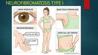
- 77. NEUROFIBROMA Neurofibromas can arise as solitary tumors or be a component of neurofibromatosis Solitary tumors are most common in young adults and present as slow- growing, soft, painless lesions that vary in size from small nodules to larger masses. The tongue and buccal mucosa are the most common intraoral sites The treatment for solitary neurofibromas is local surgical excision, and recurrence is rare.
- 78. NEUROFIBROMA
- 80. MELANOTIC NEUROECTODERMAL TUMOUR OF INFANCY This rare tumour, which arises from the neural crest, may appear in the anterior maxilla in the first few months of life. It is usually painless, and slowly expansive, but occasionally grows rapidly. Radiograpically, there is an area of bone destruction, frequently with ragged margins, and displacement of the developing teeth Conservative treatment is usually curative but, even when excision is incomplete, recurrence is rare
- 81. MELANOTIC NEUROECTODERMAL TUMOUR OF INFANCY
- 82. INFANTILE HEMANGIOMA & VASCULAR MALFORMATION hemangiomas are considered to be benign tumors of infancy that display a rapid growth phase with endothelial cell proliferation, followed by gradual involution Hemangiomas are the most common tumors of infancy, occurring in 5% to 10% of 1-year-old children During the first few weeks of life, the tumor will demonstrate rapid development The proliferative phase usually lasts for 6 to 10 months, after which the tumor slows in growth and begins to involute
- 84. INFANTILE HEMANGIOMA & VASCULAR MALFORMATION About half of all hemangiomas will show complete resolution by 5 years of age, with 90% resolving by age 9 On the other hand, vascular malformations are structural anomalies of blood vessels without endothelial proliferation. By definition, vascular malformations are present at birth and persist throughout life. They can be categorized according to the type of vessel involved (capillary, venous, arteriove- nous) and according to hemodynamic features (low flow or high flow).
- 86. ARTEROVENOUS VASCULAR MALFORMATION (FAST FLOW)
- 87. EMBOLIZATION
- 88. METASTASIS TUMORS TO THE ORAL CAVITY The mechanism by which tumors can spread to the oral cavity is poorly understood. Primary malignancies from immediately adjacent tissues might be able to spread by a lymphatic route; however, such a mechanism cannot explain metastases from tumors from lower parts of the body One possible explanation for blood-borne metastases to the head and neck, especially in the absence of pulmonary metastases, is Batson’s plexus, a valve less vertebral venous plexus that might allow retrograde spread of tumor cells, bypassing filtration through the lungs
- 89. METASTASIS TUMORS TO THE ORAL CAVITY
- 91. Q1 : name the cyst with have the higher tendency to cause root resorption of adjacent teeth ? Q2: name the odontogenic tumor that occur commonly in anterior part of maxilla ?