Lesson 2- Bacteria structuresLesson 2- Bacteria structuresLesson 2- Bacteria structuresLesson 2- Bacteria structuresLesson 2- Bacteria structuresLesson 2- Bacteria structuresLesson 2- Bacteria structures.pptx
Download as PPTX, PDF0 likes417 views

The document provides an overview of bacteria, highlighting their structure, function, and various types, including gram-positive and gram-negative bacteria. It describes the morphology, cell wall composition, and key organelles like the nucleoid, ribosomes, and vacuoles, as well as appendages such as flagella and pili essential for movement and adhesion. Additionally, it addresses the concepts of spores and cysts, detailing their roles in bacterial survival and reproduction under adverse conditions.


Lesson 2- Bacteria structuresLesson 2- Bacteria structuresLesson 2- Bacteria structuresLesson 2- Bacteria structuresLesson 2- Bacteria structuresLesson 2- Bacteria structuresLesson 2- Bacteria structures.pptx
- 2. INTRODUCTION Bacteria are single-celled prokaryotes ubiquitous in nature. They can be found in different types of environments. The bacteria contains a well-developed cell structure, which is responsible for its biological structures and pathogenicity. Many structural features are unique to bacteria and are not found among archaea or eukaryotes. bacteria may exist as free-living organisms, symbiosis with other organisms, as parasitic organisms that can cause harm and disease in their hosts.
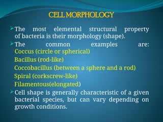
- 3. CELL MORPHOLOGY The most elemental structural property of bacteria is their morphology (shape). The common examples are: Coccus (circle or spherical) Bacillus (rod-like) Coccobacillus (between a sphere and a rod) Spiral (corkscrew-like) Filamentous(elongated) Cell shape is generally characteristic of a given bacterial species, but can vary depending on growth conditions.
- 4. ARRANGEMENT OF BACTERIA CELLS
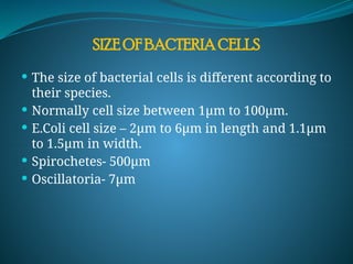
- 5. The size of bacterial cells is different according to their species. Normally cell size between 1µm to 100µm. E.Coli cell size – 2µm to 6µm in length and 1.1µm to 1.5µm in width. Spirochetes- 500µm Oscillatoria- 7µm SIZE OF BACTERIA CELLS
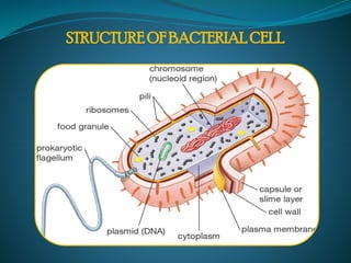
- 6. STRUCTURE OF BACTERIAL CELL
- 7. Bacterial cells mostly two types. Gram Positive bacterial cell Gram negative Bacterial cell TYPES OF BACTERIAL CELL
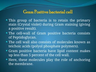
- 8. This group of bacteria is to retain the primary stain (Crystal violet) during Gram staining (giving a positive result). The cell-wall of Gram positive bacteria consists of Peptidoglycan. The cell wall also consists of molecules known as teichoic acids (polyol phosphate polymers). Gram positive bacteria have lipid content makes up less than 5 percent of the cell wall. Here, these molecules play the role of anchoring the membrane. Gram Positive bacterial cell
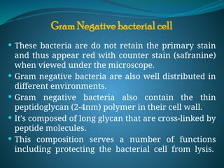
- 9. These bacteria are do not retain the primary stain and thus appear red with counter stain (safranine) when viewed under the microscope. Gram negative bacteria are also well distributed in different environments. Gram negative bacteria also contain the thin peptidoglycan (2-4nm) polymer in their cell wall. It's composed of long glycan that are cross-linked by peptide molecules. This composition serves a number of functions including protecting the bacterial cell from lysis. Gram Negative bacterial cell
- 10. Gram negative bacteria has the presence of different forms of a complex macromolecule known as lipopolysaccharide (LPS). They are particularly important for bacteria in that they contribute to the pathogenesis of the organisms. The other components of Gram negative bacteria include: Cytoplasmic membrane Mesosomes Cytoplasm Nucleoid Endospores
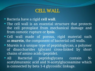
- 11. CELL WALL Bacteria have a rigid cell wall. The cell wall is an essential structure that protects the cell protoplast from mechanical damage and from osmotic rupture or lysis. Cell wall made of porous, rigid material such as murein, the component of bacterial cell walls. Murein is a unique type of peptidoglycan, a polymer of disaccharides (glycan) cross-linked by short chains of amino acids (peptide). All Bacterial peptidoglycans contain N- acetylmuramic acid and N-acetylglucosamine which is connected by beta 1-4 glycosidic linkage.
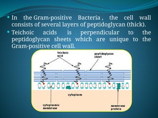
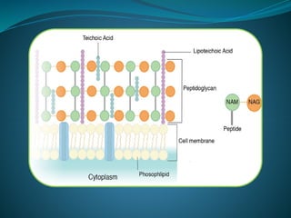
- 13. In the Gram-positive Bacteria , the cell wall consists of several layers of peptidoglycan (thick). Teichoic acids is perpendicular to the peptidoglycan sheets which are unique to the Gram-positive cell wall.
- 15. In the Gram-negative Bacteria, the cell wall is composed of a single layer of peptidoglycan (thin) surrounded by a membranous structure called the outer membrane. The outer membrane of Gram-negative bacteria contains a component, lipopolysaccharide (LPS or endotoxin), which is toxic to animals.
- 16. PLASMA MEMBRANE The plasma membrane, also called the cytoplasmic membrane. Its main function is as a selective permeability barrier that regulates the passage of substances into and out of the cell. The plasma membrane separating the cytoplasm from the environment. The bacterial membrane allows passage of water and small uncharged molecules, but does not allow passage of larger molecules or any charged substances.
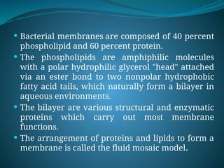
- 17. Bacterial membranes are composed of 40 percent phospholipid and 60 percent protein. The phospholipids are amphiphilic molecules with a polar hydrophilic glycerol "head" attached via an ester bond to two nonpolar hydrophobic fatty acid tails, which naturally form a bilayer in aqueous environments. The bilayer are various structural and enzymatic proteins which carry out most membrane functions. The arrangement of proteins and lipids to form a membrane is called the fluid mosaic model.
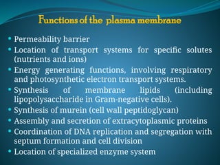
- 19. Functions of the plasma membrane Permeability barrier Location of transport systems for specific solutes (nutrients and ions) Energy generating functions, involving respiratory and photosynthetic electron transport systems. Synthesis of membrane lipids (including lipopolysaccharide in Gram-negative cells). Synthesis of murein (cell wall peptidoglycan) Assembly and secretion of extracytoplasmic proteins Coordination of DNA replication and segregation with septum formation and cell division Location of specialized enzyme system
- 20. Bacterial flagella are long, thin, whip-like appendages that move the bacteria towards nutrients and other attractants. Flagella are free at one end and attached to the cell at the other end. Flagellum can never be seen directly with the light microscope but only after staining with special flagella stains. Flagella are usually found in gram-negative bacilli. Gram-positive rods and cocci also have flagella. FLAGILLA
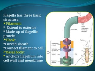
- 21. Flagella has three basic structure: Filament: Extend to exterior Made up of flagellin protein Hook: Curved sheath Connect filament to cell Basal body: Anchors flagellum into cell wall and membrane
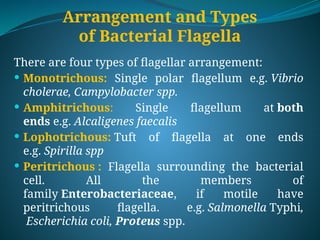
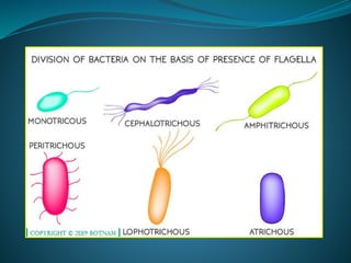
- 22. Arrangement and Types of Bacterial Flagella There are four types of flagellar arrangement: Monotrichous: Single polar flagellum e.g. Vibrio cholerae, Campylobacter spp. Amphitrichous: Single flagellum at both ends e.g. Alcaligenes faecalis Lophotrichous: Tuft of flagella at one ends e.g. Spirilla spp Peritrichous : Flagella surrounding the bacterial cell. All the members of family Enterobacteriaceae, if motile have peritrichous flagella. e.g. Salmonella Typhi, Escherichia coli, Proteus spp.
- 24. Flagella Function They help an organism in movement. They act as sensory organs to detect temperature and pH changes. Few eukaryotes use flagellum to increase reproduction rates. Recent researches have proved that flagella are also used as a secretory organelle. For eg., in Chlamydomonas
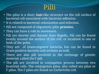
- 25. Pilli The pilus is a short, hair-like structure on the cell surface of bacterial cell associated with bacterial adhesion. It is related to bacterial colonization and infection. Pili are composed of oligomeric pilin proteins. They can have a role in movement. Pili are shorter and thinner than flagella. Pili can be found evenly around the surface of the cell, or localized to one or both of the poles. They are of Gram-negative bacteria, but can be found in Gram-positive bacteria and archaea as well. There are also pili involved in the exchange of genetic material, called the F pili. The pili are involved in conjugation process between two bcaterial cells. The conjugation pilus, also called sex pilus or F pilus. The F pilus are found on Escherichia coli.
- 26. Nucleoid region The nucleoid (meaning nucleus-like) is an irregularly-shaped region within the cell of a prokaryote. It contains all or most of the genetic material. The genome of prokaryotic organisms is a circular, double-stranded piece of DNA, called genophore. The nucleoid, also has no membrane around it. The nucleoid doesn’t take a uniform shape and has no specific size. The nucleoid is mainly composed of multiple copies of DNA with the addition of some RNA and proteins.
- 27. The nucleoid is essential for controlling the activity of the cell and reproduction. It is the site where transcription and replication of DNA take place. It help the formation of DNA, facilitating cell growth, and regulating the genetic material of the cell. Function of Nucleoid
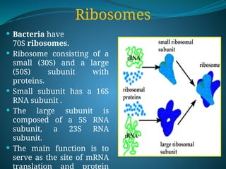
- 28. Ribosomes Bacteria have 70S ribosomes. Ribosome consisting of a small (30S) and a large (50S) subunit with proteins. Small subunit has a 16S RNA subunit . The large subunit is composed of a 5S RNA subunit, a 23S RNA subunit. The main function is to serve as the site of mRNA translation and protein
- 29. Metachromatic Granules Metachromatic granules, sometimes termed as Volutin granules because of their colour reaction with the dyes used in light microscopy. It contain polymerized inorganic phosphate, an energy- rich compound that acts as a reserve store of energy. Polyphosphate granules display the metachromatic effect, appearing red when stained with methylene blue. They can also be found in the cytoplasm of Saccharomyces. Metachromatic granules will stain pink with methylene blue. They are composed of complex polyphosphate, lipid, and n ucleoprotein molecules (volutin) and serve as an intracell ular phosphate reserve.
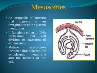
- 30. Mesosomes An organelle of bacteria that appears as an invagination of the plasma membrane. It functions either in DNA replication and cell division or excretion of exoenzymes. Mature mesosomes formed a link between the cytoplasmic membrane and the nucleus of the cell.
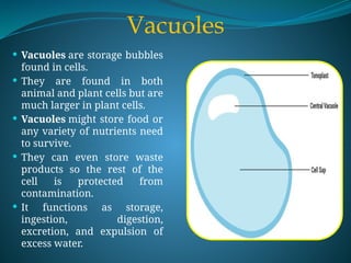
- 31. Vacuoles Vacuoles are storage bubbles found in cells. They are found in both animal and plant cells but are much larger in plant cells. Vacuoles might store food or any variety of nutrients need to survive. They can even store waste products so the rest of the cell is protected from contamination. It functions as storage, ingestion, digestion, excretion, and expulsion of excess water.
- 32. Vacuoles are essentially enclosed compartments which are filled with water containing inorganic and organic molecules including enzymes. Vacuoles has outer membrane called tonoplast. Vacuoles are formed by the fusion of multiple membrane vesicles. The organelle has no basic shape or size; its structure varies according to the requirements of the cell. It maintain an acidic internal pH. It containing small molecules. Exporting unwanted substances from the cell. Vacuoles also play a major role in autophagy.
- 33. Spores are the single-celled reproductive unit of bacteria, fungi, algae and non-flowering plants. Spores are units of asexual reproduction. They can produced a new bacteria or plants. They are able to travel over long distances. Some bacteria also produce spores as to survive in very harsh conditions. A spore is a single cell surrounded by a thick cell wall for protection. Once the spores are formed, the organism releases them into the environment to grow. Spores are often formed through a process called sporogenesis, through mitosis, or cellular reproduction. Spores
- 35. Bacterial spores serve as a resting, or dormant, stage in the bacterial life cycle, help to preserve the bacteria through unfavourable conditions. Bacillus and Clostridium bacteria generally produced spores. Spores are haploid in nature, germinate and produced haploid gametophyte. Spores are classify into endospore and exospore depending on the position. Endospore are generally produced by bacteria and exospore are mainly produced by eukaryotes, fungi and algae. Both have low metabolic rate and non- nutritive. They are highly resistant.
- 36. Cyst A microbial cyst is a resting or dormant stage of a microorganism, that helps the organism to survive in unfavorable environmental conditions. It is a state, in which the metabolic processes of the cell are slowed and the cell ceases all activities like feeding and locomotion. Unfavorable environmental conditions such as lack of nutrients or oxygen, extreme temperatures, lack of moisture and presence of toxic chemicals, which are not suitable for the growth of the microbe trigger the formation of a cyst.
- 37. The cyst walls of bacteria are formed by the thickening of the normal cell wall with added peptidoglycan layers . Cyst is a group of cells, which come together to protect the organism from harsh environmental conditions. Cysts are not reproductive cells.
- 38. Thank You