Microscope
Download as PPTX, PDF17 likes4,241 views

The document provides a comprehensive overview of microscopy, detailing its definition, history, types, and components, along with their functions. It discusses various microscopic techniques including optical, electron, and scanning probe microscopy, as well as methods for specimen preparation and staining. Additionally, it covers the principles of magnification, resolving power, and specific staining procedures to enhance the visualization of microorganisms.


Microscope
- 1. Microscopic Observations of Microorganisms MUHAMMED MAHFUZUR RAHMAN Assistant Professor Department of PHARMACY
- 2. Introduction & Definition The word Microscope comes from Greek word “mikrós” which means “small” & “skopeîn” which means “to look” or “see”. Microscopes are instruments designed to produce magnified visual or photographic images of small objects. A microscope is an instrument used to see objects that are too small for the naked eye.
- 3. Introduction & Definition The science of investigating small objects using such an instrument is called microscopy. Microscopic means invisible to the eye unless aided by a microscope. Zacharias Janssen invented the microscope in 1590. The microscope must accomplish three tasks- Produce a magnified image of the specimen Separate the details in the image Render the details visible to the human eye or camera
- 5. Parts of Microscope and their functions Eyepiece Ocular lens Nosepiece Objective lens Stage Stage clip Light switch Light intensity knob Fine adjustment Coarse adjustment Stage manipulator knobs Condenser Light source Iris diaphragm knob Cord holder Microscope body
- 6. Parts of Microscope and their functions Arm Light Source Diaphragm Stage Stage Clips Revolving Nosepiece Objective Lenses Ocular Lens Fine adjustment knob Coarse adjustment knob
- 7. Parts of Microscope and their functions Eyepiece and Body Tube The eyepiece contains the ocular lens which magnifies objects a given amount that is listed on the eyepiece. The body tube supports the eyepiece and objectives Nosepiece, Objectives, and Stage Clips The nosepiece holds the 3 objectives. The objective lenses range in magnification from 4X, 10X, and 40X. The stage clips holds the slide in place.
- 9. Parts of Microscope and their functions Stage, Light & Diaphragm The stage supports the slide being viewed. The light source projects upward through the diaphragm, the specimen and the lenses. The diaphragm regulates the amount of light on the specimen.
- 10. Parts of Microscope and their functions Arm and Base The arm is used to support the microscope when it is carried. The base supports the microscope.
- 11. Parts of Microscope and their functions Coarse Adjustment Knob Moves the stage up and down for focusing. Fine Adjustment Knob Moves the stage slightly to sharpen the image. Used with the 10X and 40X objective to focus.
- 12. Types of Microscopes Optical Microscope This is an optical instrument using visible light and a system of lenses to magnify images of small samples. Typical magnification is up to 1250x with a theoretical resolution limit of around 0.250 micrometres or 250 nanometres. It includes: Bright-field microscopy Dark-field microscopy Fluorescence microscopy Phase contrast microscopy
- 13. Types of Microscopes Electron Microscope Passes electrons through the sample or looks at the surface of bulk objects by scanning the surface with a fine electron beam. Types: Transmission electron microscope (TEM) & Scanning electron microscope (SEM) Objective Lens
- 14. Types of Microscopes Scanning probe Microscope Scanning probe microscopes also analyze a single point in the sample and then scan the probe over a rectangular sample region to build up an image. Many scanning probe microscopes can image several interactions simultaneously. The manner of using these interactions to obtain an image is generally called a mode. Different types: AFM (atomic force microscopy) , BEEM (ballistic electron emission microscopy), CFM (chemical force microscopy) etc.
- 15. Preparations for Microscopic Examinations The Wet-Mount & Hanging drop technique Wet preparations permit examination of organisms in a normal living condition. A wet mount is made by placing a drop of fluid containing the organisms onto a glass slide & covering the drop with a cover slip. A special slide with a circular concave is sometimes used. A suspension of microbial specimen is placed on a cover slip, then inverted over the concave depression to produce a “hanging drop” of the specimen.
- 16. Preparations for Microscopic Examinations The Fixed, stained smears They are most frequently used for the observations of the morphological characteristics of microbes. The essential steps are: The preparations of the film or smear Fixation Application of one or more staining solutions Advantages are: More clearly visible after staining Differentiation of cells of diff. species or within the same species
- 17. Different types of Microscopy
- 18. Bright-Field Microscopy Dark sample on a bright background The Microscopic field is brightly lighted & the microbes appear dark because they absorb some of the light Usually, microbes do not absorb much light, but staining them with a dye greatly increases their light absorbing capacity Bright field illumination is useful for samples which have an intrinsic colour, for example chloroplasts in plant cells. Some of the light is absorbed by stains, pigmentation, or dense areas of the sample and this contrast allows us to see the specimen.
- 20. Dark-Field Microscopy Dark background against which objects are brilliantly illuminated. This is accomplished by equipping with a special condenser that transmits a hollow cone of light. Most of the light directed through the condenser does not enter the objective, the field is dark. However, some of the light rays will be scattered if the medium contains objects. The diffracted light will enter the objective & reach the eye, thus the object will appear bright in an dark background. Best for observing pale objects, unstained microbes.
- 23. Fluorescence Microscopy After absorbing light of a particular wavelength & energy, some substances will then emit light of a longer wavelength & a lesser energy content. The phenomenon is termed fluorescence. Application of this phenomenon is the basis of fluorescence microscopy. In practice, microbes are stained with fluorescent dye & then illuminated with blue light; the blue light is absorbed & green light is emitted by the dye. Example of fluorescent dyes: Fluorochrome Excitation Emission Fluorescein Isothiocyanate (FITC) ~480 nm (Blue) ~520 nm (Green) Rhodamine Isothiocyanate (RITC) ~540 nm (Green) ~590 nm (Red)
- 25. Phase-Contrast Microscopy The phase contrast microscopy made it possible to study living cells and how they proliferate through cell division. Used to examine living organisms or specimens that would be damaged/altered by attaching them to slides or staining. It uses a conventional light microscope fitted with a phase-contrast objective & phase-contrast condenser. Light passing through one material & into another material of slightly different refractive index or thickness will undergo a change in phase. This change in are translated into variations in brightness of the structures.
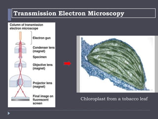
- 27. Transmission Electron Microscopy Electrons scatter when they pass through thin sections of a specimen Transmitted electrons (those that do not scatter) are used to produce image Denser regions in specimen, scatter more electrons and appear darker Allows the observation of molecules within cells
- 28. Transmission Electron Microscopy Chloroplast from a tobacco leaf
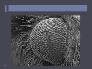
- 29. Scanning Electron Microscopy The specimen is subjected to a narrow electron beam which rapidly move over the surface of the specimen. This causes the release of a shower of a secondary electrons & other type of radiations. The intensity of these secondary electrons depends on the shape & chemical composition of the irradiated objects. The secondary electrons are collected by a detector which generates an electrical signal. These signals are then scanned in the manner of a television system to produce an image.
- 31. Scanning Electron Microscopy Images
- 32. Optical Microscope vs Electron Microscope FEATURE Optical Microscope Electron Microscope Electromagnetic spectrum used Visible light 760nm (red) – 390nm Electrons app. 4nm Maximum resolving power app. 200nm 0.2nm Maximum magnification x1000 – x1500 x500 000 Radiation source Tungsten or quartz halogen lamp High voltage (50kV) tungsten lamp Lenses Glass Magnets Interior Air-filled Vacuum Focussing screen Human eye (retina) fluorescent (TV) screen
- 33. Optical Microscope vs Electron Microscope FEATURE Optical Microscope Electron Microscope Preparation of specimens Temporary mounts living or dead Tissues must be dehydrated (dead) Fixation Alcohol OsO4 or KMnO4 Embedding Wax Resin Sectioning Hand or microtome slices 20 000nm Microtome only. Slices 50nm Stains Water soluble dyes Heavy metals Support Glass slide Copper grid
- 34. Optical Microscope vs Electron Microscope Image produced by an optical microscope Image produced by an electron microscope
- 35. The limits of resolution
- 36. Some Important Terminals used in Microscopy
- 37. Resolving power The ability to distinguish two adjacent points as distinct & separate is known as resolving power. Mere increase in size without the ability to distinguish structural details is not beneficial. In other words, the largest magnification produce by a microscope may not be the most useful because the image obtained may be unclear or fuzzy. The more lines or dots per unit area that can be seen separately, the greater is the resolving power. It is a function of the wavelengths of lights used & the numerical aperture of the lens system.
- 38. Numerical Aperture In optics, the numerical aperture (NA) of an optical system is a dimensionless number that characterizes the range of angles over which the system can accept or emit light. The sine value of half-aperture angle multiplied by the refractive index n of the medium gives the numerical aperture (NA) Thus, NA= n sin θ
- 39. The limit of Resolution The limit of resolution is the smallest distance by which two objects can be separated & still be distinguished as two separate objects. The greatest resolution in optical microscopy can be obtained with the shortest wavelength of visible light & an objective with maximum NA. The relationship between NA & limit of resolution can be expressed as follows: d= 2NA λ Here, d= Resolution λ= Wavelength of light
- 40. Magnification Magnification beyond the resolving power is of no value since the larger image will be less distinct in detail & fuzzy in appearance. The situation is analogous to the of a movie screen: If we move closer to the screen the image is larger but is also less sharp than when viewed from distance. Most laboratory microscopes are equipped with three objectives, each capable of a different degree of magnification. The total magnification of the system is determined by magnification of the objective by that of eyepiece.
- 41. Lets play a GAME!! Look at the following micrographs (a picture made by a microscope) and try to determine what the object is!
- 42. DENTIST’S DRILL
- 43. PAPER
- 45. EYE OF A NEEDLE WITH THREAD RUNNING THROUGH IT
- 46. ORANGE JUICE
- 47. SNOW
- 48. BROCOLLI
- 49. SMALL INTESTINE
- 51. SURFACE OF THE TONGUE
- 53. FINGERPRINT
- 54. SALT
- 55. HUMAN TOOTH
- 56. BANANA
- 57. CLOT OF BLOOD
- 58. MASCARA BRUSH
- 59. GUITAR STRING
- 60. ANT
- 61. BLACK FLY
- 62. MOSQUITO
- 63. MITE FEEDING
- 64. POLLEN GRAIN
- 65. ANT EYE
- 66. APHID ON A LEAF
- 67. EYELASHES
- 68. VELCRO
- 69. POLLEN
- 70. CHALK
- 71. HEAD OF A BUTTERFLY
- 72. CHEEK CELL
- 73. FLEA
- 74. A HAIR FOLICLE
- 75. RUST
- 77. A HUMAN CELL INFECTED WITH HIV VIRUS
- 79. Staining of Specimens Increases contrast & resolution by coloring specimens with stains/dyes. Smear of microorganisms (thin film) made prior to staining. Microbiological stains contain chromophore. Acidic dyes stain alkaline structures; more commonly, basic dyes stain acidic structures. Types of staining: Simple staining Differential staining o Gram stain o Acid fast stain o Endospore stain Special staining o Negative (capsule) stain o Flagellar stain
- 80. Fixation Process by which internal and external structures are preserved and fixed in position Process by which organism is killed and firmly attached to microscope slide Heat fixing o preserves overall morphology but not internal structures Chemical fixing o protects fine cellular substructure and morphology of larger, more delicate organisms
- 81. Dyes and Simple Staining Dyes Make internal and external structures of cell more visible by increasing contrast with background Have two common features: o Chromophore groups Chemical groups with conjugated double bonds which give dye its color o Ability to bind cells Simple staining A single staining agent is used Basic dyes are frequently used o Dyes with positive charges o E.g. crystal violet
- 82. Dyes and Simple Staining
- 83. Differential Staining Use a primary stain & a counter stain to distinguish cell types or parts. Divides microorganisms into groups based on their staining properties E.g., Gram stain, acid-fast stain, Schaffer-Fulton endospore stain etc. Endospore Stain Gram Stain Acid-fast Stain
- 84. Gram Staining Most widely used differential staining procedure. The name comes from its inventor, Hans Christian Gram. Gram staining differentiates bacteria by the chemical & physical properties of their cell walls by detecting peptidoglycan, which is present in a thick layer in gram-positive bacteria. Primary stain Positive Negative Mordant Decolorization Counter stain
- 85. Gram Staining
- 86. Gram Staining A Gram stain of mixed Staphylococcus aureus (gram-positive cocci, in purple) and Escherichia coli (gram-negative bacilli, in red)
- 87. Acid-fast Staining Particularly useful for staining members of the genus Mycobacterium. E.g., Mycobacterium tuberculosis – causes tuberculosis E.g., Mycobacterium leprae – causes leprosy High lipid content in cell walls is responsible for their staining characteristics
- 88. Schaeffer-Fulton endospore Staining Double staining procedure. Bacterial endospore is one color & vegetative cell is a different color.
- 89. Negative Staining Often used to visualize capsules surrounding bacteria Capsules are colorless against a stained background. Bacteria Capsule Background stain
- 90. Flagellar Staining Bacterial flagella are normally too thin to be seen under normal microscopic conditions. The flagella stains employs a mordant to coat the flagella with stain until they are thick enough to be seen. Flagella