SlideShare a Scribd company logo
OPEN FRACTURE
MANAGEMENT
BY DR ANIL KUMAR P(junior resident)
MODERATOR : DR NAGAKUMAR J S
PROFESSOR
DEPT OF ORTHOPAEDICS
SDUMC
Definition
• Open fracture by definition is communication
between external environment and the
fracture.
• A soft tissue injury complicated by a broken
bone.
• Egyptians 1sttime recognised he need for coverage over
fracture wounds to minimise morbidity.
• Hippocrates favoured the debridementof purulent
material.
• Ambroise Parèalso favoured the debridement asthe
mainstay of treatment ofopen
HISTORY
• Desault (coined the term ‘debridement) in18th century
reiterated this belief by advising debridement of dead
necroticmaterial.
• Still in the 19thcentury, prior to aseptic technique &
antibiotics, emergency amputation wasthe life saving
measureafter open fracture.
• Lastcentury, high mortality with openfractures of longbones
• WWI, mortality of open femur fractures>70%
• Bythe WWI the main principle of treatmentwas debridement and
stabilization & all healing by secondary intention.
• 1939 Trueta “closed treatment of warfractures”
– Included open wound treatment and thenenclosure of the extremity ina
cast
– “Greatest danger of infection lay in muscle, not bone”
• During the WW-II useof local antibioticslike
sulfonamides wasstarted for wound treatment.
•Useof penicillinon the battlefield quicklyreduced the
rate of woundsepsis.
• Advances shifted the focus
Preservation of life and limb
preservation of function and
prevention of complications
• However, amputation rates still exceed 50% in the most
severe open tibial fractures associated with vascular
injury.
Epidemiology
• Highest rate of fractures are seen in tibia followed femur
• Diaphyseal > metaphyseal
• Generally aresult of high energy mechanisms which cause greater soft
tissue disruption that leaves the wound more susceptible to infection by
contaminatingbacteria.
• Most Common Causes of Death in the United States Among
Individuals 25 to 44 Years Old
• Fracture pattern
• Local factors
– Amount of foreign debris and contaminant
material
– Extent of soft tissue and bonedevitalization.
• Systemicfractures
– Host nutrition
– Medications
– Nicotine abuse
Prognosis
COMPONENTS OF OPENFRACTURE
10
• Fracture
• Soft-tissue damage
• Neurovascularcompromise
• Contamination.
Open fracture classification
• Allows comparison of results
• Provides guidelines on prognosis and treatment
– Fracture healing, infection and amputation rate correlate
with the degree of soft tissue injury
• Gustilo upgraded to Gustilo and Anderson
• AO open fracture classification
Gustilo andAnderson
12
Gustilo andAnderson
13
14
15
16
17
Nerve
Sensate No major nerve 1
injury
Dorsal Deep peroneal 2
nerve
Plantar partial Tibial nerveinjury 3
Plantar complete Sciaticnerve 4
Ischemia
None Good to fair pulses, 0
no ischemia
Mild Decreasedpulses 1
perfusion
Moderate Prolongedcapillary2
refill,Doppler
pulses fill
Severe Pulseless, cool, 3
ischemic,no
doppler
SoftTissue
GradeI Minimal 0
contamination
GradeII Moderatesoft 1
tissue injury, low
velocity
GradeIIIA Moderatecrush 2
injury, high velocity
with Considerable
contamination
GradeIIIB Massivecrush 3
injury severe
contamination
Skeletal
Spiral or oblique 0
fracture
Transverse 1
fracture-minimal
contamination
Moderate 2
displacement and
communitionwith
high velocity
Segmental 3
fracture, severe
communition,bony
loss
Shock
Normotensive 0
Transient 1
hypotensive
Persistent 2
hypotensive
18
• Gradesof soft tissue injury correlates withinfection
and fracture healing
Grade 1 2 3A 3B 3C
Infection Rates
0-2% 2-7% 10-25% 10-50% 25-50%
Fracture
Healing (weeks) 21-28 28-28 30-35 30-35
Amputation Rate
50%
Tscherne Classification for Open Tibial
Fractures
• This system includes soft-
tissue damage and
compartment syndrome,
which are not included in
other grading schemes
AO-ASIF Soft-Tissue Injury
Classification
• The AO-ASIF(Association for
the Study of Internal Fixation)
group added to their
extensive fracture
classification a soft-tissue
classification scheme that
closely follows that of
Tscherne and Gotzen.
• This classification includes
both closed and open injuries,
musculotendinous injury, and
neurovascular injury
Proposed Classification of Open
Fractures
(OTA 2010)
This new classification uses five
categories of assessment: skin injury,
muscle injury, arterial injury,
contamination, and bone
loss.
This provides a systemic approach to
classification at the time of arrival
before treatment has occurred.
• Number of other trauma scoring systems have been proposed,
including
• The trauma score (TS),
• Revised trauma score (RTS),
• Injury severity score (ISS),
• Modified abbreviated injury severity scale (MISS),
• Pediatric trauma score (PTS),
• Nerve injury, ischemia, soft-tissue injury, skeletal injury, shock, and
age of patient score (NISSSA), and the
• Hanover fracture scale-97 (HFS-97)
Common bacteria encountered with
open fractures
• Blunt Trauma, LowEnergy trauma
• Farm Wounds
• Fresh Water SeaWater wounds
• War Wounds,
• High Energy gun shot wounds
Staph,Strept
Clostridia
Pseudomonas,Aeromonas
Aeromonas, Vibrios
GramNegative
MANGLED EXTREMITY SEVERITYSCORE
• Given by Johansenin 1990.
• Astrong weightage wasgiven for the presence of warm
ischemia time and anage above 30 years.
• Johansenreported that ascore of 7 ormore predicted
amputation with 100%accuracy.
Mangled Extremity SeverityScore
Ganga Hospital Open InjurySeverity Score
• GHOISS was proposed by Rajasekaran
in 2006 as a score specifically to
assess severe Grade IIIB limb injuries
without a vascular injury.
23
Open fracture management
• Injuries with ascoreof
14 and below should be attemptedfor salvage,
17 and above should be consideredfor primary
amputation,
in between(15,16) must be assessedbyan
experienced team on acase-to-casebasis.
Injury Severity Score for Gustilo Type
IIIA and IIIB Open Tibial Fractures
Management
Goalsof treatment
•
•
•
1. preserve life
2. preserve limb
3. preserve function
– Prevent infection
– Fracture stabilization
– Soft tissue coverage
Stagesof care for openfractures
Initial assessment
traumatized
• Important components in assessing
extremity are
1. History and mechanism of injury
2. Neurovascular status
3. Sizeof skin wound
4. Muscle crush or loss
5. Periosteal stripping or boneloss
6. Fracture pattern, fragmentation
7. Contamination
8. Compartment syndrome.
Open fracture management
Irrigation
• Supplements systemic debridement by
removing foreign material anddecreasing
bacterial load.
• Simpulse irrigation system(HPPL-high
pressure pulsatile lavage).
• Pressure must be lessthan 50psiunits.
Fracturetype Volof fluid usedfor irrigation
Type I 3 L
Type 2 6L
Type 3 9L
Irrigation
NS normally used for
irrigation.
Antibiotic solution has no
advantage than soap for
irrigation.
Surfactant(non sterile soap):
same effectiveness, less
tissue damage and more
economical.
Open fracture management
Timing of debridement and
irrigation
• Most guidelines recommenddebridement within 6
hrs and If clean Primaryclosure.
• Serial debridement may be necessaryevery
24-72hrs ifdebrima is delayed until the wound viability
isensured.
• If clean within 2-3 daysdelayed Primary Closure.
• Later Secondary closure (Flaps)or healingby Secondary
intentions (scarring )
Open fracture management
ANTIBIOTICS AND INFECTION
• 24-70% of open fractures are contaminated with bacteria
• 14-15% of open complicated fracture with infection in the
absence of antibiotic prophylaxis
Antibiotics
• Systemicadministration:
Antibiotics
• Localantibiotics:-In gustilo type III fractures additional
useof local aminoglycoside impregnated
polymethylmethacrylate(PMMA) beads reduces overall
infection rate.
Antibiotic bead pouchtechnique
Tetanus prophylaxis
• Tetanus Toxoid(TT),dose is 0.5ml i.m.
regardless of age.
• Immunoglobulin:-
75IU <5yrs of age
125IU 5-10yrs
250IU >10yrs.
Open fracture management
Typesof fracture stabilization
Splint
– Good option if operative fixation not
required
Internal fixation
– Wound is clean and softtissue coverage
available
External fixation
– Dirty wounds or extensivesoft tissue injury
Primary surgery
Timing :
•Surgical emergency
•Operating within 6-8hrs of injury – contaminated
wounds not treated within this time will have sustained
bacterialmultiplication to result in earlyinfection.
Primary surgery
Fracture stabilization:
•Assoon asprimary wound care is completed,
treatment should proceed to fracturereduction and
fixation.
•Surgeon should rescrub and regown.
•Different set of instruments thanthose used for
debridement isnecessary.
Fixation Options
External Fixation VsInternal Fixation
Indications for External Fixation in OpenFractures
1) Severecontamination anysite
2) Periarticular fractures
– Definitive
• Distal radius
• Elbow dislocation
– Relative
• Knee
• Ankle
• Elbow
• Wrist
External fixation
• Advantages of ExtFixation:-
• Canbe applied relatively easily and quickly,
• It provides relatively stable fracturefixation,
• There is no further damagedone if applied correctly,
• Itavoids implantation of hardware in open wound.
External Fixator
• Disadvantages:
• Major problems with external fixation are
related to pin tract infection, malalignent ,
delayed union, poor patientscompliance.
• Ringfixators may be an optionin open
diaphyseal fractures like ilizarov.
Indications for Internal Fixation in Open
Fractures
1)Periarticular fractures
– Distal/proximal tibia
– Distal/proximal
femur
– Distal/proximal
humerus
– Proximal ulnar radius
– Selected distal
radius/ulna
– Acetabulum/pelvis
2) Diaphyseal fractures
– Femur
– Tibia
– Humerus
– Radius/ulna
Plates
• Open diaphyseal fractures of the radius andulna as
well asthe humerus are best managedwith plate
fixation.
• The plate fixation of lower extremity diaphyseal
fractures is generally not recommended due to
higher rate of infections.
Open fracture management
INTRAMEDULLARY NAILING
• Locked intramedullary nailing has been established as
the treatment of choice for most diaphyseal fractures
in lower extremity.
• Intramedullary nails can be inserted with no further
disruption of the already injured soft-tissue envelope
and preserves the remaining extra osseous blood
supply to corticalbone.
IM NAILING
• Data showing that solid intramedullary nails inserted without
reaming have a lower risk of infection.
• On the other hand reamed intramedullary nails can reliably
maintain fracture reduction with regards to angulation, rotation,
displacement, and length.
• Prospective randomised trails that compared reamed with
unreamed interlocked IM nails did not show any significant
difference concerning outcome and risk of complication.
Open fracture management
Open wound coverage after primary
surgery
Primary Closure
The following criteria must be met:-
1.The original wound must have been fairly clean, and not have occurred in
a highly contaminated environment.
2.All necrotic tissue and foreign material have been debrided.
3.Circulation to the limb is essentially normal.
4.Nerve supply to the limb is intact.
5.The patient's general condition is satisfactory and allows careful
postoperative assessment.
6.The wound can be closed without tension.
7.Closure will not create a dead space.
8.The patient does not have multisystem injuries.
Delayed primary closure
• Closure before the 5th day is termed delayed primary
closure.
• As long as closure is achieved before the fifth day, wound
strengths at 14 days are comparable with those in wounds
closed on the first day.
• Leaving the wound open minimizes the risk of anaerobic
infection, and the delay allows the host to mount local
wound defensive mechanisms that permit safer closure than
is possible on the first day.
• Current standard of care for all open fracture wounds
is to be left open initially.
• Delayed closure is accomplished within 2- 7days
• VAC assisted wound closure is presently
recommended for temporary management of open
fracture wounds.
VAC
• Thewound bed is exposed to mechanically induced negative
pressure in aclosed system.
• Thesystem removes fluid from extravascular space,reduces
edema, improves micro circulation and enhancesthe
proliferation of preparative granulation tissue.
• Polyurethane foam dressing is placed in wound and ensures
an even distribution of negative pressure.
VAC
Contraindications to primary
closure
• Inadequate debridement
• Grosscontamination
• Farmrelated or freshwater immersioninjuries
• Delay in treatment >12hours
• Delay in givingabx
• Compromised host or tissueviability
Dressings
• Temporary closures – rubber bands
• Wet to dry dressings
• Semi-permeable membranes
• Antibiotic bead pouch
• VAC
Reconstructive ladder: options for wound
coverage
Type1open fx
Type3Bopen
fx
Type2/3A open fx
FLAPCOVERAGE FOR TYPE 3B
Gunshot injuries
• Energydissipated at impact =damage severity
• High velocity rifles and closerange shotguns
– Worst, high energy of impact
– Huge secondary cavitation
– Secondary effects of shatteredbone
fragments
• Bullets lodged in joints should beremoved
– avoid lead arthropathy and systemiclead
poisoning
Low velocity GSW<2000ft/sec
• Lowvelocity handguns
– Lesssevere, not treated like openfractures
– Cavitation is not significant
– Secondary missile effects are minimal
– Bone fragments rarely stripped of softtissue
attachments and blood supply
– Soft tissue injuries not severe and skin woundsare
small
ADVANCES
• BMPs
– 40%decreased infection rate with BMPin type3 open tibia
fractures
• Antibiotic LadenBoneGraft
– Tobramycin-impregnated calcium sulfate pellets with
demineralized bonematrix
– successful in preventinginfection
*BESTTStudyGroup, Govender S,CsimmaC,Genant H, Valentin-OpranA. “Recombinant HumanBoneMorphogenetic Protein-2 for Treatment of OpenTibial Fractures:
Aprospective, controlled, randomized study of four hundred andfifty patients.” JBJS-A2002. 84(12): 2123-2134.
**Beardmore AA,BrooksDE,WenkeJC,ThomasDB.“Effectiveness of local antibiotic delivery with an osteoinductive and osteoconductive bone-graft
substitute.” JBJS-A2005. 87(1): 107-112.
S
ummary
Complications
• Hypovolemic shock
• Compartment syndrome
• Fat embolism
• ARDS
• Neurovascular injuries
• Infection
• Morel-Lavallee syndrome
Hypovolemic shock
Hypovolemic shock - management
• Two large-bore IV lines should be started.
• Once IV access is obtained, initial fluid
resuscitation is performed with an isotonic
crystalloid, such as Ringer lactate solution or
normal saline.
• An initial bolus of 1-2 L is given in an adult (20
mL/kg in a pediatric patient), and the patient's
response is assessed.
HYPOVOLEMIC SHOCK
• Type of fluid:-
• Colloid – albumin, dextran, plasma. Crystalloid – NS, D5, RL.
• Blood – uncrossed ‘O’ –ve.
• Basic Rule:-
• 3:1 rule when using crystalloids.Eg. If blood
loss is 100cc the patient should receive
300cc of normal saline or Ringer lactate.
• 1:1 rule for colloids.
Open fracture management
Morel-Lavallee syndrome
• Skin is separated from the fascia.
• This creates a pocket under which considerable bleeding can occur.
• Usually this is a subcutaneous hematoma, but the hematoma can
become so large that it seriously threatens the viability of the skin
above it .
• This syndrome occurs frequently in patients with pelvic fractures,
especially in obese individuals.
• MRI and ultrasonography have been recommended to confirm the
diagnosis.
• Multiple treatments present but PERCUTANEOUS DRAINAGE is
mostly accepted and done.
Compartment syndrome
34
• Classically 5 "Ps" associated with
compartment syndrome:—
1)Pain out of proportion to what is expected
2)Paresthesia
3)Pallor
4)Paralysis
5)Palpable pulse absent.
Compartment syndrome
• Treatment -Fasciotomy
Fat embolism
• Definition - Occlusion of small vessels by fat globules.
• Types:-
1.cerebral – drowsy, restless and disoriented.
2.pulmonary – tachypnea, tachycardia, petechial
rash(in front of neck, ant axillary fold, chest and
conjunctiva )
Fat embolism
• Diagnosis:–
• Triad : hypoxia, neurological abnormalities , petechial
rash
• signs of retinal artery emboli(striate hemorrhages
and exudate) may be present.
• Sputum and urine may reveal presence of fat
globules.
• X-ray of chest shows patchy pulmonary
infarcts.
Fat embolism
• Treatment:-
• Respiratory support
• Heparinization
• Intravenous low-molecular weightdextran and
corticosteriods.
ARDS
• Is caused by release of inflammatory
mediators which cause disruption of
pulmonary vasculature.
• Signs and symptoms– Tachypnea, low BP,
Cyanosis.
• Treatment – 100% oxygen inhalation.
Vascularinjury
• Absent peripheral pulses in an injured limb should be
considered to be due to vascular damage unless
proved otherwise.
• Classical signs of arterial injury:-
(a)absent pulses
(b)active hemorrhage
(c)expanding hematoma, and
(d)bruit or thrill.
Vascularinjury
•
•
• Investigations:-
Colour doppler study
Arteriography.
Treatment:-
Arterial reconstruction
Bypassgrafts.
Timing:- loss of total blood
supply to the limb for >8
hours nearly always results
in amputation.
Nerve injury
• Nerve repair should be done within 3 weeksof
injury for betterresults
THANKING YOU
Open fracture management

More Related Content

PPTX
Principles of management of open fracture
PPTX
Open fracture
PPTX
Management of open fracture true ppt
PPT
Open fractures
PPT
management of open fracture
PPTX
Open Fractures
PPTX
Management of open fractures final
PPTX
Open fracture
Principles of management of open fracture
Open fracture
Management of open fracture true ppt
Open fractures
management of open fracture
Open Fractures
Management of open fractures final
Open fracture

What's hot (20)

PPT
Open Fractures Classification and Management.
PPTX
Management of open fractures
PPTX
Distal femur fracture
PPTX
Open Fracture
PPTX
Open fractures
PPTX
management of closed fracture
PPT
External fixator
PPTX
Pelvic and acetabular fractures
PPT
Physeal injuries
PPTX
Management of Nonunion
PPTX
Shaft of humerus fracture
PPTX
Calcaneal fractures
PPTX
Proximal humerus fractures
PPTX
Subtrochanteric fractures
PPTX
Open fractures
PPT
Compound fractures
PPTX
Inra medullary nailing - basic concepts
PDF
Principles of external fixation
PPTX
Principles of amputation
Open Fractures Classification and Management.
Management of open fractures
Distal femur fracture
Open Fracture
Open fractures
management of closed fracture
External fixator
Pelvic and acetabular fractures
Physeal injuries
Management of Nonunion
Shaft of humerus fracture
Calcaneal fractures
Proximal humerus fractures
Subtrochanteric fractures
Open fractures
Compound fractures
Inra medullary nailing - basic concepts
Principles of external fixation
Principles of amputation

Similar to Open fracture management (20)

PDF
Management of open fracture
PPTX
principlesofmanagementofopenfracture-200603225250-1.pptx
PPTX
open fracture management.pptxrtgghjjhhyyyg
PPT
Openfracture
PPTX
EVALUATION AND MANAGEMENT OF OPEN FRACTURE
PPT
Open fractures
PDF
c60076df263dc8684a6ac74307b1f94a.pdf
PPTX
[TRAUMATOLOGY] SOFT TISSUE MANAGEMENT AND RECONSTRUCTION IN ORTHOPAEDICS EMER...
PPTX
Advanced Treatment of Open Fractures by Habeeb Almajidi.pptx
PPT
openfracture management
PPTX
Open fractures ppt
PPTX
Ppt sghj.pptx
PPTX
Open Fractures.pptx
PPTX
Open Fracture management 2-.........1.pptx
PPTX
management_of_open_fractures.pptx
PDF
KIRU OPEN FRACTURES from Bapras guideline
PPTX
Open Fracture, materi orthopedi .pptx
PPTX
4.Open Fracture management 2..-1 (1).pptx
PPTX
Openfracture
PPTX
open fractures final presentation ortho dept
Management of open fracture
principlesofmanagementofopenfracture-200603225250-1.pptx
open fracture management.pptxrtgghjjhhyyyg
Openfracture
EVALUATION AND MANAGEMENT OF OPEN FRACTURE
Open fractures
c60076df263dc8684a6ac74307b1f94a.pdf
[TRAUMATOLOGY] SOFT TISSUE MANAGEMENT AND RECONSTRUCTION IN ORTHOPAEDICS EMER...
Advanced Treatment of Open Fractures by Habeeb Almajidi.pptx
openfracture management
Open fractures ppt
Ppt sghj.pptx
Open Fractures.pptx
Open Fracture management 2-.........1.pptx
management_of_open_fractures.pptx
KIRU OPEN FRACTURES from Bapras guideline
Open Fracture, materi orthopedi .pptx
4.Open Fracture management 2..-1 (1).pptx
Openfracture
open fractures final presentation ortho dept

More from Anil Kumar Prakash (20)

PPTX
Fat embolism
PPTX
Osteomyelitis investigation profile
PPTX
Sequestrum and its types
PPTX
Xray findings of tb spine
PPTX
maliganancy in osteomyeitis
PPTX
Tb spine and pott’s paraplegia
PPTX
TB HIP JOINT
PPTX
dupuytrens contracture
PPTX
Tarsal coalition
PPTX
Vac therapy
PPTX
Physeal healing
PPTX
Biparatite patella
PPTX
Epiphysis and apophysis
PPTX
Treatment scaphoid nonunion
PPTX
Heterotrophic ossification
PPTX
Supracondylar humerus fracture percutaneous pinning video demo
PPTX
Prescription writing
PPTX
structure of physis
PPTX
PPTX
COMPARTMENT SYNDROME
Fat embolism
Osteomyelitis investigation profile
Sequestrum and its types
Xray findings of tb spine
maliganancy in osteomyeitis
Tb spine and pott’s paraplegia
TB HIP JOINT
dupuytrens contracture
Tarsal coalition
Vac therapy
Physeal healing
Biparatite patella
Epiphysis and apophysis
Treatment scaphoid nonunion
Heterotrophic ossification
Supracondylar humerus fracture percutaneous pinning video demo
Prescription writing
structure of physis
COMPARTMENT SYNDROME

Recently uploaded (20)

PPTX
Human Reproduction: Anatomy, Physiology & Clinical Insights.pptx
PPTX
anal canal anatomy with illustrations...
PPT
Management of Acute Kidney Injury at LAUTECH
PPT
Obstructive sleep apnea in orthodontics treatment
PDF
Therapeutic Potential of Citrus Flavonoids in Metabolic Inflammation and Ins...
PDF
Human Health And Disease hggyutgghg .pdf
PPT
genitourinary-cancers_1.ppt Nursing care of clients with GU cancer
PPT
ASRH Presentation for students and teachers 2770633.ppt
PPTX
antibiotics rational use of antibiotics.pptx
PPTX
Acid Base Disorders educational power point.pptx
PPTX
ca esophagus molecula biology detailaed molecular biology of tumors of esophagus
DOC
Adobe Premiere Pro CC Crack With Serial Key Full Free Download 2025
PPT
STD NOTES INTRODUCTION TO COMMUNITY HEALT STRATEGY.ppt
PDF
Oral Aspect of Metabolic Disease_20250717_192438_0000.pdf
PPTX
Chapter-1-The-Human-Body-Orientation-Edited-55-slides.pptx
PPTX
post stroke aphasia rehabilitation physician
PPTX
Stimulation Protocols for IUI | Dr. Laxmi Shrikhande
PPTX
ACID BASE management, base deficit correction
DOCX
RUHS II MBBS Microbiology Paper-II with Answer Key | 6th August 2025 (New Sch...
PPTX
CME 2 Acute Chest Pain preentation for education
Human Reproduction: Anatomy, Physiology & Clinical Insights.pptx
anal canal anatomy with illustrations...
Management of Acute Kidney Injury at LAUTECH
Obstructive sleep apnea in orthodontics treatment
Therapeutic Potential of Citrus Flavonoids in Metabolic Inflammation and Ins...
Human Health And Disease hggyutgghg .pdf
genitourinary-cancers_1.ppt Nursing care of clients with GU cancer
ASRH Presentation for students and teachers 2770633.ppt
antibiotics rational use of antibiotics.pptx
Acid Base Disorders educational power point.pptx
ca esophagus molecula biology detailaed molecular biology of tumors of esophagus
Adobe Premiere Pro CC Crack With Serial Key Full Free Download 2025
STD NOTES INTRODUCTION TO COMMUNITY HEALT STRATEGY.ppt
Oral Aspect of Metabolic Disease_20250717_192438_0000.pdf
Chapter-1-The-Human-Body-Orientation-Edited-55-slides.pptx
post stroke aphasia rehabilitation physician
Stimulation Protocols for IUI | Dr. Laxmi Shrikhande
ACID BASE management, base deficit correction
RUHS II MBBS Microbiology Paper-II with Answer Key | 6th August 2025 (New Sch...
CME 2 Acute Chest Pain preentation for education

Open fracture management

  • 1. OPEN FRACTURE MANAGEMENT BY DR ANIL KUMAR P(junior resident) MODERATOR : DR NAGAKUMAR J S PROFESSOR DEPT OF ORTHOPAEDICS SDUMC
  • 2. Definition • Open fracture by definition is communication between external environment and the fracture. • A soft tissue injury complicated by a broken bone.
  • 3. • Egyptians 1sttime recognised he need for coverage over fracture wounds to minimise morbidity. • Hippocrates favoured the debridementof purulent material. • Ambroise Parèalso favoured the debridement asthe mainstay of treatment ofopen HISTORY
  • 4. • Desault (coined the term ‘debridement) in18th century reiterated this belief by advising debridement of dead necroticmaterial. • Still in the 19thcentury, prior to aseptic technique & antibiotics, emergency amputation wasthe life saving measureafter open fracture.
  • 5. • Lastcentury, high mortality with openfractures of longbones • WWI, mortality of open femur fractures>70% • Bythe WWI the main principle of treatmentwas debridement and stabilization & all healing by secondary intention. • 1939 Trueta “closed treatment of warfractures” – Included open wound treatment and thenenclosure of the extremity ina cast – “Greatest danger of infection lay in muscle, not bone”
  • 6. • During the WW-II useof local antibioticslike sulfonamides wasstarted for wound treatment. •Useof penicillinon the battlefield quicklyreduced the rate of woundsepsis.
  • 7. • Advances shifted the focus Preservation of life and limb preservation of function and prevention of complications • However, amputation rates still exceed 50% in the most severe open tibial fractures associated with vascular injury.
  • 8. Epidemiology • Highest rate of fractures are seen in tibia followed femur • Diaphyseal > metaphyseal • Generally aresult of high energy mechanisms which cause greater soft tissue disruption that leaves the wound more susceptible to infection by contaminatingbacteria. • Most Common Causes of Death in the United States Among Individuals 25 to 44 Years Old
  • 9. • Fracture pattern • Local factors – Amount of foreign debris and contaminant material – Extent of soft tissue and bonedevitalization. • Systemicfractures – Host nutrition – Medications – Nicotine abuse Prognosis
  • 10. COMPONENTS OF OPENFRACTURE 10 • Fracture • Soft-tissue damage • Neurovascularcompromise • Contamination.
  • 11. Open fracture classification • Allows comparison of results • Provides guidelines on prognosis and treatment – Fracture healing, infection and amputation rate correlate with the degree of soft tissue injury • Gustilo upgraded to Gustilo and Anderson • AO open fracture classification
  • 14. 14
  • 15. 15
  • 16. 16
  • 17. 17
  • 18. Nerve Sensate No major nerve 1 injury Dorsal Deep peroneal 2 nerve Plantar partial Tibial nerveinjury 3 Plantar complete Sciaticnerve 4 Ischemia None Good to fair pulses, 0 no ischemia Mild Decreasedpulses 1 perfusion Moderate Prolongedcapillary2 refill,Doppler pulses fill Severe Pulseless, cool, 3 ischemic,no doppler SoftTissue GradeI Minimal 0 contamination GradeII Moderatesoft 1 tissue injury, low velocity GradeIIIA Moderatecrush 2 injury, high velocity with Considerable contamination GradeIIIB Massivecrush 3 injury severe contamination Skeletal Spiral or oblique 0 fracture Transverse 1 fracture-minimal contamination Moderate 2 displacement and communitionwith high velocity Segmental 3 fracture, severe communition,bony loss Shock Normotensive 0 Transient 1 hypotensive Persistent 2 hypotensive 18
  • 19. • Gradesof soft tissue injury correlates withinfection and fracture healing Grade 1 2 3A 3B 3C Infection Rates 0-2% 2-7% 10-25% 10-50% 25-50% Fracture Healing (weeks) 21-28 28-28 30-35 30-35 Amputation Rate 50%
  • 20. Tscherne Classification for Open Tibial Fractures • This system includes soft- tissue damage and compartment syndrome, which are not included in other grading schemes
  • 21. AO-ASIF Soft-Tissue Injury Classification • The AO-ASIF(Association for the Study of Internal Fixation) group added to their extensive fracture classification a soft-tissue classification scheme that closely follows that of Tscherne and Gotzen. • This classification includes both closed and open injuries, musculotendinous injury, and neurovascular injury
  • 22. Proposed Classification of Open Fractures (OTA 2010) This new classification uses five categories of assessment: skin injury, muscle injury, arterial injury, contamination, and bone loss. This provides a systemic approach to classification at the time of arrival before treatment has occurred.
  • 23. • Number of other trauma scoring systems have been proposed, including • The trauma score (TS), • Revised trauma score (RTS), • Injury severity score (ISS), • Modified abbreviated injury severity scale (MISS), • Pediatric trauma score (PTS), • Nerve injury, ischemia, soft-tissue injury, skeletal injury, shock, and age of patient score (NISSSA), and the • Hanover fracture scale-97 (HFS-97)
  • 24. Common bacteria encountered with open fractures • Blunt Trauma, LowEnergy trauma • Farm Wounds • Fresh Water SeaWater wounds • War Wounds, • High Energy gun shot wounds Staph,Strept Clostridia Pseudomonas,Aeromonas Aeromonas, Vibrios GramNegative
  • 25. MANGLED EXTREMITY SEVERITYSCORE • Given by Johansenin 1990. • Astrong weightage wasgiven for the presence of warm ischemia time and anage above 30 years. • Johansenreported that ascore of 7 ormore predicted amputation with 100%accuracy.
  • 27. Ganga Hospital Open InjurySeverity Score • GHOISS was proposed by Rajasekaran in 2006 as a score specifically to assess severe Grade IIIB limb injuries without a vascular injury.
  • 28. 23
  • 30. • Injuries with ascoreof 14 and below should be attemptedfor salvage, 17 and above should be consideredfor primary amputation, in between(15,16) must be assessedbyan experienced team on acase-to-casebasis.
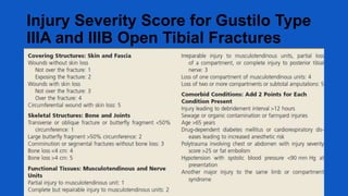
  • 31. Injury Severity Score for Gustilo Type IIIA and IIIB Open Tibial Fractures
  • 33. Goalsof treatment • • • 1. preserve life 2. preserve limb 3. preserve function – Prevent infection – Fracture stabilization – Soft tissue coverage
  • 34. Stagesof care for openfractures
  • 35. Initial assessment traumatized • Important components in assessing extremity are 1. History and mechanism of injury 2. Neurovascular status 3. Sizeof skin wound 4. Muscle crush or loss 5. Periosteal stripping or boneloss 6. Fracture pattern, fragmentation 7. Contamination 8. Compartment syndrome.
  • 37. Irrigation • Supplements systemic debridement by removing foreign material anddecreasing bacterial load. • Simpulse irrigation system(HPPL-high pressure pulsatile lavage). • Pressure must be lessthan 50psiunits. Fracturetype Volof fluid usedfor irrigation Type I 3 L Type 2 6L Type 3 9L
  • 38. Irrigation NS normally used for irrigation. Antibiotic solution has no advantage than soap for irrigation. Surfactant(non sterile soap): same effectiveness, less tissue damage and more economical.
  • 44. Timing of debridement and irrigation • Most guidelines recommenddebridement within 6 hrs and If clean Primaryclosure. • Serial debridement may be necessaryevery 24-72hrs ifdebrima is delayed until the wound viability isensured. • If clean within 2-3 daysdelayed Primary Closure. • Later Secondary closure (Flaps)or healingby Secondary intentions (scarring )
  • 46. ANTIBIOTICS AND INFECTION • 24-70% of open fractures are contaminated with bacteria • 14-15% of open complicated fracture with infection in the absence of antibiotic prophylaxis
  • 48. Antibiotics • Localantibiotics:-In gustilo type III fractures additional useof local aminoglycoside impregnated polymethylmethacrylate(PMMA) beads reduces overall infection rate.
  • 50. Tetanus prophylaxis • Tetanus Toxoid(TT),dose is 0.5ml i.m. regardless of age. • Immunoglobulin:- 75IU <5yrs of age 125IU 5-10yrs 250IU >10yrs.
  • 53. Typesof fracture stabilization Splint – Good option if operative fixation not required Internal fixation – Wound is clean and softtissue coverage available External fixation – Dirty wounds or extensivesoft tissue injury
  • 54. Primary surgery Timing : •Surgical emergency •Operating within 6-8hrs of injury – contaminated wounds not treated within this time will have sustained bacterialmultiplication to result in earlyinfection.
  • 55. Primary surgery Fracture stabilization: •Assoon asprimary wound care is completed, treatment should proceed to fracturereduction and fixation. •Surgeon should rescrub and regown. •Different set of instruments thanthose used for debridement isnecessary.
  • 56. Fixation Options External Fixation VsInternal Fixation
  • 57. Indications for External Fixation in OpenFractures 1) Severecontamination anysite 2) Periarticular fractures – Definitive • Distal radius • Elbow dislocation – Relative • Knee • Ankle • Elbow • Wrist
  • 58. External fixation • Advantages of ExtFixation:- • Canbe applied relatively easily and quickly, • It provides relatively stable fracturefixation, • There is no further damagedone if applied correctly, • Itavoids implantation of hardware in open wound.
  • 59. External Fixator • Disadvantages: • Major problems with external fixation are related to pin tract infection, malalignent , delayed union, poor patientscompliance. • Ringfixators may be an optionin open diaphyseal fractures like ilizarov.
  • 60. Indications for Internal Fixation in Open Fractures 1)Periarticular fractures – Distal/proximal tibia – Distal/proximal femur – Distal/proximal humerus – Proximal ulnar radius – Selected distal radius/ulna – Acetabulum/pelvis 2) Diaphyseal fractures – Femur – Tibia – Humerus – Radius/ulna
  • 61. Plates • Open diaphyseal fractures of the radius andulna as well asthe humerus are best managedwith plate fixation. • The plate fixation of lower extremity diaphyseal fractures is generally not recommended due to higher rate of infections.
  • 63. INTRAMEDULLARY NAILING • Locked intramedullary nailing has been established as the treatment of choice for most diaphyseal fractures in lower extremity. • Intramedullary nails can be inserted with no further disruption of the already injured soft-tissue envelope and preserves the remaining extra osseous blood supply to corticalbone.
  • 64. IM NAILING • Data showing that solid intramedullary nails inserted without reaming have a lower risk of infection. • On the other hand reamed intramedullary nails can reliably maintain fracture reduction with regards to angulation, rotation, displacement, and length. • Prospective randomised trails that compared reamed with unreamed interlocked IM nails did not show any significant difference concerning outcome and risk of complication.
  • 66. Open wound coverage after primary surgery
  • 67. Primary Closure The following criteria must be met:- 1.The original wound must have been fairly clean, and not have occurred in a highly contaminated environment. 2.All necrotic tissue and foreign material have been debrided. 3.Circulation to the limb is essentially normal. 4.Nerve supply to the limb is intact. 5.The patient's general condition is satisfactory and allows careful postoperative assessment. 6.The wound can be closed without tension. 7.Closure will not create a dead space. 8.The patient does not have multisystem injuries.
  • 68. Delayed primary closure • Closure before the 5th day is termed delayed primary closure. • As long as closure is achieved before the fifth day, wound strengths at 14 days are comparable with those in wounds closed on the first day. • Leaving the wound open minimizes the risk of anaerobic infection, and the delay allows the host to mount local wound defensive mechanisms that permit safer closure than is possible on the first day.
  • 69. • Current standard of care for all open fracture wounds is to be left open initially. • Delayed closure is accomplished within 2- 7days • VAC assisted wound closure is presently recommended for temporary management of open fracture wounds.
  • 70. VAC • Thewound bed is exposed to mechanically induced negative pressure in aclosed system. • Thesystem removes fluid from extravascular space,reduces edema, improves micro circulation and enhancesthe proliferation of preparative granulation tissue. • Polyurethane foam dressing is placed in wound and ensures an even distribution of negative pressure.
  • 71. VAC
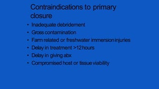
  • 72. Contraindications to primary closure • Inadequate debridement • Grosscontamination • Farmrelated or freshwater immersioninjuries • Delay in treatment >12hours • Delay in givingabx • Compromised host or tissueviability
  • 73. Dressings • Temporary closures – rubber bands • Wet to dry dressings • Semi-permeable membranes • Antibiotic bead pouch • VAC
  • 74. Reconstructive ladder: options for wound coverage Type1open fx Type3Bopen fx Type2/3A open fx
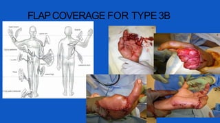
  • 76. Gunshot injuries • Energydissipated at impact =damage severity • High velocity rifles and closerange shotguns – Worst, high energy of impact – Huge secondary cavitation – Secondary effects of shatteredbone fragments • Bullets lodged in joints should beremoved – avoid lead arthropathy and systemiclead poisoning
  • 77. Low velocity GSW<2000ft/sec • Lowvelocity handguns – Lesssevere, not treated like openfractures – Cavitation is not significant – Secondary missile effects are minimal – Bone fragments rarely stripped of softtissue attachments and blood supply – Soft tissue injuries not severe and skin woundsare small
  • 78. ADVANCES • BMPs – 40%decreased infection rate with BMPin type3 open tibia fractures • Antibiotic LadenBoneGraft – Tobramycin-impregnated calcium sulfate pellets with demineralized bonematrix – successful in preventinginfection *BESTTStudyGroup, Govender S,CsimmaC,Genant H, Valentin-OpranA. “Recombinant HumanBoneMorphogenetic Protein-2 for Treatment of OpenTibial Fractures: Aprospective, controlled, randomized study of four hundred andfifty patients.” JBJS-A2002. 84(12): 2123-2134. **Beardmore AA,BrooksDE,WenkeJC,ThomasDB.“Effectiveness of local antibiotic delivery with an osteoinductive and osteoconductive bone-graft substitute.” JBJS-A2005. 87(1): 107-112.
  • 80. Complications • Hypovolemic shock • Compartment syndrome • Fat embolism • ARDS • Neurovascular injuries • Infection • Morel-Lavallee syndrome
  • 82. Hypovolemic shock - management • Two large-bore IV lines should be started. • Once IV access is obtained, initial fluid resuscitation is performed with an isotonic crystalloid, such as Ringer lactate solution or normal saline. • An initial bolus of 1-2 L is given in an adult (20 mL/kg in a pediatric patient), and the patient's response is assessed.
  • 83. HYPOVOLEMIC SHOCK • Type of fluid:- • Colloid – albumin, dextran, plasma. Crystalloid – NS, D5, RL. • Blood – uncrossed ‘O’ –ve. • Basic Rule:- • 3:1 rule when using crystalloids.Eg. If blood loss is 100cc the patient should receive 300cc of normal saline or Ringer lactate. • 1:1 rule for colloids.
  • 85. Morel-Lavallee syndrome • Skin is separated from the fascia. • This creates a pocket under which considerable bleeding can occur. • Usually this is a subcutaneous hematoma, but the hematoma can become so large that it seriously threatens the viability of the skin above it . • This syndrome occurs frequently in patients with pelvic fractures, especially in obese individuals. • MRI and ultrasonography have been recommended to confirm the diagnosis. • Multiple treatments present but PERCUTANEOUS DRAINAGE is mostly accepted and done.
  • 87. Compartment syndrome 34 • Classically 5 "Ps" associated with compartment syndrome:— 1)Pain out of proportion to what is expected 2)Paresthesia 3)Pallor 4)Paralysis 5)Palpable pulse absent.
  • 89. Fat embolism • Definition - Occlusion of small vessels by fat globules. • Types:- 1.cerebral – drowsy, restless and disoriented. 2.pulmonary – tachypnea, tachycardia, petechial rash(in front of neck, ant axillary fold, chest and conjunctiva )
  • 90. Fat embolism • Diagnosis:– • Triad : hypoxia, neurological abnormalities , petechial rash • signs of retinal artery emboli(striate hemorrhages and exudate) may be present. • Sputum and urine may reveal presence of fat globules. • X-ray of chest shows patchy pulmonary infarcts.
  • 91. Fat embolism • Treatment:- • Respiratory support • Heparinization • Intravenous low-molecular weightdextran and corticosteriods.
  • 92. ARDS • Is caused by release of inflammatory mediators which cause disruption of pulmonary vasculature. • Signs and symptoms– Tachypnea, low BP, Cyanosis. • Treatment – 100% oxygen inhalation.
  • 93. Vascularinjury • Absent peripheral pulses in an injured limb should be considered to be due to vascular damage unless proved otherwise. • Classical signs of arterial injury:- (a)absent pulses (b)active hemorrhage (c)expanding hematoma, and (d)bruit or thrill.
  • 94. Vascularinjury • • • Investigations:- Colour doppler study Arteriography. Treatment:- Arterial reconstruction Bypassgrafts. Timing:- loss of total blood supply to the limb for >8 hours nearly always results in amputation.
  • 95. Nerve injury • Nerve repair should be done within 3 weeksof injury for betterresults