ORAL RED
AND WHITE
LESIONS
By SAFA AYAD
Supervised by DR.MUSTAFA IBRAHIM
Introduction
• Definition: White and red lesions refer to
abnormal changes in the oral mucosa color and
texture.
• Importance: May indicate benign conditions,
infections, or potentially malignant disorders.
White appearance of oral
mucosa
1. Hyperkeratosis
2. Abnormal but benign thickening of stratum
spinosum (acanthosis).
3. Intra and extracellular fluid accumulation
(edema).
4. Whitish pseudomembranes by microbes
(plaque).
5. Fibrosis
Red appearance of oral
mucosa
1. Atrophy of epithelium.
2. Increased vascularization.
Classification
INFECTIOUS DISEASE PREMALIGNANT
DISORDER
IMMUNOPATHOLOGI
C DISEASE
ALLERGIC
REACTIONS
TOXIC REACTIONS REACTION TO
MECHANICAL
TRAUMA
OTHERS
01 02 03 04
05 06 07
I.Infectous disease
I.ORAL CANDIDIASIS
■The most prevalent opportunistic infection affecting
the oral mucosa.
■majority of cases, the lesions are caused by Candida
albican.
■Divided into primary &secondary infections
■number of predisposing factors have been
shown to convert C. albicans from the normal
commensal flora (saprophytic stage) to a
pathogenic organism:
Denture wearing
Smoking
Quality and quantity of saliva
Immunosuppressive diseases
Immunosuppressive medications
Endocrine disorders
Types of primary candidiasis
a.pseudomembranous :creamy white, removable plaques or
patches on the oral mucosa,may resemble cottage cheese
and
can be scraped off, often revealing a red, inflamed, or
bleeding
surface underneath.
●soreness, burning sensation, difficulty swallowing (if it
spreads
to the throat). asymptomatic in mild cases.
b.erthematous:doesn't present with white plaques
but
instead appears as red, inflamed patches on the oral
mucosa. It's often associated with a smooth, shiny
surface and can be painful.
C.Chronic Hyperplastic Candidiasis
■(Chronic Plaque Type)
•Also known as candidal leukoplakia. •thick, white,
adherent plaques that cannot be easily scraped off
•Usually asymptomatic but may cause mild discomfort
or a rough texture
■(Chronic Nodular Type)
rare, small, raised, nodular lesions
rather than flat plaques
or diffuse redness.
d.candida assossiated lesion
(Denture stomatitis )
Type I minor erthematous site duo to denture
trauma
Type II major denture covered mucosa
Type III granular mucosa
(Angular cheilitis)
■ infected fissures of the commissures of the
mouth
surrounded by erythema
■The lesions are frequently infected with both
Candida
albicans and Staphylococcus aureus.
■Causes :Vitamin B12 deficiency, iron deficiencies,
and loss of vertical dimension and Dry skin
(Median Rhomboid Glossitis)
■ asymptomatic erythematous lesion in the
center
of the posterior part of the dorsum of the
tongue
with an oval configuration.
■This area of erythema results
from atrophy of the filiform papillae
and the surface may be lobulated.
■ the lesion frequent shows a mixed
bacterial/fungal microflora
SECONDARY CANDIDIASIS
●group of disorders, which, in addition to
oral
candidiasis, also affect the skin, nail, genital
Mucosa.
● The face and scalp may be involved.
●Causes : endocrine disorder, severe
combined
immunodeficiency , thymoma and HIV .
MANAGEMENT
☆Reduction of predisposing factor like improving
denture hygiene , reduction denture porosity , smoke
cessation.
☆pseudomembranous ,erythematous__topical
antifungal
☆chronic hyperplastic __systemic antifungal
☆type III denture stomatitis __surgical removal
☆Angular cheilitis __topical antifungal like miconazole
and moisturizing cream.
☆secondary candidiasis __systemic antifungal
II.Oral hairy leukoplakia
It is caused by the Epstein-Barr virus (EBV).
▪︎
white, corrugated, or "hairy" patches that cannot be
▪︎
scraped off.
On the lateral borders of the tongue, though it can
▪︎
occasionally appear on other oral mucosal surfaces like
the buccal mucosa or palate.
The lesions are typically asymptomatic
▪︎
associated with immunocompromised individuals like
▪︎
HIV/AIDS, organ transplant recipients.
■Treatment:
Focuses on improving immune function. For example,
in HIV patients, antiretroviral therapy can resolve
OHL by boosting immunity.
Antiviral medications (e.g., acyclovir) may be used
▪︎
to target EBV directly
2.premalignant disease
I.LEUKOPLAKIA
white patches or plaques on the mucous
■
membranes of the mouth that cannot be wiped
off
it has the potential to develop into oral cancer,
■
particularly squamous cell carcinoma
risk factors (smoking , excessive alcohol
■
consumption, chronic irritation (e.g., from sharp
cusps or ill-fitting dentures).
main types include:
1. Homogeneous Leukoplakia
• Uniformly white, flat, and smooth plaques
with a consistent texture.
• lower risk of malignant transformation.
2. Non-Homogeneous Leukoplakia
• Irregular, mixed white and red patches
(sometimes called erythroleukoplakia), with a
nodular, verrucous (warty), or speckled
surface.
• higher risk of progressing to cancer 15-40%.
• Subtypes:
Nodular: Small, raised bumps on the surface.
■
Verrucous: Warty or corrugated appearance.
■
Speckled: Mixed red and white areas, often with
■
an uneven texture.
3. Proliferative Verrucous Leukoplakia (PVL)
• A rare, aggressive subtype with multiple,
widespread warty lesions that tend to recur and
spread over time.
• Highly resistant to treatment and has a strong
tendency to transform into squamous cell
carcinoma (up to 70-100%).
MANAGEMENT
a.homogenous __eliminate the irritant +follow up
every (3_6)weeks.
b.non homogenous__eliminate the
irritant+surgical removal+follow up biopsy to role
out cancer.
C.PVL__wide surgical excision +biopsy follow up
every (1_3)months.if biopsy show malignant
transformation then chemotherapy or
radiotherapy needed.
II. ERYTHROPLAKIA
• red lesion or patch that has appearance of
Smooth, velvety, or granular red areas, often
sharply defined.
• Commonly found on the floor of the mouth,
tongue, or soft palate
• Rare, less frequent than leukoplakia.
• Risk Factors: smoking or , alcohol consumption,
and sometimes human papillomavirus (HPV)
infection.
•Malignant Potential: High risk-up to 90%
•asymptomatic or report mild discomfort, burning, or
soreness.
•Unlike leukoplakia, erythroplakia rarely causes
noticeable
thickening.
•Duration: Lesions persisting beyond 2-3 weeks despite
removing potential irritants
• Management:
• Eliminate Risk Factors.
• Surgical Removal.
• Follow-Up
III.ORAL SUBMUCUS FIBROSIS
•A progressive, precancerous fibrosis of the
submucosal tissues leading to stiffness and
restricted mouth opening.
• Cause: chronic use of areca nut (betel quid),
nutritional deficiencies, genetic predisposition, and
autoimmune processes.
•Pale, marble-like mucosa with palpable fibrous
bands in the cheeks, lips, or palate. May develop ulcers
.
•Reduced mouth opening (trismus), stiff cheeks,
difficulty swallowing or speaking, and sometimes
hearing loss due to Eustachian tube involvement.
MANAGEMENT
EARLY __cessation habit +Intralesional steroids
+physiotherapy (excersice to adjust mouth opening).
ADVANCED __ surgical intervation.
3.lmmunopathologic disease
I. LICHEN PLANUS (LP)
is a common chronic immunologic inflammatory
mucocutaneous disorder.
(Skin lesion )
characterized by Kobner's phenomena (development of
new
lesion on normal looking skin following trauma as
scratching).
(Oral lesion)
•Bilateral esions surrounded by a network of bluish
white lines called Wickham's striae radiating from
periphery of the lesion.
•pain or discomfort, which interferes with function.
FORMS OF THE LESION
RETICULAR
PAPULAR
PLAQUE
ATROPHIC
EROSIVE
BULLOUS
Causes
. Autoimmune reaction
. Stress
. Medications
. Hepatitis c
. Diabetes millitus
MANAGEMENT
Reticular, plaque, papules __No treatment and
follow up
Atrophic, erosive__ Corticosteroids + Retinoids
II.LICHENOID REACTION
associated with the administration of a drug,
contact with a metal, the use of a food flavoring, or
systemic disease.
resolution when the drug or other factor was
eliminated
III.LUPUS ERYTHEMATOUS
•Systemic lupus erythematosus (SLE)
multi-organ damage with disk shaped
skin scar .
• Discoid lupus erythematosus (DLE) is
affecting the oral cavity+skin lesion has
butterfly distribution on tip and bridge
of the nose.
• Subacute cutaneous lupus
erythematosus intermediate between SLE
Causes
1-Immunologic formation.
2- Genetic factors.
3-Infectious (EBV, CMV, VZV).
4. environmental (e.g.sun exposure).
5- Endocrine factors: (hormones).
(Oral lesion)
•central atrophy, scar formation, and occasional loss
of surface pigmentation.
• The primary locations for these lesions include the
buccal mucosa, palate, tongue
MANAGEMENT
Improve oral hygiene
DLE__Topical corticosteroids
SLE __ Systemic corticosteroids
4.Allergic reactions
Dentifrices and mouthwashes allergy
•The Lesions are usually located on the
Mucobuccal folds and Gingiva.
•irregular in shape, white, covered with a
pseudomembrane, and very painful.
5.Toxic reactions
I.SMOKLESS TOBACCO INDUCED
KERATOSIS
•The surface of mucosa appears white and granular
or
wrinkled.
•These lesions are accepted as precancerous.
• area of gingival recession with periodontal-tissue
destruction in the area of contact
on facial aspect of the teeth .
•The lesion is asymptomatic .
II.NECOTINE STOMATITIS
Due to the chronic insult, the palatal mucosa
becomes diffusely gray or white. Numerous
slightly elevated papuleswith punctate red
centers that represent inflamed altered minor
salivary gland ducts are noted.
6.Reaction to mechanical trauma
I.LINEA ALBA
It is a horizontal white streak on the buccal Mucosa
bilaterally at the level of the occlusal plane from the
commissure of mouth to the posterior teeth.
II. FRICTIONAL KERATOSIS
• Frictional (traumatic) keratosis is defined as
a white plaque with a rough surface that is
clearly related to a source of mechanical
irritation .
•Frictional keratosis is frequently associated
with rough or maladjusted dentures and with
sharp cusps .
7.others
I.LEUKOEDEMA
• Diffuse grayish-white milky appearance of thebuccal
mucosa
• Appearance will disappear when cheek is stretched.
Managment
• No treatment is indicated
II. BENIGN MIGRATORY GLOSSITIS
asymptomatic red, smooth patches on the tongue
with irregular white borders.
• Patches change in size, shape, and location over time
(hence "migratory").
•papillae are absent or flattened in certain areas,
creating smooth, red patches.
•The loss of papillae in these patches is temporary and
reversible.
III.WHITE SPONGY NEVUS
• rare autosomal dominant disorder. It present from
birth and appear during childhood.
• Presents as bilateral white, soft, "spongy," or velvety
thick plaques of the buccal mucosa.
• Other sites in the oral cavity may be involved,
including the ventral tongue, floor of the mouth, labial
mucosa, soft palate, and alveolar mucosa.
• asymptomatic
• require no treatment
THANK
YOU

More Related Content

PPTX
Oral cavity ppt- college seminar
PPTX
Candidiasis
PPTX
Lesions of oral cavity
PPTX
Fungal infections part II
PPTX
Oral candidiasis
PPTX
Oral mycotic infections
PPT
White lesions ppt
PPTX
RED AND WHITE LESIONS OF THE ORAL MUCOSA.pptx
Oral cavity ppt- college seminar
Candidiasis
Lesions of oral cavity
Fungal infections part II
Oral candidiasis
Oral mycotic infections
White lesions ppt
RED AND WHITE LESIONS OF THE ORAL MUCOSA.pptx

Similar to Oral red and white lesionss in dentistry (20)

PPTX
Part i head and neck pathology
PPTX
presentation Red and white lesions edt.ppt
PPTX
red and white part1 in oral medicine.pptx
PPT
Cheilitis
PPTX
candidal infection.pptx
PPTX
Non plaque induced gingivitis
PPTX
Oral cavity and salivary gland diseases
PPTX
1 FUNGAL DISEASES OF ORAL CAVITY.pptx
PPTX
Premalignant & malignant diseases of oral cavity ii n
PPTX
Oral mucosal ulceration
PPTX
white lesions of oral mucosa........pptx
PPTX
Desquamative gingivitis
PPTX
Oral candidiosis: A Review
PPTX
Oral diseases - Benign and premalignant lesions
DOC
A)benign oral & tg,atypical facial pain,burning pain syndrome
PPTX
(Potentially)Malignant Disorders of OC.pptx
PDF
Diseases of lip ( oral medicine ) dentistry 4th year
PPTX
Lesions of oral cavity
PPTX
Red and White Lesions of the.pptx
PPTX
Developmental disturbances of soft tissues-1,I.pptx
Part i head and neck pathology
presentation Red and white lesions edt.ppt
red and white part1 in oral medicine.pptx
Cheilitis
candidal infection.pptx
Non plaque induced gingivitis
Oral cavity and salivary gland diseases
1 FUNGAL DISEASES OF ORAL CAVITY.pptx
Premalignant & malignant diseases of oral cavity ii n
Oral mucosal ulceration
white lesions of oral mucosa........pptx
Desquamative gingivitis
Oral candidiosis: A Review
Oral diseases - Benign and premalignant lesions
A)benign oral & tg,atypical facial pain,burning pain syndrome
(Potentially)Malignant Disorders of OC.pptx
Diseases of lip ( oral medicine ) dentistry 4th year
Lesions of oral cavity
Red and White Lesions of the.pptx
Developmental disturbances of soft tissues-1,I.pptx
Ad

Recently uploaded (20)

PDF
heliotherapy- types and advantages procedure
PDF
FMCG-October-2021........................
PPTX
Ocular Drug Delivery Systems: Advances, Challenges, and Pharmaceutical Applic...
PDF
NCCN CANCER TESTICULAR 2024 ...............................
PPTX
Computed Tomography: Hardware and Instrumentation
PDF
communicable diseases for healthcare - Part 1.pdf
PPTX
Approch to weakness &paralysis pateint.pptx
PDF
11 Antiepileptic Drugs.pdf file for pharmacy
PPTX
Nutrition needs in a Surgical Patient.pptx
PPTX
LIVER DIORDERS OF PREGNANCY in detail PPT.pptx
PDF
neonatology-for-nurses.pdfggghjjkkkkkkjhhg
PDF
periodontaldiseasesandtreatments-200626195738.pdf
PPTX
ENT-DISORDERS ( ent for nursing ). (1).p
PPTX
Geriatrics_(0).pptxxvvbbbbbbbnnnnnnnnnnk
PPTX
FORENSIC MEDICINE and branches of forensic medicine.pptx
PPTX
Surgical anatomy, physiology and procedures of esophagus.pptx
PPTX
A Detailed Physiology of Endocrine System.pptx
PDF
Seizures and epilepsy (neurological disorder)- AMBOSS.pdf
PPTX
This book is about some common childhood
PPTX
ANTI BIOTICS. SULPHONAMIDES,QUINOLONES.pptx
heliotherapy- types and advantages procedure
FMCG-October-2021........................
Ocular Drug Delivery Systems: Advances, Challenges, and Pharmaceutical Applic...
NCCN CANCER TESTICULAR 2024 ...............................
Computed Tomography: Hardware and Instrumentation
communicable diseases for healthcare - Part 1.pdf
Approch to weakness &paralysis pateint.pptx
11 Antiepileptic Drugs.pdf file for pharmacy
Nutrition needs in a Surgical Patient.pptx
LIVER DIORDERS OF PREGNANCY in detail PPT.pptx
neonatology-for-nurses.pdfggghjjkkkkkkjhhg
periodontaldiseasesandtreatments-200626195738.pdf
ENT-DISORDERS ( ent for nursing ). (1).p
Geriatrics_(0).pptxxvvbbbbbbbnnnnnnnnnnk
FORENSIC MEDICINE and branches of forensic medicine.pptx
Surgical anatomy, physiology and procedures of esophagus.pptx
A Detailed Physiology of Endocrine System.pptx
Seizures and epilepsy (neurological disorder)- AMBOSS.pdf
This book is about some common childhood
ANTI BIOTICS. SULPHONAMIDES,QUINOLONES.pptx
Ad

Oral red and white lesionss in dentistry

  • 1. ORAL RED AND WHITE LESIONS By SAFA AYAD Supervised by DR.MUSTAFA IBRAHIM
  • 2. Introduction • Definition: White and red lesions refer to abnormal changes in the oral mucosa color and texture. • Importance: May indicate benign conditions, infections, or potentially malignant disorders.
  • 3. White appearance of oral mucosa 1. Hyperkeratosis 2. Abnormal but benign thickening of stratum spinosum (acanthosis). 3. Intra and extracellular fluid accumulation (edema). 4. Whitish pseudomembranes by microbes (plaque). 5. Fibrosis
  • 4. Red appearance of oral mucosa 1. Atrophy of epithelium. 2. Increased vascularization.
  • 5. Classification INFECTIOUS DISEASE PREMALIGNANT DISORDER IMMUNOPATHOLOGI C DISEASE ALLERGIC REACTIONS TOXIC REACTIONS REACTION TO MECHANICAL TRAUMA OTHERS 01 02 03 04 05 06 07
  • 6. I.Infectous disease I.ORAL CANDIDIASIS ■The most prevalent opportunistic infection affecting the oral mucosa. ■majority of cases, the lesions are caused by Candida albican. ■Divided into primary &secondary infections
  • 7. ■number of predisposing factors have been shown to convert C. albicans from the normal commensal flora (saprophytic stage) to a pathogenic organism: Denture wearing Smoking Quality and quantity of saliva Immunosuppressive diseases Immunosuppressive medications Endocrine disorders
  • 8. Types of primary candidiasis a.pseudomembranous :creamy white, removable plaques or patches on the oral mucosa,may resemble cottage cheese and can be scraped off, often revealing a red, inflamed, or bleeding surface underneath. ●soreness, burning sensation, difficulty swallowing (if it spreads to the throat). asymptomatic in mild cases.
  • 9. b.erthematous:doesn't present with white plaques but instead appears as red, inflamed patches on the oral mucosa. It's often associated with a smooth, shiny surface and can be painful.
  • 10. C.Chronic Hyperplastic Candidiasis ■(Chronic Plaque Type) •Also known as candidal leukoplakia. •thick, white, adherent plaques that cannot be easily scraped off •Usually asymptomatic but may cause mild discomfort or a rough texture ■(Chronic Nodular Type) rare, small, raised, nodular lesions rather than flat plaques or diffuse redness.
  • 11. d.candida assossiated lesion (Denture stomatitis ) Type I minor erthematous site duo to denture trauma Type II major denture covered mucosa Type III granular mucosa
  • 12. (Angular cheilitis) ■ infected fissures of the commissures of the mouth surrounded by erythema ■The lesions are frequently infected with both Candida albicans and Staphylococcus aureus. ■Causes :Vitamin B12 deficiency, iron deficiencies, and loss of vertical dimension and Dry skin
  • 13. (Median Rhomboid Glossitis) ■ asymptomatic erythematous lesion in the center of the posterior part of the dorsum of the tongue with an oval configuration. ■This area of erythema results from atrophy of the filiform papillae and the surface may be lobulated. ■ the lesion frequent shows a mixed bacterial/fungal microflora
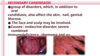
  • 14. SECONDARY CANDIDIASIS ●group of disorders, which, in addition to oral candidiasis, also affect the skin, nail, genital Mucosa. ● The face and scalp may be involved. ●Causes : endocrine disorder, severe combined immunodeficiency , thymoma and HIV .
  • 15. MANAGEMENT ☆Reduction of predisposing factor like improving denture hygiene , reduction denture porosity , smoke cessation. ☆pseudomembranous ,erythematous__topical antifungal ☆chronic hyperplastic __systemic antifungal ☆type III denture stomatitis __surgical removal ☆Angular cheilitis __topical antifungal like miconazole and moisturizing cream. ☆secondary candidiasis __systemic antifungal
  • 16. II.Oral hairy leukoplakia It is caused by the Epstein-Barr virus (EBV). ▪︎ white, corrugated, or "hairy" patches that cannot be ▪︎ scraped off. On the lateral borders of the tongue, though it can ▪︎ occasionally appear on other oral mucosal surfaces like the buccal mucosa or palate. The lesions are typically asymptomatic ▪︎ associated with immunocompromised individuals like ▪︎ HIV/AIDS, organ transplant recipients.
  • 17. ■Treatment: Focuses on improving immune function. For example, in HIV patients, antiretroviral therapy can resolve OHL by boosting immunity. Antiviral medications (e.g., acyclovir) may be used ▪︎ to target EBV directly
  • 18. 2.premalignant disease I.LEUKOPLAKIA white patches or plaques on the mucous ■ membranes of the mouth that cannot be wiped off it has the potential to develop into oral cancer, ■ particularly squamous cell carcinoma risk factors (smoking , excessive alcohol ■ consumption, chronic irritation (e.g., from sharp cusps or ill-fitting dentures).
  • 19. main types include: 1. Homogeneous Leukoplakia • Uniformly white, flat, and smooth plaques with a consistent texture. • lower risk of malignant transformation. 2. Non-Homogeneous Leukoplakia • Irregular, mixed white and red patches (sometimes called erythroleukoplakia), with a nodular, verrucous (warty), or speckled surface. • higher risk of progressing to cancer 15-40%.
  • 20. • Subtypes: Nodular: Small, raised bumps on the surface. ■ Verrucous: Warty or corrugated appearance. ■ Speckled: Mixed red and white areas, often with ■ an uneven texture.
  • 21. 3. Proliferative Verrucous Leukoplakia (PVL) • A rare, aggressive subtype with multiple, widespread warty lesions that tend to recur and spread over time. • Highly resistant to treatment and has a strong tendency to transform into squamous cell carcinoma (up to 70-100%).
  • 22. MANAGEMENT a.homogenous __eliminate the irritant +follow up every (3_6)weeks. b.non homogenous__eliminate the irritant+surgical removal+follow up biopsy to role out cancer. C.PVL__wide surgical excision +biopsy follow up every (1_3)months.if biopsy show malignant transformation then chemotherapy or radiotherapy needed.
  • 23. II. ERYTHROPLAKIA • red lesion or patch that has appearance of Smooth, velvety, or granular red areas, often sharply defined. • Commonly found on the floor of the mouth, tongue, or soft palate • Rare, less frequent than leukoplakia. • Risk Factors: smoking or , alcohol consumption, and sometimes human papillomavirus (HPV) infection. •Malignant Potential: High risk-up to 90%
  • 24. •asymptomatic or report mild discomfort, burning, or soreness. •Unlike leukoplakia, erythroplakia rarely causes noticeable thickening. •Duration: Lesions persisting beyond 2-3 weeks despite removing potential irritants • Management: • Eliminate Risk Factors. • Surgical Removal. • Follow-Up
  • 25. III.ORAL SUBMUCUS FIBROSIS •A progressive, precancerous fibrosis of the submucosal tissues leading to stiffness and restricted mouth opening. • Cause: chronic use of areca nut (betel quid), nutritional deficiencies, genetic predisposition, and autoimmune processes.
  • 26. •Pale, marble-like mucosa with palpable fibrous bands in the cheeks, lips, or palate. May develop ulcers . •Reduced mouth opening (trismus), stiff cheeks, difficulty swallowing or speaking, and sometimes hearing loss due to Eustachian tube involvement. MANAGEMENT EARLY __cessation habit +Intralesional steroids +physiotherapy (excersice to adjust mouth opening). ADVANCED __ surgical intervation.
  • 27. 3.lmmunopathologic disease I. LICHEN PLANUS (LP) is a common chronic immunologic inflammatory mucocutaneous disorder. (Skin lesion ) characterized by Kobner's phenomena (development of new lesion on normal looking skin following trauma as scratching).
  • 28. (Oral lesion) •Bilateral esions surrounded by a network of bluish white lines called Wickham's striae radiating from periphery of the lesion. •pain or discomfort, which interferes with function. FORMS OF THE LESION RETICULAR PAPULAR PLAQUE ATROPHIC EROSIVE BULLOUS
  • 29. Causes . Autoimmune reaction . Stress . Medications . Hepatitis c . Diabetes millitus MANAGEMENT Reticular, plaque, papules __No treatment and follow up Atrophic, erosive__ Corticosteroids + Retinoids
  • 30. II.LICHENOID REACTION associated with the administration of a drug, contact with a metal, the use of a food flavoring, or systemic disease. resolution when the drug or other factor was eliminated
  • 31. III.LUPUS ERYTHEMATOUS •Systemic lupus erythematosus (SLE) multi-organ damage with disk shaped skin scar . • Discoid lupus erythematosus (DLE) is affecting the oral cavity+skin lesion has butterfly distribution on tip and bridge of the nose. • Subacute cutaneous lupus erythematosus intermediate between SLE
  • 32. Causes 1-Immunologic formation. 2- Genetic factors. 3-Infectious (EBV, CMV, VZV). 4. environmental (e.g.sun exposure). 5- Endocrine factors: (hormones). (Oral lesion) •central atrophy, scar formation, and occasional loss of surface pigmentation. • The primary locations for these lesions include the buccal mucosa, palate, tongue
  • 33. MANAGEMENT Improve oral hygiene DLE__Topical corticosteroids SLE __ Systemic corticosteroids
  • 34. 4.Allergic reactions Dentifrices and mouthwashes allergy •The Lesions are usually located on the Mucobuccal folds and Gingiva. •irregular in shape, white, covered with a pseudomembrane, and very painful.
  • 35. 5.Toxic reactions I.SMOKLESS TOBACCO INDUCED KERATOSIS •The surface of mucosa appears white and granular or wrinkled. •These lesions are accepted as precancerous. • area of gingival recession with periodontal-tissue destruction in the area of contact on facial aspect of the teeth . •The lesion is asymptomatic .
  • 36. II.NECOTINE STOMATITIS Due to the chronic insult, the palatal mucosa becomes diffusely gray or white. Numerous slightly elevated papuleswith punctate red centers that represent inflamed altered minor salivary gland ducts are noted.
  • 37. 6.Reaction to mechanical trauma I.LINEA ALBA It is a horizontal white streak on the buccal Mucosa bilaterally at the level of the occlusal plane from the commissure of mouth to the posterior teeth.
  • 38. II. FRICTIONAL KERATOSIS • Frictional (traumatic) keratosis is defined as a white plaque with a rough surface that is clearly related to a source of mechanical irritation . •Frictional keratosis is frequently associated with rough or maladjusted dentures and with sharp cusps .
  • 39. 7.others I.LEUKOEDEMA • Diffuse grayish-white milky appearance of thebuccal mucosa • Appearance will disappear when cheek is stretched. Managment • No treatment is indicated
  • 40. II. BENIGN MIGRATORY GLOSSITIS asymptomatic red, smooth patches on the tongue with irregular white borders. • Patches change in size, shape, and location over time (hence "migratory"). •papillae are absent or flattened in certain areas, creating smooth, red patches. •The loss of papillae in these patches is temporary and reversible.
  • 41. III.WHITE SPONGY NEVUS • rare autosomal dominant disorder. It present from birth and appear during childhood. • Presents as bilateral white, soft, "spongy," or velvety thick plaques of the buccal mucosa. • Other sites in the oral cavity may be involved, including the ventral tongue, floor of the mouth, labial mucosa, soft palate, and alveolar mucosa. • asymptomatic • require no treatment