Oxygen cascade
Download as PPTX, PDF4 likes7,080 views

Oxygen is essential for aerobic respiration in humans. It undergoes a "cascade" of decreasing partial pressure from the atmosphere into the mitochondria of cells. Key steps include uptake in the lungs (PaO2 of 100 mmHg), transport in blood bound to hemoglobin and dissolved in plasma, delivery to tissues, and cellular uptake and use. Hemoglobin's oxygen-binding curve allows for efficient oxygen loading in the lungs and unloading in tissues. Factors like pH, CO2, and 2,3-DPG regulate the curve to facilitate oxygen transport.


![ Effect of 2,3DPG
32
Tendency to bind to β chains of Hb and thereby decrease the affinity of
Hemoglobin for oxygen.
HbO2 + 2,3 DPG → Hb-2,3 DPG + O2
It promotes a rightward shift and enhances oxygen unloading at the tissues.
This shift is longer in duration than that due to [H+], PCO2 or temperature.
Level increase with Level Decrease with
– Cellular hypoxia.
– Anemia
– Hypoxemia secondary to COPD
– Congenital Heart Disease
– Ascent to high altitudes
– Septic Shock
– Acidemia
– Stored blood
• No DPG after 2 weeks of
storage.](https://image.slidesharecdn.com/oxygencascade-210604065359/85/Oxygen-cascade-29-320.jpg)

![Oxygen content (CaO2)
The oxygen content of arterial blood is the sum of the
oxygen bound to haemoglobin and oxygen dissolved in
plasma.
Total amount of O2 present in 100 ml of Arterial Blood
CaO2=Hb. Bound O2+ dissolved Hb
= [1.34 x Hb x SaO2] + 0.003 x PO2
= [1.34×15×97.5] +0.003×100
=19.9=20ml /dl approx .
=200ml/L
35](https://image.slidesharecdn.com/oxygencascade-210604065359/85/Oxygen-cascade-32-320.jpg)
![ Similarly for Venous blood
CvO2=[1.34 × Hb × SvO2] + 0.003 × PvO2
replacing with values we have
CvO2=15 ml/dl =150 ml/L
Total oxygen content:
= 200 × arterial blood vol. + 150 × venous blood vol.
= 200 × 0.25 × 5+150 × 0.75 × 5
= 250 + 562.5
=812ml
36](https://image.slidesharecdn.com/oxygencascade-210604065359/85/Oxygen-cascade-33-320.jpg)


Oxygen cascade
- 1. OXYGEN CASCADE DR BIBEK PAUDEL DEPARTMENT OF ANESTHESIOLOGY NEPALGUNJ MEDICAL COLLEGE 1
- 2. INTRODUCTION Oxygen is vital for life sustaining aerobic respiration in humans. Within the mitochondrial inner membrane , oxygen acts as the terminal electron acceptor at the end of the electron transport chain. Whereby oxidative phosphorylation results in the synthesis of adenosine triphosphate (ATP). 2
- 3. Oxygen Cascade Progressive decrease in the partial pressure of oxygen from the ambient air to the cellular level. The Po2 reaches the lowest level (4-20 mmHg) in the mitochondria 3
- 4. KEY STEPS IN OXYGEN CASCADE • Uptake in the lungs • Carrying capacity of blood • Delivery to capillaries • Delivery to interstitium • Delivery to individual cells • Cellular use of oxygen 5
- 6. Atmosphere PIO2 at Sea level Barometric pressure=760 mmHg Water vapour pressure= 3.7 mmHg Pressure of remaining gases= 760- 3.7=756 mmHg Inspired O2 concentration = 21% PIO2 =21/100*756 =159 mmHg PIO2 at MT Everest Barometric pressure=253 mmHg Water vapour pressure= 3.7 mmHg Pressure of remaining gases= 253- 3.7= 249.3 mmHg Inspired O2 concentration = 21% PIO2 =21/100*249.3 =52 mmHg 7
- 7. ALVEOLAR OXYGEN TENSION : With every breath, the inspired gas is humidified at 37°C in the upper airway. The inspired tension of oxygen (PIO2 ) is therefore reduced by the added water vapor. Water vapor pressure is 47 mm Hg at 37°C In humidified air, in the trachea the normal partial pressure of O2 at sea level is 149.7 mm Hg. (760-47)0.21=150mm Hg 8
- 8. ALVEOLAR GAS EQUATION In alveoli the inspired gases are mixed with residual alveolar gas from previous breath. O2 is taken up and Co2 is added. The final alveolar 02 tension can be estimated by PA02 = PI02 - PaC02/RQ where PIO2 is inspired oxygen tension, PaCO2 is arterial CO2 tension (assumed to equal alveolar PACO2), R is the respiratory exchange ratio (normally in the range of 0.8 to 1.0). The alveolar oxygen tension is approximately 104 mm of Hg 9
- 9. Pulmonary Ventilation Respiratory muscle activity of inspiration/expiration cycling maintains two–way airflow and averaged over several cycles, maintains a partial pressure of oxygen and carbon dioxide in the alveolar air of 100 and 40 mm Hg respectively Alveolar pO2 and pCO2 are maintained remarkably constant by complex neural regulation of the total and alveolar ventilation. 10
- 10. 11
- 11. The following are the different layers of the respiratory membrane: 1. A layer of fluid lining the alveolus and containing surfactant reduces the surface tension of the alveolar fluid. 2. The alveolar epithelium composed of thin epithelial cells. 3. An epithelial basement membrane. 4. A thin interstitial space between the alveolar epithelium and capillary membrane. 5. A capillary basement membrane that in many places fuses with the alveolar epithelial basement membrane. 6. The capillary endothelial membrane. 12
- 12. FACTORS AFFECTING DIFFUSION ACROSS RESPIRATORY MEMBRANE 13
- 13. Exchange of Gases in Alveoli Henry’s law states that “the amount of gas dissolved in a liquid will be directly proportional to the partial pressure of the gas in the liquid-gas interface”. When a liquid is exposed to air containing a particular gas, molecules of the gas will enter the liquid and dissolve in it. Diffusion equilibrium will be reached only when the PO2 in the liquid phase is equal to the PO2 in the gas phase. 15
- 14. Diffusion of Gases Through the Respiratory Membrane The alveolar walls are extremely thin, and between the alveoli is an almost solid network of interconnecting capillaries & the alveolar gases are in very close proximity to the blood of the pulmonary capillaries . Respiratory Membrane: Gas exchange between the alveolar air and the pulmonary blood occurs through the membranes of all the terminal portions of the lungs. All these membranes are collectively known as the respiratory membrane, also called the pulmonary membrane. 16
- 15. Alveolar–arterial gradient (PAO2 – PaO2) It is used in diagnosing the source of hypoxemia. It helps to assess the integrity of alveolar capillary unit normally about 5–10 mm Hg, but progressively increases with age up to 25 mm Hg A high A–a gradient could indicate a patient breathing hard to achieve normal oxygenation. 17
- 16. Pulmonary Systemic Capillaries Transport of oxygen in blood The oxygen is present in two forms: 1. dissolved in the plasma 2. reversibly combined with hemoglobin molecules in the RB Cs 18
- 17. Oxygen Transport Carried in blood in 2 forms 1. By red blood cells Bound to Hb 97-98% 2. Dissolved O2 in plasma Obeys Henry’s law 2-3% Bound to Hgb Dissolved 19
- 18. The mathematical expression is as follows: The amount of oxygen dissolved in blood can be derived from the Henry’s Law. Gas concentration= Gas solubility coefficient x Partial pressure The solubility coefficient for oxygen at normal body temperature is 0.003ml/dl/mmHg . Even with a Pao2 of 100mmHg the maximum amount of 02 dissolved is very small (0.3ml/dl) compared with that bound to hemoglobin. 20
- 19. Hemoglobin . Each hemoglobin molecule is a protein made up of four subunits bound together. Each subunit consists of a molecular group known as h eme and a polypeptide attached to the heme. The four polypeptides of a hemoglobin molecule are collectively called globin. Heme is an iron porphyrin compound that is an essential part of the oxbinding sites; only the divalent form (+2 c harge) of iron can bind O2 . Each gram of hemoglobin can theoretically carry up to 1.34ml of 02 21
- 20. Effect of PO2 on Hb Saturation: The O2- Hb Dissociation Curve The oxygen-hemoglobin dissociation curve plots the proportion of Hb in its saturated form on the vertical axis against the prevailing O2 tension on the horizontal axis. Important tool for understanding how blood carries and releases oxy gen. More specifically it relates between the percentage of O2 carrying cap acity of Hb and PaO2 It is an S-shaped curve that has 2 parts: - 1. upper flat (plateau) part. – 2. lower steep part. 23
- 22. P50: The PaO2 in the blood at which the hemoglobin is 50% saturated, typically about 26.6 mmHg for a healthy person. Increased P50 indicates a rightward shift of the standard curve, which means that a larger partial pressure is necessary to maintain a 50% oxygen saturation. This indicates a decreased affinity Conversely, a lower P50 indicates a leftward shift and a higher affinity 25
- 23. STEEP PHASE:- PO2 of 10 - 60mmHg The steep portion of the curve is the range that exist at the systemic capillaries. Significance:- Allows large quantities of O2 to be released from Hb in the tissue capillaries where a lower capillary PO2 prevails. 26
- 24. PLATEAU PHASE:- Begins to plateau at PO2 of 60mmHg and flattens at and beyond PO2 of 70mmHg. Significance:- The plateau in curve provides a safety factor through which even a significant decrease in lung function can allow normal saturation of Hb. For e.g., in situations like high altitude, pulmonary diseases, where there is moderate hypoxia (decrease in PO2 from 95 to 60mmHg), Capacity decreases only by 5- 10%. O2 saturation and content remain fairly constant inspite of wide fluctuations in alveolar PO2. For this reason, O2 content cannot be increased appreciably by hyperventilation or breathing 100% O2 at PO2 of 100mmHg in a normal person at sea level. 27
- 25. Factors that affects oxygen hemoglobin dissociation curve 28
- 26. Effect Of pH : H+ decreases the affinity of Hb molecule for O2 . It does so by combining with the globin portion of hemoglobin and altering the conformation of the Hb molecule. H+ and O2 both compete for binding to the hemoglobin molecule. Therefore, with increased acidity, the hemoglobin binds less O2 for a given PO2 (and more H+ ) An increase in blood hydrogen ion concentration (acidosis) reduces the affinity of O2 to hemoglobin therefore release O2 to the tissue (BOHR EFFECT) 29
- 27. Effect of CO2: CO2 affects the curve in two ways: Most of the CO2 content (80–90%) is transported as bicarbonate ions. The formation of a bicarbonate ion will release a proton into the plasma. Hence, the elevated CO2 content creates a respiratory acidosis and shifts the oxygen– hemoglobin dissociation curve to the right. About 5–10% of the total CO2 content of blood is transported as carbamino compounds which bind to Hb forming CarbaminoHb. Levels of carbamino compounds have the effect of shifting the curve to the left. 30
- 28. Bohr's Effect In 1904 by the Danish physiologist Christian Bohr, stating that the “oxygen binding affinity of Hb is inversely related to the concentration of carbon dioxide & H+ concentration.” - At tissues: Increased PCO2 & H+ conc shift of O2-Hb curve to the right. - At lungs: Decreased PCO2 & H+ conc shift of O2-Hb curve to the left. So, Bohr's effect facilitates i) O2 release from Hb at tissues. ii) O2 uptake by Hb at lungs. 31
- 29. Effect of 2,3DPG 32 Tendency to bind to β chains of Hb and thereby decrease the affinity of Hemoglobin for oxygen. HbO2 + 2,3 DPG → Hb-2,3 DPG + O2 It promotes a rightward shift and enhances oxygen unloading at the tissues. This shift is longer in duration than that due to [H+], PCO2 or temperature. Level increase with Level Decrease with – Cellular hypoxia. – Anemia – Hypoxemia secondary to COPD – Congenital Heart Disease – Ascent to high altitudes – Septic Shock – Acidemia – Stored blood • No DPG after 2 weeks of storage.
- 30. O2 Dissociation Curve Of Fetal Hb Fetal Hb (HbF) contains 2α and 2γ polypeptide chains and has no β chain which is found in adult Hb (HbA). So, it cannot combine with 2, 3 DPG that binds only to β chains. So, fetal Hb has a dissociation curve to the left of that of adult Hb. So, its affinity to O2 is high increased O2 uptake by the fetus from the mother. 33
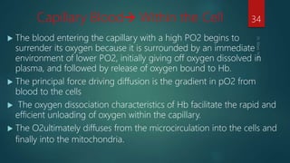
- 31. Capillary Blood Within the Cell The blood entering the capillary with a high PO2 begins to surrender its oxygen because it is surrounded by an immediate environment of lower PO2, initially giving off oxygen dissolved in plasma, and followed by release of oxygen bound to Hb. The principal force driving diffusion is the gradient in pO2 from blood to the cells The oxygen dissociation characteristics of Hb facilitate the rapid and efficient unloading of oxygen within the capillary. The O2ultimately diffuses from the microcirculation into the cells and finally into the mitochondria. 34
- 32. Oxygen content (CaO2) The oxygen content of arterial blood is the sum of the oxygen bound to haemoglobin and oxygen dissolved in plasma. Total amount of O2 present in 100 ml of Arterial Blood CaO2=Hb. Bound O2+ dissolved Hb = [1.34 x Hb x SaO2] + 0.003 x PO2 = [1.34×15×97.5] +0.003×100 =19.9=20ml /dl approx . =200ml/L 35
- 33. Similarly for Venous blood CvO2=[1.34 × Hb × SvO2] + 0.003 × PvO2 replacing with values we have CvO2=15 ml/dl =150 ml/L Total oxygen content: = 200 × arterial blood vol. + 150 × venous blood vol. = 200 × 0.25 × 5+150 × 0.75 × 5 = 250 + 562.5 =812ml 36
- 34. Oxygen delivery (DO2) The total amount of oxygen delivered or transported to the peripheral tissues is dependent on 1. The body's ability to oxygenate blood 2. The hemoglobin concentration 3. The cardiac output (QT) 37 DO2 decreases in response to: DO2 increases in response to: Low blood oxygenation that can be caused by Low PaO2 Low SaO2 Low hemoglobin concentration Low cardiac output Increased blood oxygenation caused by Increased PaO2 Increased SaO2 Increased hemoglobin concentration Increased cardiac output
- 35. Oxygen consumption (VO2) The amount of oxygen extracted by the peripheral tissues during the period of one minute is called oxygen consumption Normal resting O2 consumption ~ 200 to 300 ml/min in adult humans 38 Factors that Increase VO2 Factors that Decrease VO2 Exercise Seizures Shivering Hyperthermia Body Size Skeletal Muscle Relaxation Induced by drugs Peripheral shunting Sepsis, trauma Certain poisons Cyanide Hypothermia
- 36. 16 Arterial-Venous O2 Content Difference • The arterial-venous oxygen content difference is the difference between the CaO2 and the CvO2. Factors that increase the C(a-v)O2: Factors that decrease the C(a-v)O2: • decreased cardiac output • increased O2consumption exercise seizures shivering increased temp • Increased cardiac output • Skeletal relaxation Induced by drugs • Peripheral shunting • Sepsis, trauma • Certain poisons Cyanide - prevents tissues from using oxygen • Hypothermia 39
- 37. Pulmonary Shunting the portion of the cardiac output that moves from the right side to the left side of the heart without being exposed to alveolar oxygen (PAO2). Clinically, pulmonary shunting can be subdivided into absolute and relative shunts: Absolute Shunt, also called True Shunt (Anatomic Shunt) 40
- 38. 41 Common Causes of Absolute Shunt Common Causes of Relative Shunt Congenital heart disease Intrapulmonary fistula Vascular lung tumors Capillary shunting is commonly caused by: Alveolar collapse or atelectasis Alveolar fluid accumulation Alveolar consolidation When pulmonary capillary perfusion is in excess of alveolar ventilation, a relative or shunt-like effect is said to exist Hypoventilation Ventilation/perfusion mismatches Chronic emphysema, bronchitis, asthma Alveolar-capillary diffusion defects Alveolar fibrosis or alveolar edema Key Concept: True shunts are refractory to supplemental oxygen Key Concept: Relative or Shunt-Like effects can be corrected by supplemental oxygen
- 39. Oxygen extraction ratio The oxygen extraction ratio (O2ER) is the amount of oxygen extracted by the peripheral tissues divided by the amount of O2 delivered to the peripheral cells aka Oxygen coefficient ration / Oxygen utilization ratio It can increased to 70-80% during maximal exercise in well trained athletes. 42 Factors that increase the O2ER Factors that decrease the O2ER • decreased cardiac output • increased O2consumption exercise seizures shivering hyperthermia anemia • Increased cardiac output • Skeletal relaxation (Induced by drugs) • Peripheral shunting • Sepsis, trauma • Certain poisons(Cyanide) • Hypothermia • Increased Hb • Increased arterial Oxygenation
- 40. O2 DIFFUSION FROM INTERSTITIUM TO CELLS Intracellular PO2 < Interstitial fluid PO2 • O2 constantly utilized by the cells • Cellular metabolic rate determines overall O2 consumption intracellular req for optimal maintenance of metabolic pathways ~ 3 mm Hg 43
- 41. Pasteur point – critical mitochondrial PO2 below which aerobic metabolism cannot occur 0.15 – 0.3 kPa = 1.4 – 2.3mmHg 44
- 42. The DO2–VO2 Relationship As O2 delivery (DO2) begins to decrease below normal, the O2 uptake (VO2) initially remains constant, indicating that the O2 extraction (O2ER) is increasing as the DO2 decreases. Further decreases in DO2 eventually leads to a point where the VO2 begins to decrease. The transition from a constant to a varying VO2 occurs when the O2 extraction increases to a maximum level of 50% to 60% (O2ER = 0.5 to 0.6). Once the O2ER is maximal, further decreases in DO2 will result in equivalent decreases in VO2 because the O2ER is fixed and cannot increase further. 45
- 43. When this occurs, the VO2 is referred to as being supply-dependent, and the rate of aerobic metabolism is limited by the supply of oxygen. This condition is known as dysoxia . As aerobic metabolism (VO2) begins to decrease, the oxidative production of high energy phosphates (ATP) begins to decline, resulting in impaired cell function and eventual cell death. 46
- 44. The Critical DO2 : The DO2 at which the VO2 becomes supply- dependent is called the critical oxygen delivery (critical DO2). It is the lowest DO2 that is capable of fully supporting aerobic metabolism . 47
- 45. Conclusion Inspired O2 from the environment moves across the alveolar- capillary membrane into the blood & is then transported to the peripheral tissue & used to fuel aerobic cellular metabolism. OXYGENATION is the process of O2 diffusing passively from the alveolus to the pulmonary capillary, where it binds to hemoglobin in RBC or dissolves to plasma. OXYGEN DELIVERY is the rate of O2 transport from lungs to the peripheral tissues. OXYGEN CONSUMPTION is the rate at which oxygen is removed from the blood for use by the tissues Within the pulmonary capillaries , one hemoglobin molecule binds up to four oxygen molecules in a cooperative manner 48
- 46. Oxygen Cascade “CO2 Cascade” Thank you 49
Editor's Notes
- #3: ATP,the coenzyme that supplies energy to all active metabolic processes.
- #6: Deoxygenated venous blood becomes oxygenated on the pulmonary capillaries after diffusion down a concentration gradient across the alveolar capillary membrane.
- #8: When dealing with the gas mixture,each gas contribute seperatly to the total gas pressure k/a partial pressure of the individual gas. And the partial pressure of the gas is directly propotional to its concemtration. Haemoglobin saturation is determined by the partial pressure of oxygen. When these values are graphed they produce the Oxygen Disassociation Curve piO2= Fio2 8 BAROMETRIC PRESSURE Barometric pressure at mt everest =253 mmHg
- #9: In alveoli the inspired gases are mixed with residual alveolar gas from previous breath.o2 is taken up and co2 is added.the final alveolar 02 tension can be estimated by pA02=PI02-pac02/RQ piO2= Fio2 * BAROMETRIC PRESSURE (760-47)0.21=150mm Hg
- #10: The factors that determine the precise value of alveolar PO2 are (1) the PO2 of atmospheric air, (2) the rate of alveolar ventilation, and (3) the rate of total body oxygen consumption piO2= Fio2 * BAROMETRIC PRESSURE 0.21 *(760-47) –(40/0.8) 1000mmHg
- #11: Within the lung, oxygen diffuses from the alveoli into the pulmonary capillaries, driven by the gradient between the partial pressure of oxygen in the alveolar space and that in the deoxygenated pulmonary capillary blood. O2 moves across the alveolar membranes into the pulmonary capillaries by passive diffusion , across the alveolar-capillary membrane, through the plasma and across the erythrocyte membrane and binds to Hb.
- #12: Within the lung, oxygen diffuses from the alveoli into the pulmonary capillaries, driven by the gradient between the partial pressure of oxygen in the alveolar space and that in the deoxygenated pulmonary capillary blood. O2 moves across the alveolar membranes into the pulmonary capillaries by passive diffusion , across the alveolar-capillary membrane, through the plasma and across the erythrocyte membrane and binds to Hb.
- #13: A layer of fluid lining the alveolus and containing surfactant that reduces the surface tension of the alveolar fluid. The alveolar epithelium composed of thin epithelial cells. An epithelial basement membrane. A thin interstitial space between the alveolar epithelium and the capillary membrane. A capillary basement membrane that in many places fuses with the alveolar epithelial basement membrane. The capillary endothelial membrane.
- #14: Transit time for blood is ~ 0.75 sec (time blood remains in the lungs capillary). EQUILIBRIUM TIME –> Normally ~0.25 sec(1/3rd of total transit time) is required for Oxygen as well as Carbon Dioxide diffusion. Rest 0.50 sec is the safety margin (which helps in diffusion during exercise, high altitude or impaired diffusion) Rate of gas diffusion = Diffusion coefficient X Pressure gradient x Surface area of the membrane /Thickness of the membrane any factor that increases the thickness (eg. Fibrosis, oedema fluid) can interfere significantly with normal respiratory exchange of gases. Greater the surface area greater is the rate of diffusion. The large capillary surface area in alveoli and the 0.4 to 0.5 micrometer thickness of the alveolar-capillary membrane greatly facilitate )2 diffusion. The volume of gas transfer across the alveolar-capillary membrane per unit time is
- #16: – Henry’s Law the amount of gas dissolved in a liquid will be directly proportional to the partial pressure of the gas in the liquid-gas interface” As long as the PO2 in the gas phase is higher than the PO2 in the liquid phase, there will be a net diffusion of O2 into the blood .
- #17: Respiratory Unit is composed of a respiratory bronchiole, alveolar ducts, atria, and alveoli.
- #18: The Alveolar–arterial gradient is a measure of the difference between the alveolar concentration (A) and the arterial (a) concentration of oxygen. most common mechanism of hypoxemia is increased A-a gradient which depends on right to left shunt (directly propotional),v/q mismatch,mixed venous o2 tension (incersely propotional)
- #21: Which states that the concentration of any gas in solution is propotional to its partial pressure.
- #22: At a normal PaO2 of 100 mm Hg, however, the Hb saturation (SaO2) is only about 97% due to the following three normal physiologic shunts •Thebesian venous drainage into the left atrium •Bronchial venous drainage into pulmonary veins •Alveoli that are under ventilated, also referred to as dead space ventilation Thus, the amount of arterial oxygen in the preceding equation must be adjusted to 97 percent:
- #25: It is the percentage expression of no of oxygen binding site occupied out of maximum no of a binding site available.
- #26: At pH 7.4, temp 37, po2 50% is at 27 mmhg
- #27: A small drop is systemic capillaries po2 can result in release of large amount of oxygen from the metabolically active cells
- #32: The high co2 content of venous capillary blood by decreasing hemoglobin affinity for o2 facilitates the release of o2 to tissues. Conversely the lower co2 content in pulmonary capillaries increases hemoglobin affinity for 02 again, facilitating 02 uptake from alveoli.
- #38: DO2 decreases in response to: Low blood oxygenation that can be caused by Low PaO2 Low SaO2 Low hemoglobin concentration Low cardiac output DO2 increases in response to: Increased blood oxygenation caused by Increased PaO2 Increased SaO2 Increased hemoglobin concentration Increased cardiac output
- #39: The Critical DO2 •The DO2 at which the VO2 becomes supply-dependent is called the critical oxygen delivery (critical DO2). It is the lowest DO2 that is capable of fully supporting aerobic metabolism . • It is identified by the bend in the DO2–VO2 curve . • Despite the ability to identify the anaerobic threshold, the critical DO2 has limited clinical value. • First, the critical DO2 has varied widely in studies of critically ill patient, and it is not possible to predict the critical DO2 in any individual patient in the ICU. •Second, the DO2–VO2 curve can be curvilinear (i.e., without a single transition point from constant to changing VO2) , and in these cases, it is not possible to identify a critical DO2.
- #43: This means that only 25% of the oxygen delivered to the systemic capillaries is taken up into the tissues. The DO2–VO2 Relationship • As O2 delivery (DO2) begins to decrease below normal, the O2 uptake (VO2) initially remains constant, indicating that the O2 extraction (O2ER) is increasing as the DO2 decreases. Further decreases in DO2 eventually leads to a point where the VO2 begins to decrease. • The transition from a constant to a varying VO2 occurs when the O2 extraction increases to a maximum level of 50% to 60% (O2ER = 0.5 to 0.6). Once the O2ER is maximal, further decreases in DO2 will result in equivalent decreases in VO2 because the O2ER is fixed and cannot increase further. • When this occurs, the VO2 is referred to as being supply-dependent, and the rate of aerobic metabolism is limited by the supply of oxygen. This condition is known as dysoxia . • As aerobic metabolism (VO2) begins to decrease, the oxidative production of high energy phosphates (ATP) begins to decline, resulting in impaired cell function and eventual cell death.
- #48: With further reduction in delivery of oxygen critical point is reached where oxygen consumption becomes directly propotional to oxygen delivery
- #49: .