2
Most read
6
Most read
17
Most read
SAMPLE PREPARATION
Introduction
• Sample is a small part or quantity intended to show what the whole is like.
• Following as sample can be used for laboratory diagnosis of leishmaniasis.
i. Cells/Tissues
ii. DNA from tissues
iii. Blood or Urine.
• Tissues can either be examined under microscope for presence of parasite or DNA
from tissues can be examined, while blood can be used to test the presence of
immunoglobulins, proteins or other components.
Several steps involved in sample preparation are based on type of sample and mode of test.
(S.Sundar,Rai.M; Clin Diagn Lab Immunol, 2002. PubMed Central)
Protein sample preparation
Extraction of the entire protein from serum requires a multi –step protocol, where various sample processing steps are
performed to increase efficacy of the extraction procedure.
In case of bacterial/ animal/plant protein extraction- sample collection, stabilization, and protein extraction.
After separation of serum from whole blood or lysis of cell (bacterial/animal/plant) cell lysate, other components must
be removed by filtration, or centrifugation.
Cells or tissue are disrupted in such a way as to minimize proteolysis and other modes of protein degradation.
Lysis of these cells could be done using- Osmotic shock, Enzyme digestion, Detergent ,Grinding or UltraSonicator.
It should be performed at as low temperature as possible and with a minimum of heat generation and even using
protease inhibitors will give good results.
Separation of plasma
1) Blood is collected into purple top EDTA tubes and centrifuged (2000 rpm) at 4 degrees centigrade for 20 minutes.
2) 2) After centrifugation place 1.0ml of plasma into 1.5ml eppendorf tube and label it.
3) 3) Freeze immediately at –80 degree freezer.
Separation of Serum
1. A 10 ml tube of whole blood is collected following standard procedures using a serum separator tube (Golden, tiger top
tube) from each patient.
2. 2. Allow samples to clot for one hour at room temperature
3. 3. Centrifuge for 10 minutes at approximately 4000 rpm
4. 4. Aliquot serum into labelled cryovials.
5. 5. Immediately freeze vials of serum at –80-degree freezer
For sample proteins from Blood
Source: Rutgers Cancer institute of New Jerzy-protocol
Preparation of lysate from cell culture
1. Placed the cell culture dish on ice and wash the cells with ice-cold PBS.
2. Then ice-cold lysis buffer is added (1 mL per 107 cells/100 mm dish/150 cm2 flask; 0.5 mL per 5x10⁶ cells/60 mm dish/75 cm2 flask).
3. Gently transfer the cell suspension into a pre-cooled microcentrifuge tube.
4. Maintain constant mixing for 30 min at 4°C.
5. Centrifuge in a microcentrifuge at 4°C for 20 min at 12,000 rpm.
6. Gently remove the tubes from the centrifuge and place on ice, aspirate the supernatant and place in a fresh tube
kept on ice. Discard the pellet.
For sample proteins from Cells/Tissues
Source: Abcom protocol : protein sample preparation
Extraction methods are divided in three major types- Gentle, Moderate, and Vigorous.
For sample proteins from Cells/Tissues Contd..
Gentle
• Cell Lysis (osmotic
Shock)
• Enzyme digestion
• Detergent digestion
• Hand homogenization
• Mortar pestle grinding
Moderate
• Grinding with glass
beads
• Blade homogenization
• Freeze/Thaw
Vigorous
• Ultrasonication
• French Press
Source: G E Healthcare life sciences protein extraction
handbook
Protein Purification
Proteins can be purified using techniques based on molecular size , solubility of proteins, or charge.
Based on Molecular size-
Density gradient centrifugation,
Size-exclusion chromatography.
Based on charge-
Ion-exchange chromatography
electrophoresis
Based on Solubility of Proteins-
Using Salt (NaCl, MgCl2
) at different concentration
Density gradient centrifugation
Protein containing solution is suspended in uniform density
gradient of sucrose and sedimentation of protein occurs in
tube depending upon its rate of sedimentation.
Sedimentation depends upon weight, density and size on
the protein.
Some other centrifugation technique like differential
sedimentation centrifugation,
Source: Sigma Aldrich
Chromatography
Chromatography is a technique for separating mixture into their components in order to analyze, identify, purify
the components.
It is based on the separation of components based on their relative affinity or solubility to different phages of
chromatography. i.e. Mobile phase (M.P.) and Stationary phase(S.P.).
The different types of combination of stationary and mobile phases are used for different types of
chromatography, which is selected based on our requirement.
Types of chromatography
I. Paper chromatography – for dried liquid samples with a liquid solvent and paper as S.P.
II. Thin Layer chromatography – for dried liquid samples with liquid solvent and thin layer of stationary
phase (alumina or silica gel on glass plate)
III. Liquid chromatography - for Liquid samples with liquid solvent and solid beads.
IV. Gas chromatography - mainly used for vaporized or volatile samples with carrier gas and S.P. of
Liquid or solid beads.
V. HPLC – mainly for already purified protein, used for analytical purposes.
Gel-filtration chromatography
Porous particles are used to separate molecules of different
size.
Proteins passed over a column filled with a hydrated porous
beads made of a carbohydrate or polyacrylamide polymer.
Mixture of protein dissolved in suitable buffer, is allowed to
flow by gravity down the column.
Very large molecules cannot penetrate into the pores of the
beads without entering the pores, the small molecules enter
the pores.
Large molecules are excluded and small proteins are retarded
due to pores.
This way separation on the basis of size and weight is done.
Source: www.mikeblaber.org/lecture 31
Ion-exchange chromatography
Separation of proteins in a column filled with charged polmer
beads.
Positively charged beads- anion exchange chromatography
Negatively charged beads- cation exchange chromatography
Proteins with opposite charge binds to the beads respectively.
More charged protein will bound well and so less charged
proteins will be eluted first.
Finally bound proteins can be eluted using salt.
Source: mastering-biochemistry.com
Gel Electrophoresis
Macromolecules ( proteins, nucleic acids) are separated under the electric field on the basis of their size and charge.
Voltage of approx. 70- 100 volts is used, which may vary depending upon the sample and gel size.
Different types of electrophoresis are performed on the basis of molecules.
Molecules move towards opposite charged electrode and hence get resolved.
The movement of molecule also depend on size, shape, and applied voltage.
Polyacrylamide or agarose gels are used for caring sample and for resolution of their components.
SDS (sodium dodecyl sulfate) is anionic detergent which bind to peptide chain and provides uniform negative charge,
and so cause proteins move to cathode.
PAGE
Poly Acrylamide gel electrophoresis is mainly performed for protein
samples.
It is mainly of two types- Native PAGE and, SDS-PAGE.
In Native PAGE the protein is separated in their native form or
conformation, however in SDS-PAGE.
As SDS is an anionic detergent which breaks polymeric proteins to
their monomeric subunits and linearize them.
Image Source: www.bio-rad.com- literature bulletin
Gel preparation
Stacking Gel Resolving/Separating Gel
4% 7.5% 12%
30% Acrylamide/bis 1.98ml 3.75ml 6.0ml
0.5M Tris HCl (pH6.8) 3.78ml - -
1.5M Tris HCl (pH8.8) - 3.75ml 3.75ml
10%SDS 150µl 150µl 150µl
dH2O 9ml 7.28ml 5.03ml
TEMED 15µl 7.5µl 7.5µl
10%APS 75µl 75µl 75µl
Total Volume 15ml 15ml 15ml
Buffer preparation
Components/L volume
Tris Base 30.4 g
Glycine 144.2 g
SDS 10.0g
The volume was made up to 1L with water
Running Buffer (10X)
Components/L Concentration
Tris HCL pH 8 50 mM
NaCl 150 mM
NP-40 1%
Sodium deoxycholate 0.5 %
SDS 0.1 %
Lysis Buffer
Laemmli Buffer
Tris-Cl 60mM
Glycerol 10%
SDS 3%
Bromophenol blue 0.05%
ᵝ- mercaptoethanol 10%
TAE Buffer
Ccomponents Main Composition 50X 1X
Tris Base 2 M 242.2 g/l 4.844 g/l
Acetic Acid 1 M 60.5 ml/l 1.21 ml/l
EDTA Sodium salt
dihydrate
50 mM 18.612 g/l 0.372 g/l
DNA Isolation
DNA is a very long molecule made up of a chain of nucleotides and the order of these nucleotides is what makes
organisms similar to others of their species and yet different as individuals. Genes are sections within this long
DNA molecule.
In eukaryotic cells, DNA is organized as chromosomes in an organelle called the nucleus.
Bacterial cells have no nucleus, so their DNA is organized in rings or circular plasmids, which are in the
cytoplasm.
The DNA extraction process frees DNA from the cell and then separates it from cellular fluid and proteins so you
are left with pure DNA.
The three basic steps of DNA extraction are
1) lysis,
2) precipitation, and
3) purification.
Protein/DNA Sample preparation methods
Extraction Protocol
1. Take 3ml of whole blood in Falcon tube.
2. Add 2ml of reagent A.
3. Mix well at room temperature.
4. Centrifuge at 10000rpm for 5 minutes at room temperature.
5. Discard supernatant carefully and add 1ml of reagent B.
6. Vortex briefly for better mixing.
7. Place tube in water bath for 15 min at 65 degree Celsius.
8. Allow to cool at room temperature and add Phenol : Chloroform : isoamyl alcohol.
9. After proper mixing, centrifuge at 12000pm for 2 min.
10. Transfer upper phase to new tube and add 2ml of ice cold ethanol and invert mix well.
11. Spool the cottony DNA using pipette.
12. Air dry and resuspend in TAE buffer for further use.
Reagents
Reagent A- for RBC lysis
0.01M Tris-HCl pH 7.4
320mM Sucrose
5mM MgCl2
1% Triton X100
Preparation- Add 10 ml of Tris,109.54g of sucrose, 0.47g of MgCl2,and 10 ml of Triton X-100 to 800ml of
distilled water and adjust pH to 8.0. make final volume to 1 L.
Reagent B- for cell lysis
0.4M tris-HCl,
150 mM NaCl,
0.06M EDTA,
1% sodium dodecyl sulfate ,
pH 8.0
preparation- Take 400 mL of Tris, 120 mL of EDTA, 8.76g of NaCl, and adjust pH, final volume to 1L.
The obtained DNA can be then used for PCR, or Electrophoresis.
For electrophoresis the DNA sample is mixed with Loading dye and added to well for allowing it to resolve on gel.
For PCR-(for amplification of DNA ) A mix of templet DNA , dNTPs, Polymerase Enzyme and Primes, and allow it to
step by step Denaturation, Annealing, and Elongation.
The PCR product could be checked for amplification on Agarose gel electrophoresis.
Thank You !

More Related Content

PPTX
FPLC - Fast Protein Liquid Chromatography
PPTX
SDS PAGE
PPTX
Cell culture
PDF
Flow cytometry
PDF
Flow cytometry
PPTX
Centrifugation
PPTX
Proteomics
FPLC - Fast Protein Liquid Chromatography
SDS PAGE
Cell culture
Flow cytometry
Flow cytometry
Centrifugation
Proteomics

What's hot (20)

PPTX
Ultra centrifuge
PPTX
Enzyme assay methods
PPTX
Animal cell culture
PPTX
Sds page electrophoresis
PPTX
ISOELECTRIC FOCUSING
PPTX
Ppt of centrifugation
PPTX
Analytical Ultracentrifugation
PPT
centrifugation
PPT
Fplc(fast protein liquid chromatography )
PPTX
Paper electrophoresis
PPT
5. Cell culture media
PPTX
Biology and characterization of the cell culture
PPTX
2 d gel electrophoresis
PDF
Electrophoresis
PDF
Electrophoresis
PPTX
Cell Culture.pptx
PPT
Liquid chromatography
PPTX
ICAT Technique in Proteomics
PPTX
Protein separation
Ultra centrifuge
Enzyme assay methods
Animal cell culture
Sds page electrophoresis
ISOELECTRIC FOCUSING
Ppt of centrifugation
Analytical Ultracentrifugation
centrifugation
Fplc(fast protein liquid chromatography )
Paper electrophoresis
5. Cell culture media
Biology and characterization of the cell culture
2 d gel electrophoresis
Electrophoresis
Electrophoresis
Cell Culture.pptx
Liquid chromatography
ICAT Technique in Proteomics
Protein separation
Ad

Similar to Protein/DNA Sample preparation methods (20)

PPTX
02 Protein Isolation02 Protein Isolation
PPTX
MUGDHA's SEMINAR MSc sem 1.pptx
PDF
Electrophoresis
PPT
96481966-Electrophoresis.ppt
PPT
Electrophoresis techniques and its types.ppt
PPTX
Electrophoresis & its types
PPT
Purification techniques
PPTX
electrophoresis
PPT
Electrophoresis presentation
PPTX
Protein purification-and-characterization-techniques (1).pptx
PPTX
Protein purification-and-characterization-techniques (1).pptx
PPT
Plasma proteins
PPTX
Protein analysis
PPTX
Electrophoresis
PPTX
Electrophoresis for pgs by Dr siva kumar reddy
PPTX
Electrophoresis ppt.pptx
PPT
Gel electrophoresis
PPTX
SDS PAGE.pptx sodium dodecyl sulfate PAGE
PPTX
Global analysis of protein by kk sahu sir
02 Protein Isolation02 Protein Isolation
MUGDHA's SEMINAR MSc sem 1.pptx
Electrophoresis
96481966-Electrophoresis.ppt
Electrophoresis techniques and its types.ppt
Electrophoresis & its types
Purification techniques
electrophoresis
Electrophoresis presentation
Protein purification-and-characterization-techniques (1).pptx
Plasma proteins
Protein analysis
Electrophoresis
Electrophoresis for pgs by Dr siva kumar reddy
Electrophoresis ppt.pptx
Gel electrophoresis
SDS PAGE.pptx sodium dodecyl sulfate PAGE
Global analysis of protein by kk sahu sir
Ad

Recently uploaded (20)

PPTX
A powerpoint on colorectal cancer with brief background
PPT
Presentation of a Romanian Institutee 2.
PPTX
INTRODUCTION TO PAEDIATRICS AND PAEDIATRIC HISTORY TAKING-1.pptx
PPTX
ap-psych-ch-1-introduction-to-psychology-presentation.pptx
PPTX
Microbes in human welfare class 12 .pptx
PPT
veterinary parasitology ````````````.ppt
PPT
Heredity-grade-9 Heredity-grade-9. Heredity-grade-9.
PPT
1. INTRODUCTION TO EPIDEMIOLOGY.pptx for community medicine
PDF
Wound infection.pdfWound infection.pdf123
PDF
Social preventive and pharmacy. Pdf
PPTX
gene cloning powerpoint for general biology 2
PPTX
Presentation1 INTRODUCTION TO ENZYMES.pptx
PDF
S2 SOIL BY TR. OKION.pdf based on the new lower secondary curriculum
PDF
CHAPTER 2 The Chemical Basis of Life Lecture Outline.pdf
PDF
Assessment of environmental effects of quarrying in Kitengela subcountyof Kaj...
PDF
BET Eukaryotic signal Transduction BET Eukaryotic signal Transduction.pdf
PPTX
Seminar Hypertension and Kidney diseases.pptx
PPT
Computional quantum chemistry study .ppt
PDF
Packaging materials of fruits and vegetables
PPT
Enhancing Laboratory Quality Through ISO 15189 Compliance
A powerpoint on colorectal cancer with brief background
Presentation of a Romanian Institutee 2.
INTRODUCTION TO PAEDIATRICS AND PAEDIATRIC HISTORY TAKING-1.pptx
ap-psych-ch-1-introduction-to-psychology-presentation.pptx
Microbes in human welfare class 12 .pptx
veterinary parasitology ````````````.ppt
Heredity-grade-9 Heredity-grade-9. Heredity-grade-9.
1. INTRODUCTION TO EPIDEMIOLOGY.pptx for community medicine
Wound infection.pdfWound infection.pdf123
Social preventive and pharmacy. Pdf
gene cloning powerpoint for general biology 2
Presentation1 INTRODUCTION TO ENZYMES.pptx
S2 SOIL BY TR. OKION.pdf based on the new lower secondary curriculum
CHAPTER 2 The Chemical Basis of Life Lecture Outline.pdf
Assessment of environmental effects of quarrying in Kitengela subcountyof Kaj...
BET Eukaryotic signal Transduction BET Eukaryotic signal Transduction.pdf
Seminar Hypertension and Kidney diseases.pptx
Computional quantum chemistry study .ppt
Packaging materials of fruits and vegetables
Enhancing Laboratory Quality Through ISO 15189 Compliance

Protein/DNA Sample preparation methods

  • 2. Introduction • Sample is a small part or quantity intended to show what the whole is like. • Following as sample can be used for laboratory diagnosis of leishmaniasis. i. Cells/Tissues ii. DNA from tissues iii. Blood or Urine. • Tissues can either be examined under microscope for presence of parasite or DNA from tissues can be examined, while blood can be used to test the presence of immunoglobulins, proteins or other components. Several steps involved in sample preparation are based on type of sample and mode of test. (S.Sundar,Rai.M; Clin Diagn Lab Immunol, 2002. PubMed Central)
  • 3. Protein sample preparation Extraction of the entire protein from serum requires a multi –step protocol, where various sample processing steps are performed to increase efficacy of the extraction procedure. In case of bacterial/ animal/plant protein extraction- sample collection, stabilization, and protein extraction. After separation of serum from whole blood or lysis of cell (bacterial/animal/plant) cell lysate, other components must be removed by filtration, or centrifugation. Cells or tissue are disrupted in such a way as to minimize proteolysis and other modes of protein degradation. Lysis of these cells could be done using- Osmotic shock, Enzyme digestion, Detergent ,Grinding or UltraSonicator. It should be performed at as low temperature as possible and with a minimum of heat generation and even using protease inhibitors will give good results.
  • 4. Separation of plasma 1) Blood is collected into purple top EDTA tubes and centrifuged (2000 rpm) at 4 degrees centigrade for 20 minutes. 2) 2) After centrifugation place 1.0ml of plasma into 1.5ml eppendorf tube and label it. 3) 3) Freeze immediately at –80 degree freezer. Separation of Serum 1. A 10 ml tube of whole blood is collected following standard procedures using a serum separator tube (Golden, tiger top tube) from each patient. 2. 2. Allow samples to clot for one hour at room temperature 3. 3. Centrifuge for 10 minutes at approximately 4000 rpm 4. 4. Aliquot serum into labelled cryovials. 5. 5. Immediately freeze vials of serum at –80-degree freezer For sample proteins from Blood Source: Rutgers Cancer institute of New Jerzy-protocol
  • 5. Preparation of lysate from cell culture 1. Placed the cell culture dish on ice and wash the cells with ice-cold PBS. 2. Then ice-cold lysis buffer is added (1 mL per 107 cells/100 mm dish/150 cm2 flask; 0.5 mL per 5x10⁶ cells/60 mm dish/75 cm2 flask). 3. Gently transfer the cell suspension into a pre-cooled microcentrifuge tube. 4. Maintain constant mixing for 30 min at 4°C. 5. Centrifuge in a microcentrifuge at 4°C for 20 min at 12,000 rpm. 6. Gently remove the tubes from the centrifuge and place on ice, aspirate the supernatant and place in a fresh tube kept on ice. Discard the pellet. For sample proteins from Cells/Tissues Source: Abcom protocol : protein sample preparation
  • 6. Extraction methods are divided in three major types- Gentle, Moderate, and Vigorous. For sample proteins from Cells/Tissues Contd.. Gentle • Cell Lysis (osmotic Shock) • Enzyme digestion • Detergent digestion • Hand homogenization • Mortar pestle grinding Moderate • Grinding with glass beads • Blade homogenization • Freeze/Thaw Vigorous • Ultrasonication • French Press Source: G E Healthcare life sciences protein extraction handbook
  • 7. Protein Purification Proteins can be purified using techniques based on molecular size , solubility of proteins, or charge. Based on Molecular size- Density gradient centrifugation, Size-exclusion chromatography. Based on charge- Ion-exchange chromatography electrophoresis Based on Solubility of Proteins- Using Salt (NaCl, MgCl2 ) at different concentration
  • 8. Density gradient centrifugation Protein containing solution is suspended in uniform density gradient of sucrose and sedimentation of protein occurs in tube depending upon its rate of sedimentation. Sedimentation depends upon weight, density and size on the protein. Some other centrifugation technique like differential sedimentation centrifugation, Source: Sigma Aldrich
  • 9. Chromatography Chromatography is a technique for separating mixture into their components in order to analyze, identify, purify the components. It is based on the separation of components based on their relative affinity or solubility to different phages of chromatography. i.e. Mobile phase (M.P.) and Stationary phase(S.P.). The different types of combination of stationary and mobile phases are used for different types of chromatography, which is selected based on our requirement. Types of chromatography I. Paper chromatography – for dried liquid samples with a liquid solvent and paper as S.P. II. Thin Layer chromatography – for dried liquid samples with liquid solvent and thin layer of stationary phase (alumina or silica gel on glass plate) III. Liquid chromatography - for Liquid samples with liquid solvent and solid beads. IV. Gas chromatography - mainly used for vaporized or volatile samples with carrier gas and S.P. of Liquid or solid beads. V. HPLC – mainly for already purified protein, used for analytical purposes.
  • 10. Gel-filtration chromatography Porous particles are used to separate molecules of different size. Proteins passed over a column filled with a hydrated porous beads made of a carbohydrate or polyacrylamide polymer. Mixture of protein dissolved in suitable buffer, is allowed to flow by gravity down the column. Very large molecules cannot penetrate into the pores of the beads without entering the pores, the small molecules enter the pores. Large molecules are excluded and small proteins are retarded due to pores. This way separation on the basis of size and weight is done. Source: www.mikeblaber.org/lecture 31
  • 11. Ion-exchange chromatography Separation of proteins in a column filled with charged polmer beads. Positively charged beads- anion exchange chromatography Negatively charged beads- cation exchange chromatography Proteins with opposite charge binds to the beads respectively. More charged protein will bound well and so less charged proteins will be eluted first. Finally bound proteins can be eluted using salt. Source: mastering-biochemistry.com
  • 12. Gel Electrophoresis Macromolecules ( proteins, nucleic acids) are separated under the electric field on the basis of their size and charge. Voltage of approx. 70- 100 volts is used, which may vary depending upon the sample and gel size. Different types of electrophoresis are performed on the basis of molecules. Molecules move towards opposite charged electrode and hence get resolved. The movement of molecule also depend on size, shape, and applied voltage. Polyacrylamide or agarose gels are used for caring sample and for resolution of their components. SDS (sodium dodecyl sulfate) is anionic detergent which bind to peptide chain and provides uniform negative charge, and so cause proteins move to cathode.
  • 13. PAGE Poly Acrylamide gel electrophoresis is mainly performed for protein samples. It is mainly of two types- Native PAGE and, SDS-PAGE. In Native PAGE the protein is separated in their native form or conformation, however in SDS-PAGE. As SDS is an anionic detergent which breaks polymeric proteins to their monomeric subunits and linearize them. Image Source: www.bio-rad.com- literature bulletin
  • 14. Image Source: www.bio-rad.com- literature bulletin
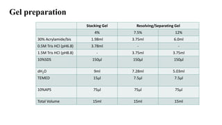
  • 15. Gel preparation Stacking Gel Resolving/Separating Gel 4% 7.5% 12% 30% Acrylamide/bis 1.98ml 3.75ml 6.0ml 0.5M Tris HCl (pH6.8) 3.78ml - - 1.5M Tris HCl (pH8.8) - 3.75ml 3.75ml 10%SDS 150µl 150µl 150µl dH2O 9ml 7.28ml 5.03ml TEMED 15µl 7.5µl 7.5µl 10%APS 75µl 75µl 75µl Total Volume 15ml 15ml 15ml
  • 16. Buffer preparation Components/L volume Tris Base 30.4 g Glycine 144.2 g SDS 10.0g The volume was made up to 1L with water Running Buffer (10X) Components/L Concentration Tris HCL pH 8 50 mM NaCl 150 mM NP-40 1% Sodium deoxycholate 0.5 % SDS 0.1 % Lysis Buffer Laemmli Buffer Tris-Cl 60mM Glycerol 10% SDS 3% Bromophenol blue 0.05% ᵝ- mercaptoethanol 10% TAE Buffer Ccomponents Main Composition 50X 1X Tris Base 2 M 242.2 g/l 4.844 g/l Acetic Acid 1 M 60.5 ml/l 1.21 ml/l EDTA Sodium salt dihydrate 50 mM 18.612 g/l 0.372 g/l
  • 17. DNA Isolation DNA is a very long molecule made up of a chain of nucleotides and the order of these nucleotides is what makes organisms similar to others of their species and yet different as individuals. Genes are sections within this long DNA molecule. In eukaryotic cells, DNA is organized as chromosomes in an organelle called the nucleus. Bacterial cells have no nucleus, so their DNA is organized in rings or circular plasmids, which are in the cytoplasm. The DNA extraction process frees DNA from the cell and then separates it from cellular fluid and proteins so you are left with pure DNA. The three basic steps of DNA extraction are 1) lysis, 2) precipitation, and 3) purification.
  • 19. Extraction Protocol 1. Take 3ml of whole blood in Falcon tube. 2. Add 2ml of reagent A. 3. Mix well at room temperature. 4. Centrifuge at 10000rpm for 5 minutes at room temperature. 5. Discard supernatant carefully and add 1ml of reagent B. 6. Vortex briefly for better mixing. 7. Place tube in water bath for 15 min at 65 degree Celsius. 8. Allow to cool at room temperature and add Phenol : Chloroform : isoamyl alcohol. 9. After proper mixing, centrifuge at 12000pm for 2 min. 10. Transfer upper phase to new tube and add 2ml of ice cold ethanol and invert mix well. 11. Spool the cottony DNA using pipette. 12. Air dry and resuspend in TAE buffer for further use.
  • 20. Reagents Reagent A- for RBC lysis 0.01M Tris-HCl pH 7.4 320mM Sucrose 5mM MgCl2 1% Triton X100 Preparation- Add 10 ml of Tris,109.54g of sucrose, 0.47g of MgCl2,and 10 ml of Triton X-100 to 800ml of distilled water and adjust pH to 8.0. make final volume to 1 L. Reagent B- for cell lysis 0.4M tris-HCl, 150 mM NaCl, 0.06M EDTA, 1% sodium dodecyl sulfate , pH 8.0 preparation- Take 400 mL of Tris, 120 mL of EDTA, 8.76g of NaCl, and adjust pH, final volume to 1L.
  • 21. The obtained DNA can be then used for PCR, or Electrophoresis. For electrophoresis the DNA sample is mixed with Loading dye and added to well for allowing it to resolve on gel. For PCR-(for amplification of DNA ) A mix of templet DNA , dNTPs, Polymerase Enzyme and Primes, and allow it to step by step Denaturation, Annealing, and Elongation. The PCR product could be checked for amplification on Agarose gel electrophoresis.