3. Definitions
• Infertility; Inability to conceive after
one year of regular and unprotected
sex.
• Fecundability; Is the probability of
conceiving in 1 menstrual cycle.
Average is 20 %, maximum is 35%.
At the end of one year trial 85 to 90%
will conceive.
• Fecundity; physiologic potential to
produce offspring
• Fertility; Actual number of children
born to a woman.
Earlier evaluation done when;
• History of prior infertility
• Presence of obvious risk factors
• Age of woman greater than 35
4. Epidemiology
In Uganda;
• 10-15 % of the couples cannot
have children.
• 75% is due to STIs.
World;
• In the U.S, it affects 9% and 10%
of men and women respectively
between 15 to 44 yrs of age.
• 48million couples and 186
million individuals live with
infertility globally (WHO).
• 1 in 4 healthy women in their 20s
and 30s will get pregnant in any
single menstrual cycle1 in 10 healthy
women in their 40s will get pregnant
in any single menstrual cycle.
• Fertility begins to decrease for
women in their 20s and 30s and
declines more quickly after the age
of 35(American college of Obstetrics
and gyneacologists).
• Couples in which the male partner is
40 years or older are more likely to
have difficulty conceiving(CDC,
2019).
5. Classification
• Primary infertility; A pregnancy has never been attained before.
• Secondary infertility; At least one prior pregnancy has been attained
before regardless of the outcome.
Basic evaluation of an infertile couple
• History and examination
• Husband semen analysis
• Test of ovulation
• Test of tubal patency
• Atleast a TVS
6. Overview of infertility
Female 40-55%, Male 40%, Unexplained in 10%
• Female causes
Ovulatory 30%
Anovovulatory disorders
Group 1-Hypogonadotropic hypogonadism
Group 2-Eugonadotropic Eugonadism eg PCOS
Group 3-Hypergondotropic Hypogonadism eg
POI
Group 4-Hyperprolactinemia; decreases GnRH
Uterine 15%
-Submucous fibroids
-Asherman syndrome
-Septate uterus
Tubal 20-30%
-PID
-Genital TB
Cervical 5%
-Antisperm
7. History and examination
Female history
Age
length of time spent trying for pregnancy
Any previous pregnancies and their outcomes if any
Any abortions
History of contraceptive use
Coital frequency (frequency, dyspareunia)
Occupation (exposure to radiation or chemotherapy, stress)
Menstrual history (menarche, LMP, regularity, length of cycles, amount)
Previous surgical and medical history (STDs, PID, fibroids, endometriosis, PCOS) (pelvic surgery, tubal surgery,
uterine instrumentation)
Previous fertilization treatment
Cervical smear history
History of galactorrhea.
Female examination
General exam;- BP, Pulse, height and weight, signs of virilisation and hirsutism, visual field testing for
bitemporal hemianopia
Breast examination;- nipple discharge (hyperprolactinemia)
Pelvic exam e.g. asses cervix, uterine size, version, tenderness and adnexal masses, fibroids, anteverted fixed
uterus
8. Male history
Length of time spent trying for pregnancy
Sexual history;- frequency and timing.
Fathered any pregnancies
History of mumps or measles
History of testicular trauma or surgery to the testes
Occupation;- nature of job and prolonged absence from home, radiations,
chemicals and heat
Medical and surgical history e.g. chronic disease like DM, renal disease
Male examination
Testicular examination;- site, volume, consistency, masses, absence of the vas
deferens, varicocele, evidence of surgical scars.
Gynacomastia
Hypospadius
PR for prostate
9. INVESTIGATIONS
Tests for ovulation
• Basal body temperature-3 to 4 days after ovulation, rise by 0.4 degrees F
• Cervical mucus changes
estrogen; mucus is thin and stretchable (spinnbarkeit phenomenon), ferning pattern
progesterone- mucus thick not stretchable, breaks apart
• Vaginal cytology; sample taken from lateral vaginal walls near fornix
Kinds of cells; Superficial cells- eosinophillic cells with small pyknotic nuclei, they predominate under influence of estrogen
Intermediate cells; predominate under progesterone
Parabasal and basal cells- round, large nucleus, small cytoplasm( stains blue)- when there's no hormone
stimulus.
Karyopyknotic index/cornification index—superficial cells/(intermediate + parabasal cells)
Maturation index– Parabasal: intermediate: superficial; during ovulation—0:30:70; the index shifts to the right
• Endometrial sampling; done 1 week prior to expected menses in luteal/secretory phase (D21 in 28 day cycle), if on D21
there proliferative changes, it means ovulation didn’t occur.
• D21 serum progesterone; less than 3ng/ml implies anovulation
greater than 10ng/ml denotes normal corpus luteum function
• Urinary LH; ovulation expected to occur 14- 26 hrs after detection of urinary LH surge, helps in timing of sex.
• Follicular monitoring on ultra sound
10. • Follicular monitoring on ultrasound;
Dominant follicle grows at 2mm/day, max 20-25 mm
After ovulation, it shrinks in size, creates margins, fluid seen in pouch of
Douglas.
Advantages; monitor treatment ie ovulation induction, see the growth of
the endometrium
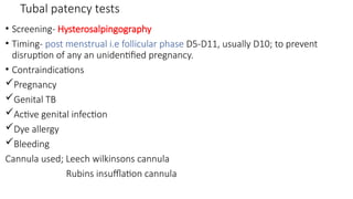
11. Tubal patency tests
• Screening- Hysterosalpingography
• Timing- post menstrual i.e follicular phase D5-D11, usually D10; to prevent
disruption of any an unidentified pregnancy.
• Contraindications
Pregnancy
Genital TB
Active genital infection
Dye allergy
Bleeding
Cannula used; Leech wilkinsons cannula
Rubins insufflation cannula
12. • Sonosalpingography; SSG or sion test
oNormal saline into the uterine cavity via Foley catheter. An inflated bulb of
the catheter prevents leakage of fluid outside uterine cavity.
oBy visualizing the flow of saline along the tube and observing it as a shower
at fimbrial end, tubal patency can be tested. Presence of free fluid in pouch
of Douglas also confirms tubal patency.
oAdvantages; no radiation
Assess the uterus
Disadvantages; Difficult to tell which tube is locked
• Laparascopic chromopertubation (gold standard)
Laparascope through the abdomen, methylene blue dye introduced through
a cannula into fallopian tube. Direct visualization of dye draining through
tube.
Advantages: silmutaneous assessment of pelvis.
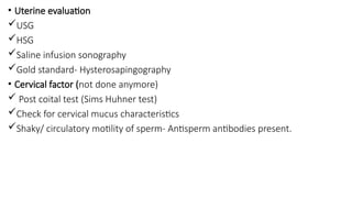
13. • Uterine evaluation
USG
HSG
Saline infusion sonography
Gold standard- Hysterosapingography
• Cervical factor (not done anymore)
Post coital test (Sims Huhner test)
Check for cervical mucus characteristics
Shaky/ circulatory motility of sperm- Antisperm antibodies present.
14. • What happens with reproductive
ageing?
Ovarian reserve tests
D3 serum inhibin
D3 serum FSH ; FSH >10IU/L is poor
reserve
D3 serum estradiol; Basal estradiol >60-
80pg/ml is poor reserve
D3 antral follicle count; <4 in both ovaries
is poor reserve
Serum AMH; derived from preantral and
small antral follicles which make the
ovarian pool hence a direct reflection of
residual ovarian pool
• Testing done anytime of the cycle
• Low AMH (0.2-0.7ng/ml) is poor reserve.
AMH > 1 is normal
Clomiphene citrate challenge test
15. Treatment methods
Anovovulatory disorders- Ovulation Iinduction
• Group 1-Hypogonadotropic hypogonadism- Pulsatile GnRH Therapy
• Group 2-Eugonadotropic Eugonadism eg PCOS- Clomiphene Citrate, Letrozole
• Group 3-Hypergondotropic Hypogonadism eg POI- Ovum donation
• Group 4-Hyperprolactinemia; decreases GnRH- Bromocriptine, Cabergoline
Clomiphene Citrate
Nonsteroidal selective Estrogen Receptor Modulator (SERM)
• Anti estrogenic action-Blocks estrogen receptors at the pituitary gland
• This creates a false signal of low estrogen levels
• In turn FSH increases, promote multi follicular growth-Risk of multiple pregnancy-7-
10%
• No teratogenic effects
• Dose; 50mg-100mg/day
16. • Letrozole
• Aromatase Inhibitor
• Inhibits conversion of androgen to estrogen
• Hence creating an estrogen like deficiency
state hence increase FSH and more
follicular growth
• DOC for ovulation induction in PCOS
women
• Dose 2.5 to 7.5 mg/day *5/7
Gonadotropins
Earlier- Human Menopausal Gonadotropins
(mix of FSH and LH)
Now- Recombinant Gonadotropins-purer
forms
Are IM injectables
Risk of multiple pregnancy is higher-30%
GnRH analogues like Leuprolide, used in a
pulsatile manner to induce ovulation
• Ovulation induction protocol
• Ovulation occurs in 36hrs after giving the
trigger
• IUI-36 hrs after giving ovulation trigger
17. What is in vitro fertilization?
Stimulation of ovaries
Retrieval of oocytes
Fertilization- on culture media
Embryo formed grows in culture media
Embryo transfer-Day 5 embryo transferred in the uterus
Indications of IVF
• Tubal factor infertility
• Severe male factor infertility
• Treatment failure
• Unexplained infertility
• IVF with donor oocyte
• IVF with surrogacy
• IVF with pre implantation genetic diagnosis
18. Infertility in males
❖ Introduction
❖ Definition
❖ Causes of infertility
❖ Investigations
❖ Management
19. introduction
• Infertility definition
• As above, in approximately 35% of couples with infertility, a male
factor is identified along with a female factor; in approximately 10%, a
male factor is the only identifiable cause
20. Male reproduction summary
• Anatomy of testis (p testes): Outer coverings, internal
structures(lobules, seminiferous tubules), interstitial tissue, rete
testis, efferent ductules
• Spermatogenesis (sermatogonial phase, meiotic phase,
spermiogenesis)
• Role of supporting cells( Sertoli cells , Leydig cells) and hormonal
control(FSH, LH, testosterone)
• Hypothalamic-pituitary-gonadal axis
21. Male reproduction summary part 2
• Male infertility is due to defect in either;
• Production of healthy sperms
• Transportation of healthy sperms to site of fertilization
• Emission( movement of sperm and glandular secretions into urethra in preparation for ejaculation with the end
result that urethra is loaded with semen):sexual stimulation of SNS, peristaltic contractions in Vas deferens,
seminal vesicles, prostate gland, ejaculatory ducts that deliver sperm and fluids into the prostatic urethras,
contraction of internal urethral sphincter
• Ejaculation( forceful expulsion of semen from urethra to outside):sexual stimulation and somatic reflexes via
pudendal nerve, rhythmic contractions of pelvic floor muscles (BS,IC) and urethral muscle, ejection of semen,
relaxation of external urethral sphincter) PLUS orgasmic sensations and temporary loss of voluntary control
• Semen: spermatozoa(5-10%), seminal fluid(about 60%), prostatic fluid(20-30%), bulbourethral (5%) and others.
• Healthy semen: pH(7.2-8.0),2-6ml in volume, sperm count is 15-200 million sperm/ml, whitish-gray, smells
chlorine-like
22. Useful terminology
CONDITION TERMINOLOGY
No semen Aspermia
No sperms Azoospermia
Less sperms
➢ Less than 15 million /ml
➢ Less than 5 million/ml
Oligospermia
Severe Oligospermia
Non motile sperms Asthenospermia
Dead sperms Necrospermia
Abnormal morphology Teratospermia
Increased WBC in sperms Leucocytospermia
23. Causes of male infertility
Congenital disorders Acquired disorders Systemic disorders
Congenital GnRH deficiency
(Kallmann syndrome)
Pituitary and hypothalamic tumors(pituitary
macroadenoma ,craniopharyngioma)
Severe systemic illness
Iron overload syndromes Pituitary and hypothalamic infiltrative
disorders( sarcoidosis, histiocytosis, tuberculosis, fungal
infections)
Nutritional deficiencies
Multiorgan genetic
disorders(Prader-Willi syndrome,
familial cerebellar ataxia, etc.)
Head trauma, intracranial radiation, surgery Morbid obesity
Vascular(pituitary infarction, aneurysm)
Hormonal(hyperprolactinemia, androgen excess, estrogen
excess, cortisol excess)
Drugs(exogenous androgens, opioids and psychotropic
drugs, GnRH agonists or antagonists
1. Endocrine and systemic disorders( hypogonadotropic hypogonadism 2% to 5%)
VITAMINSABCDEK
SPERM COUNT
Can be pre-testicular(mainly hormonal),testicular( mainly structural, genetic,
infective), post-testicular (obstructions, ejaculations issues)
24. Congenital disorders Acquired disorders Systemic illness Genetic causes of
dysspermatogenesi
s
Klinefelter syndrome
(XXY) and its
variants(XXY/XY, XXXY)
Varicocele(large, palp
able without Valsalva maneuver)
Idiopathic
dysspermatogenesis
Y-chromosome
microdeletions and
related disorders
cryptorchidism Infections; viral orchitis(mumps,
echovirus,
arbovirus),granulomatous
orchitis(leprosy, tuberculosis),
epididymo-orchitis(gonorrhea,
chlamydia)
Renal failure, hepatic
cirrhosis, cancer, sickle cell
disease, amyloidosis,
vasculitis, celiac disease
Autosome and X-
chromosome
defects
2. Primary testicular defects in spermatogenesis(65% to 80%)
25. Congenital disorders Acquired disorders Systemic illness Genetic causes of
dysspermatogenesi
s
Myotonic dystrophy Drugs :Alkylating agents, alcohol,
marijuana,
antiandrogens,ketoconazole,spironol
actone,histamine-2 receptor
antagonists, ionizing radiation
Mutations causing
severe defects in
sperm morphology
Functional prepubertal castrate
syndrome(congenital anorchia)
Environmental toxins: Di
bromochloropropane, carbon
disulfide, cadmium, lead, mercury,
environmental estrogens, and
phytoestrogens ;
smoking ;hyperthermia
Androgen insensitivity syndromes Immunological disorders, including
polyglandular autoimmune disease
and angiosperm antibodies
5-alpha reductase deficiency Trauma
Estrogen receptor or synthesis
disorders
Testicular torsion
26. • 3. sperm transport disorders( about 5%)
• epididymal dysfunction (drugs, infection)
• abnormalities of the vas deferens (congenital absence, Young syndrome,
infection, vasectomy)
• ejaculatory duct disorders
• seminal vesicles and prostate
4.Sexual dysfunction(infrequent vaginal intercourse, ED, premature
ejaculation)
5. Idiopathic male infertility(different from idiopathic
dysspermatogenesis. Here, the infertile man has normal semen analysis
and no apparent cause for infertility
27. Evaluation of male infertility
history
• Duration of infertility
• Fertility in other relationships
• Medical and surgical history( chronic severe systemic illness, head trauma, surgery)
• Infections (STIs, Guts, mumps orchitis)
• Medications
• Sexual development history(testicular descent, Tanners staging, etc)
• History of chemotherapy or radiation
• Cigarette smoking, alcohol, marijuana and drug use; environmental and occupational exposures
• Sexual dysfunction or impotence
• Frequency of intercourse, use of lubricants toxic to sperm
• Previous infertility testing and therapies
• Family history of birth defects, intellectual disability , or reproductive failures
28. Physical examination
• GENERAL APPEARANCE(sexual development, gynecomastia, obesity,
pubic hair). Skin for iron overload, Cushing, longstanding testosterone
deficiency
• EXTERNAL GENITALIA( Tanner stage, Scrotal exam( size, consistency,
nodularity of testicles, palpation of cord for presence of vas deferens,
DRE, Valsalva for varicocele). Testicular volume determination by
Prader orchidometry, ultrasound
29. Semen analysis
• Semen sample is collected after 2 to 7 days of ejaculatory abstinence
either by masturbation at health facility or at home and delivered
within an hour of collection to lab. Semen analysis is performed by
standardized methods in accordance with WHO laboratory manual for
examination and processing of human semen.
• Standard semen analysis has;
• Semen volume and pH
• Microscopy for sperm concentration, count, motility, and
morphology; debris and agglutination, leukocyte count, immature
germ cells
30. PARAMETER VALUE
Volume Greater than 1.4 ml (95% CI 1.3 -1.5)
PH > 7.2
Sperm concentration Greater than 15 million/ml
Sperm count Greater than 39 million/ejaculate (CI 35-40)
Sperm morphology Greater than 4% normal most imp (3-9)
Total motility > 42%(40-43)
Active motility > 30%(29-31)
Viability 58%
WBC Count, < 1 million/ml
Total Round Cells < 5 million/ml
32. Follow up evaluation
Endocrine testing: especially for man wil sperm conc <5 million/ml. serum total testosterone, LH, FSH
Low testosterone, high FSH and LH ( primary hypergonadotropic hypogonadism (both spermatogenesis and Leydig
function affected))
Normal testosterone, normal LH, high FSH( primary hypergonadotropic hypogonadism with seminiferous tubule damage,
intact Leydig cells
Low testosterone, FSH and LH low or normal( secondary hypogonadotropic hypogonadism. Look for cause e.g. prolactin,
iron overload, brain mass hypothyroidism, hypoadrenalism
High testosterone, LH, normal FSH; partial androgen resistance
Normal testosterone, LH,FSH( majority) further analysis depends on semen analyss
Low sperm count, very low LH, very muscular; androgen abuse
Scrotal and transrectal ultrasound; especially obstructive azoospermia( normal testicular volumes, serum
testosterone,FSH and LH with azoospermia. Also done if vasa deferens not palpable, generally for obstructions, anomalies
vasography
Genetic tests: Karyotyping and testing for Y chromosome microdeletions with very low sperm cone. Klinefelter syndrome,
cystic fibrosis
33. Semen analysis
Low volume Low concentration Abnormal
morphology(length,
width, width ratio,
area occupied by
acrosome, neck and
tail defects)
Poor motility
NORMAL CONCENTRATION; incomplete
collection, partial retrograde ejaculation
AZOOSPERMIA
Retrograde
ejaculation,
congenital absence of
vas deferens,
obstructive
azoospermia
Immature germ cels
indicate disorders of
spermatogenesis
Increased white
blood cells in
ejaculate
=inflammation/infec
tion, poor semen
quality
Very high percentage of
immobile sperm in ejaculate
=ICSI
LOW SPERM CONCWENTRATION:
Testosterone deficiency
AZOOSPERMIA OR SEVERE OLIGOSPERMIA
Genital tract obstruction or vas deferens,
seminal vesicles absent (physical exam or
scrotal/transrectal ultrasound)or obstructed
34. Treatment
• Having identified cause of infertility, therapy is aimed at correcting reversible
etiologies and overcoming irreversible factors
• Generally, therapeutic interventions for treatment of infertility are drug therapy,
surgery and/or procedures such as intrauterine insemination or in vitro fertilization
• Absolute contraindications to infertility therapy are contraindication to pregnancy or
contraindication to the use of the drugs or surgery to enhance fertility
• The couple is counseled on lifestyle modifications to improve infertility, such as
smoking cessation, reducing excessive caffeine and alcohol consumption , and
appropriate timing and frequency of coitus
• Increasing physical activity and avoiding tobacco, marijuana, excessive alcohol intake,
and obesity might be useful in optimizing spermatogenesis. Reduce scrotal heat
• Dietary supplements such as fish oil, antioxidants
35. • Endocrine and systemic disorders
The underlying disorder is managed to result in eugonadism and improved
spermatogenesis and fertility, otherwise gonadotropin replacement therapy is used.
For example, if hyperprolactinemia is cause, the hypogonadism might be corrected
by lowering serum prolactin conc.
NB: isolated use of exogenous testosterone is advised against among hypo-gonadal
men especially if still interested in fertility preservation, but otherwise pursue
treatments that result in increase of endogenous serum testosterone production
Medical therapy only benefits patients with secondary (hypogonadotropic)
hypogonadism but not those with primary (hypogonadotropic )hypogonadism who
otherwise require ART such as surgical testicular sperm extraction(TESE) and
intracytoplasmic sperm injection (ICSI) into female partner's egg
36. • Primary testicular defects in sperm production
• Low serum testosterone, elevated FSH and LH( primary hypergonadotropic hypogonadism)
usually have azoospermia . ART is used
• Normal serum testosterone and LH, high FSH have variable degrees of dysspermatogenesis. ART
• Eugonadal, infertile men. Normal serum testosterone, FSH, LH. Those with high quantity of
abnormal sperms and oligospermia have no clear medical therapy and will benefit from ART.
Those with azoospermia should be evaluated for obstructive causes and treated surgically to
enhance or restore fertility e.g. surgical excision of a large varicocele or surgical correction of
ejaculatory duct obstruction
Sperm in ejaculate(vaginal intercourse, ART), spermatids or mature spermatozoa seen only in
testicular biopsies especially in primary hypergonadotropic hypogonadism and
azoospermia( surgical retrieval) OR no sperm seen in testicular biopsies( no known therapy)
Leukospermia is managed with antibiotics, NSAIDs, mast cell blockers, antioxidants
37. Treatment of sperm transport disorders
• Optimizing sperm transportation to female partners vagina such as
optimizing sexual intercourse, treating ED, ejaculatory disorders, obstruction
of epididymis and ejaculatory duct. All can also be treated with ART
• Retrograde ejaculation with oral sympathomimetics
• Obstructive azoospermia due to obstruction of epididymis or ejaculatory
duct can be managed with microsurgical end to end anastomosis of the
epididymal duct to epididymal duct or to the vas deferens. Also, ART with
ICSI is an option. Vasectomy reversal. Congenital bilateral absence of the
vasa differentia is managed with ART
38. Assisted reproductive technologies
• Intrauterine insemination +/- gonadotropin stimulation of female partner
• IVF with ICSI(intracytoplasmic sperm injection)
• Retrieval of sperm( non obstructive azoospermia, Klinefelter syndrome, longstanding
azoospermia after chemotherapy), this is by multiple open testicular biopsies with
TESE(testicular sperm extraction) or microTESE, testicular or epididymal aspiration.
Genetic counseling and testing is important as there is an increased risk of
chromosomal abnormalities and congenital malformations in live births following ICSI.
Also infertile men with sperm conc. <10 mil,testing for Y chromosomal microdeletion
in men with sperm conc <5 mil/ml, and screening for gene mutations associated with
infertility
• ART with donor semen