


skeletal system
- 2. Content •Introduction •Development of bone •Types of bone cell •Davison of bone •Types of bone • Physiology of skeleton
- 3. Introduction The musculoskeletal system consists of the two different systems 1) skeleton ( bone ) 2) muscular system( muscles ) The Human Skeletal system is the body system composed of bones , tendons, and ligaments and other tissues that perform essential function for the human body.
- 4. This skeletal system can be divided into the axial and appendicular systems. In an adult body, it is mainly composed of 206 individual bones. • BONE DEVELOPMENT AND REMODELING TAKES PLACE IN A CONTINUOUS BASIS. FROM PRENATAL PERIOD TO EARLY CHILDHOOD TO ADULTHOOD.
- 5. Development of bone ( osteogenesis / ossification) Osteogenesis /ossification :- Formation of bone Corresponding Word is -> assify Bone is Osseous tissue. Os' is a synonym of bone Process of Converting hyaline cartilage and fibrous connective tissue, fibrous membrane into bone
- 6. Types of bone cell:- • Osteoblasts- Bone farming cells • Osteocyts-Mature Osteoblasts that maintain bone • Osteoclasts :-Bone breaking cells (Maintain Shope) Osteoclasts are one of the types of bone cells that break down and reabsorb bone tissue. These are important cells as they are used to initiate bone remodelling.
- 7. Davison of bones Axial skeleton Appendicular skeleton
- 8. Axial skeleton •The axial skeleton runs along the body’s central axis, therefore it is called the central core of the human body. The axial skeleton is composed of 80 bones and it consists of: •Skull Bone – It includes 8 cranial bones, 14 facial bones, 6 auditory ossicles, and the Hyoid Bone
- 9. •The bone of the Thoracic Cage – It includes 25 bones of the thorax- a breastbone and 24 ribs. •The bone of the Vertebral column- It includes 24 vertebrae bones, the sacrum bone, and the coccyx bone. Cranium Thoracic cage Vertebrae bones Sacrum Coccyx
- 10. Skull:- Cranium :- The portion of the skull that encloses the brain. It includes 8 cranial bones, which enclose and protect the brain.8 cranial bones ure 2 parietal, 2 temporal, 1 frontal, I occipital. 1 ethmoid, I sphenoid bone. Frontal bone (forehead bone) forms the forehead, parietal bone forms roof & sides, temporal bone forms inferior & lateral part and occipital bone forms posterior and most of the base of cranium.
- 11. • The opening in the occipital bone through which the spinal cord passes from the brain called foramen magnum
- 12. • Sutures of skull bone :- There are 4 type of suture • Coronal suture-A suture unite frontal bone with both parietal bones: • Sagittal suture-A suture unite both parietal bones at superior midline of the skull. Lambdoid suture-A suture unite both parietal bones with occipal bone framed hit is like lambda
- 13. • Squamous suture-A suture unite parietal booo with temporal bone in the lateral aspects of the skull Picture
- 14. Face:- The skeleton of face is formed by 14 bones Nasal Zygomatic bone (2)-It also called cheekbone or malar bone. It forms prominence of the cheeks Lacrimal bone (2)-These are smallest bone of face. It is posterior to nasal bone and form medial wall of each obital cavity Vomer (5)-It forms the lower and posterior portion of the nasal septum,
- 16. Thoracic cavity :- •The thoracic cavity also known as the chest cavity is the hollow space in the body that protects the lungs, heart, tracheobronchial tree, and other organs. •The thoracic cavity consists of the thoracic cage that is made up of the sternum and 12 pairs of ribs that support and protect all the organs contained within the thoracic cavity.
- 17. Mandible (1)-It also called lower jaw bone. It is largest or strongest facial bone. It is the only movable skull bone bone Inferior nasal conchae (2)-It forms part of lateral wa of nasal cavity. Maxillae (2) - Parrod maxillac units to form the upper jaw bone. It articulates with every hone of face except mandible Palatine bone (2)-it forms the posterior part of the hard palate and lateral nasul wall
- 19. Vertebrae column j • It also called spine, back bone or spinal columns • Vertebral column composed by a series of bones called vertebrae • The length of the column in average adult male is about 71cm and 61 cm in average adult female. The sacral and coccygeal vertebrae that fuse to form the sacrum and coccyx called fixed vertebra (false vertebra).
- 20. The vanchrae that remain unfused through life the cervical, thoracic, and lumbar called true vertebra Adul vertebral column contami 26 vertebrae infant and children have 33 vertebrae)
- 21. • 7 Cervical vertebrae-It form neck • 12 Thoracic vertebrae It forms posterio part of thoracic cavity • 3 Lumbar vertebrae-It support the lower back. 1 Sacrum-It consist of 5 fused sacral vertebrae. • 1 Coccyx-It consist of 4 fasod coccygeal vertebrae
- 22. Appendicular skeleton • The appendicular skeleton is composed of 126 bones and it comprises of the- • Pelvic girdle • Upper Limbs • Lower Limbs • Shoulder Girdle
- 24. Pelvic girdle:- Female pelvis consist of 4 pelvic bones:- 2 innomated bone 1 sacrum 1 coccyx 2 innomated bone:- consist of ilium, Ischium, Pubis 1 Sacrum :- sacrum consist of 5 fused vertebrae 1 coccyx:- 4 fused vertebrae
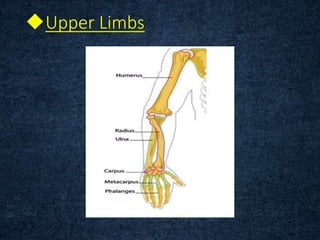
- 26. Upper Limbs
- 27. Humerus • This is the bone of the upper arm. The head sits within the glenoid cavity of the scapula, forming the shoulder joint. Distal to the head are two projections of bone, the greater and lesser tubercles, and between them there is a deep groove, the bicipital groove , occupied by one of the tendons of the biceps muscle. • The distal end of the bone presents two surfaces that articulate with the radius and ulna to form the elbow joint.
- 28. Radius and ulna • These are the two bones of the forearm. The ulna is longer than and medial to the radius and when the arm is in the anatomical position, with the palm of the hand facing forward, the two bones are parallel. They articulate with the humerus at the elbow joint, the carpal bones at the wrist joint and with each other at the proximal and distal radioulnar joints.
- 29. Wrist ( carpal bones ) • There are eight carpal bones arranged in two rows of four • proximal row: scaphoid, lunate, triquetrum, pisiform • distal row: trapezium, trapezoid, capitate, hamate.
- 30. • These bones are closely fitted together and held in position by ligaments that allow a limited amount of movement between them. The bones of the proximal row are associated with the wrist joint and those of the distal row form joints with the metacarpal bones. Tendons of muscles lying in the forearm cross the wrist and are held close to the bones by strong fibrous bands, called retinacula
- 31. Metacarpals • These five bones form the palm of the hand. They are numbered from the thumb side inwards. The proximal ends articulate with the carpal bones and the distal ends with the phalanges.
- 32. Phalanges ( fingerbones) • There are 14 phalanges, three in each finger and two in the thumb. They articulate with the metacarpal bones and with each other, by hinge joints.
- 33. Lower Limbs
- 34. Femur • The femur is the longest and heaviest bone of the body. The head is almost spherical and fits into the acetabulum of the hip bone to form the hip joint. The neck extends outwards and slightly downwards from the head to the shaft and most of it is within the capsule of the hip joint.
- 35. • The distal extremity has two articular condyles, which, with the tibia and patella, form the knee joint.
- 36. Tibia • The tibia is the medial of the two bones of the lower leg. The proximal extremity is broad and flat and presents two condyles for articulation with the femur at the knee joint. The head of the fibula articulates with the inferior aspect of the lateral condyle, forming the proximal tibiofibular joint.
- 37. • The distal extremity of the tibia forms the ankle joint with the talus and the fibula. The medial malleolus is a downward projection of bone medial to the ankle joint.
- 38. fibula • The fibula is the long slender lateral bone in the leg. The head or upper extremity articulates with the lateral condyle of the tibia, forming the proximal tibiofibular joint, and the lower extremity articulates with the tibia, and projects beyond it to form the lateral malleolus. This helps to stabilise the ankle joint.
- 39. Patela This is a roughly triangular- shaped sesamoid bone associated with the knee joint. Its posterior surface articulates with the patellar surface of the femur in the knee joint and its anterior surface is in the patellar tendon, i.e. the tendon of the quadriceps femoris muscle.
- 40. Tarsal The seven tarsal bones forming the posterior part of the foot (ankle) are the talus, calcaneus, navicular, cuboid and three cuneiform bones. The talus articulates with the tibia and fibula at the ankle joint. The calcaneus forms the heel of the foot. The other bones articulate with each other and with the metatarsal bones.
- 41. Metatarsals • which form the greater part of the dorsum of the foot. At their proximal ends they articulate with the tarsal bones and at their distal ends, with the phalanges. The enlarged distal head of the 1st metatarsal bone forms the ‘ball’ of the foot.
- 42. Phalanges The phalanges are long bones in the foot located distal to the metatarsals. Like in the hand,
- 43. Shoulder Girdle • The shoulder girdle consists of two scapulae and two clavicles. Clavicle (collar bone) • The clavicle is an S-shaped long bone. It articulates with the manubrium of the sternum at the sternoclavicular joint and forms the acromioclavicular joint with the acromion process
- 44. Scapula The scapula is a flat triangular- shaped bone, lying on the posterior chest wall superficial to the ribs and separated from them by muscles. At the lateral angle is a shallow articular surface, the glenoid cavity, which, with the head of the humerus, forms the shoulder joint.
- 45. • On the posterior surface runs a rough ridge called the spine, which extends beyond the lateral border of the scapula and overhangs the glenoid cavity. The prominent overhang, which can be felt through the skin as the highest point of the shoulder, is called the acromion process and forms a joint with the clavicle, the acromioclavicular joint, a slightly movable synovial joint that contributes to the mobility of the shoulder girdle. The coracoid process, a projection from the upper border of the bone, gives attachment to muscles that move the shoulder joint.
- 46. Types of bone Long bone Flat bone Short bone Irregular bone Sesamoid bone
- 47. Long bone Long bones are bones that are longer than they are wide. The mid section of the long bone is also contains bone marrow Examples of the long bones in humans include the femur, tibia, and fibula of the legs; humerus, radius and ulna of the arms; clavicle (collar bone), metatarsals and metacarpals of the feet.
- 49. Short bone Short bones are the ones that are as long as they are wide. They are cube shaped bones that have a thin cortical layer and a thick spongy interior. Examples of short bones include tarsals and carpals in the foot and hand, respectively.
- 50. Flat bone Flat bones are thin and curved bones that are composed of spongy cancellous tissue sandwiched between two thin layers of cortical bone. They usually form broad, flat plates as in the sternum, cranium (skull), rib cage and ilium (pelvis).
- 51. Sesamoid bone Sesamoid bones are the bones that are embedded in the tendons or muscles. The patella of the knee and pisiform of the wrist are two examples of sesamoid bones.
- 52. Irregular bone Irregular bones vary in shape and structure and therefore do not fit into any other category (flat short, long, or sesamoid). They often have a fairly complex shape, which helps protect internal organs.
- 53. Physiology of skeleton system • The primary functions of the skeletal system include movement, support, protection production of blood cells, storage of minerals.
- 54. Shape AND Support The primary function of the skeletal system is to provide a solid framework to support and safeguard the human body and its organs. This helps in maintaining the overall shape of the human body.
- 55. Protection The skeletal system also helps to protect our internal organs and other delicate body organs, including the brain, heart, lungs and spinal cord by acting as a buffer. Our cranium (skull) protects our brain and eyes, the ribs protect our heart and lungs and our vertebrae (spine, backbones) protect our spinal cord.
- 56. Movement The skeletal system also helps to protect our internal organs and other delicate body organs, including the brain, heart, lungs and spinal cord by acting as a buffer. Our cranium (skull) protects our brain and eyes, the ribs protect our heart and lungs and our vertebrae (spine, backbones) protect our spinal cord.
- 57. Storage and mineralization The bone matrix of the skeletal system is mainly involved in storing or preserving different types of essential minerals which are required to facilitate growth and repair of the body cells and tissues. The cell-matrix acts as our calcium bank by storing and releasing calcium ions into the blood cell when required.
- 58. Questions and answers How many bones present in adult ? What is meaning of Ossification ? Types of bone cell ? How many bone spresent in axial skeleton ??
- 59. Sacrum consist of how many bones ? Listout the name of skull bone ? which are the example of long bone ?? Examples of sesamoid bone ??
- 60. Thenk you !