UPTAKE AND TRANSPORT IN ORGANISMS-ADVANCED LEVEL
Download as DOCX, PDF0 likes308 views

This document discusses water and mineral uptake and transport in plants. It covers the three main types of transpiration (stomatal, cuticular, lenticular), factors that affect transpiration rate, and how transpiration is measured using a potometer or mass loss method. Stomatal transpiration accounts for 90% of water loss and occurs through stomata. Transpiration rate is affected by environmental factors like humidity, temperature, wind, and light intensity. It also discusses the internal structure of roots and the mechanism of water uptake through root hairs via osmosis.


![54 ADVANCED LEVEL TRANSPORT NOTES 2019 (modelled by king SOYEKWO ROGERS 07012119966
pressure above ventricular pressure; forcing
bloodflow from the leftatrium throughtheopen
bicuspid value; into the relaxed left ventricular;
From 0.14 seconds to 0.38 seconds represents
ventricular systole; There is more powerful
contraction of left ventricular cardiac muscle
than the atrial cardiac muscle; which decreases
ventricular volume while increasing ventricular
pressureabove atrialpressure;forcing closureof
bicuspid valve to prevent backflow of blood into
the left atrium; Ventricular pressure increases
furtherexceeding aortic pressureat0.16seconds
to force open semilunar/aortic valves; hence
allowing left ventricular blood flow into aorta;
From 0.38 seconds to 0.6 seconds represents
diastole; The left ventricular cardiac muscle
relaxes to increase ventricular volume and
decrease ventricular pressure below aortic
pressure; forcing closure of semilunar / aortic
valve to prevent backflow of blood; At 0.4
seconds ventricular pressure decreases further
below atrial pressure to force opening of
bicuspid valve; and flow of oxygenated blood
from the left atrium into the left ventricle;
1. (d) (i) Pattern of electrical activity
P wave corresponds to the wave of electrical
excitation spreading over the atria during atrial
systole/contraction;
QRS wave corresponds to the wave of electrical
excitation spreading over the ventricles during
ventricular systole/contraction;
T wave corresponds to the wave of electrical
excitation spreading over the ventricles during
ventricular diastole/relaxation;
(ii) Pattern of Sounds on the phonocardiogram
1 is the first heart sound produced by the sudden
closure of the atrioventricular valves; [described
as the ‘lub’]
2 is the second heart sound produced by the
sudden closure of the semilunar valves of the
aorta and pulmonary artery; [described as the
‘dub’]
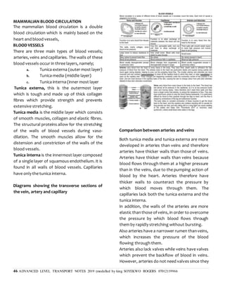
(e) Explain how the internal heart structure is
related to its functioning
- Cardiac muscle fibres interconnected to form a
network of fibre to ensure rapid and uniform
spread of excitation throughout the walls of the
heart;
- Heart divided into 4 chambers to enable
differential generation of pressure;
- Ventricles have thicker walls than auricles to
generatehigher pressuretodrive bloodover long
distance into more elaborate circulation/to the
lungs and to all body tissues;
- Walls of left ventricles are thicker than those of
right ventricles to generate more pressure to
pump
blood to longer distance in the systemic
circulation/rest of the body;
- Longitudinal septum which separates the heart
into two halves to prevent mixing of oxygenated
and deoxygenated blood;
- Valves to prevent back flow of blood;
- Valves have strands of connecting tissue
(chordas tendinae) to prevent them from being
pushed
inside out when ventricles contract;
- Sino Atrial Node (S.A.N) acts a pacemaker
regulatingrate of beating and excitation of heart;
- Heart located in the thoracic cavity where it is
protected from any external mechanical damage;
- Atrio Ventricular node (A.V.N) which delays
depolarization wave from Sino Atrial Node to
ensure thatauricles empty completely before the
ventricles contract;
- Purkinje tissues to relay waves from A.V.N to
ventricular myocardium;](https://image.slidesharecdn.com/uptakeandtransportinplantsupdated2020-210724145252/85/UPTAKE-AND-TRANSPORT-IN-ORGANISMS-ADVANCED-LEVEL-54-320.jpg)


UPTAKE AND TRANSPORT IN ORGANISMS-ADVANCED LEVEL
- 1. 1 ADVANCED LEVEL TRANSPORT NOTES 2019 (modelled by king SOYEKWO ROGERS 07012119966 UPTAKE AND TRANSPORT IN PLANTS Water and mineral salts are necessary for photosynthesis reactions and other metabolic processes; hence they must be absorbed in sufficient quantities by using the root system and transportingthemthroughthexylem to the mesophyll cells of leaves where photosynthesis takes place. Water however can be lost from the mesophyll cells into sub-stomatal air chambers and then eventually lost into the atmosphere of water vapourthroughtinypores called “stomata”by a process known as transpiration. TRANSPIRATION This is the process of water loss inform of water vapourto the atmospherefrom the plantmainly through the stomata pores. Types of transpiration There are three types of transpiration which include the following; i. Stomatal transpiration ii. Cuticular transpiration iii. Lenticular transpiration Stomatal transpiration This is the loss of water vapour to the atmosphere through the stomatal pores of the leaves. This contributes90% of the totalwater lossfrom a leafy shoot. This is because leaves contain a large number of stomata for gaseous exchange where this water vapour can pass and also there’s little resistance to the movement of water vapour through the stomatal pores. In addition, leaves also have a large surface area over which water vapour can evaporate rapidly to the atmosphere. Cuticular transpiration This is the loss of water vapour to the atmosphere directly through the epidermis coated with a cuticle layer. It contributes 5% to the totalwater lossfrom the leafy shoot. This is because the cuticle is hard, waxy and less permeable to most diffusing molecules including water vapour molecules. Lenticular transpiration This is the loss of water vapour through a mass of loosely packed cells known as lenticels found scattered on the stems. It also contributes 5% of the total water loss to theatmospherein a leafy shoot.It is because the lenticels are usually few in number and not directly exposed to environmental conditions. Lenticular transpiration is the main source of water loss from deciduous plants after shading off theirleaves.Because there aremore stomata on the leaves than elsewhere in the shoot system, it is evidence that most of the water vapour is lost from the leaves. In order to establish that transpiration occurs mostly in the leaves, an experiment using absorptive paper, dipped Cobalt II Chloride solution or Cobalt II thiocynate solution is carried out. The paper is covered on the surface of both sides of the leaves and then clamped with glass slides. After some time, the blue cobalt thiocynate paper changes to pink, indicating the evaporation of water molecules from the leaf by transpiration. The rate of change from blue to pink is higher at the lower epidermis than the upper epidermis. This is because structurally there are more stomata on the lower epidermis to prevent excessive loss of water by transpiration due to direct solar radiation. Measuring the rate of transpiration
- 2. 2 ADVANCED LEVEL TRANSPORT NOTES 2019 (modelled by king SOYEKWO ROGERS 07012119966 The rate of transpiration can be measured by either determining the rate of transpiration at which the plant loses mass due to water loss or the rate atwhich the planttakesin water (water uptake), using an instrument called a potometer. Determining the rate of transpiration using a) the weighing method The rate of mass loss by the plant can be determined by using the potted plant placed on an automatic weighing balance whereby the change in mass is noted over a given period of time. Using this method, it is assumed that the mass loss is only due to water loss by transpiration. However, the whole pot must be enclosed in a polythene bag to prevent water from evaporating from the soil. In addition, the soil must be well watered before the beginning of the experiment so that the plant has enough water throughout the experiment. The rate of transpirationis then expressed in terms of mass lost per unit time a) the potometer The potometer is used to measure the rate of water uptake by the shoot of the leafy plant. However, since most of the water taken up is lost by transpiration, it is assumed that water uptake ≈ water loss. The leafy shoot is cut under water to prevent the air bubbles from entering and blocking the xylem vessels. The cut leafy shootis immediately fixed in the sealed vesselof connected to the capillary tube. The rate of water uptake is then measured by introducing an air bubble at the end of the graduated capillary tube and the distance moved by the air bubble per unit time is noted. To drive the air bubble back to the original position, water is introduced into the capillary tube from the reservoir by opening the tap on the reservoir. The leafy area is also established by tracing the outline of the leaves on a squared graph paper and then counting the number of complete and incomplete squares enclosed in the outline The rate of transpiration is therefore expressed in terms of the volume of water taken up by the leafy shoot per unit time per unit leaf area. The structure of a potometer is shown in the diagram below. Precautions taken when using a potometer 1. The leafy shoot used should have a significant water loss by having very many leaves 2. The stem of the leaf shoot must be cut underwaterto preventair fromentering and blocking the xylem vessels 3. The setup must have plenty of water 4. Ensure that only one bubble is present in the capillary tube 5. A well graduatedscale must be used e.g. a ruler, so that clear readings are taken
- 3. 3 ADVANCED LEVEL TRANSPORT NOTES 2019 (modelled by king SOYEKWO ROGERS 07012119966 6. The air bubble should always be reset to zero mark before the potometer is used again under different conditions 7. The water reservoir should be filled with water whensettingtheair bubbleatthe zero mark 8. The cut leafy shoot must be in contact with water in the sealed vessel How to use a potometer The leafy shootis cut underwater to preventair bubbles from entering and blocking the xylem vessels. The cut leafy shoot is immediately fixed in the sealed vessel of water connected to a capillary tube. Allow time (5 minutes) for the apparatus to equilibrate. The rate of water uptakeis measuredbyintroducingtheair bubble at the end of the graduated capillary tube and the distance moved by the air bubble per unit time is noted. To drive theair bubbleback to the original point, water is introduced into the capillary tube from the reservoir by opening the tap. The leafy areais thenestablishedby tracing the outline of the leaves on squared papers and then counting the number of complete and incomplete squares in the outline of the leaves. The rate of transpiration is therefore expressed in terms of the volume of water taken up by the leafy shoot per unit time per leafy area. NOTE; since most of the water taken up by the potometer is lost by transpiration, it is assumed that water uptake = water loss. Advantages of transpiration 1. It allows the uptake of water from the roots to leaves in form of a transpiration stream. This is due to a transpiration pull created in the leaves. This ensures proper distribution of water throughout the plant to keep it alive. 2. It facilitates the uptake of the absorbed mineral salts within the xylem vessels from roots to leaves 3. It brings about the cooling of the plant since as water evaporates to the atmosphere, excessive heat is also lost as heat of vaporization, which resultsinto the cooling of the plant 4. It brings about mechanical support in non- woody or herbaceous plants, due to water uptake which provides turgidity to the parenchyma cells of the stem and leaves 5. It is important for cloud formation via evapotranspiration hence resulting into rainfall Disadvantages of transpiration i. It causes wilting of plants in case of excessive transpiration ii.It may eventually cause death of the plant, when the plant loses water excessively due to excessive transpiration NOTE: wilting is the lossof water from the plant cells. Evaporation occurs at rate greater than that at which water is absorbed, resulting into reductionin turgorpressureanddroppingof the plant. It always takes place in hot and dry areas. Wilting also results into the closure of the stomata which cuts off gaseous exchange and therefore may cause death if it persists. FACTORS AFFECTING TRANSPIRATION The potometer may be used to investigate the effect of environmental factors on the rate of transpiration i.e. it can be moved to a windy place or a place which is dark. Transpiration is affected by both environmental and non- environmental factors. ENVIRONMENTAL FACTORS
- 4. 4 ADVANCED LEVEL TRANSPORT NOTES 2019 (modelled by king SOYEKWO ROGERS 07012119966 1. Humidity The humidity of the atmosphere affects the gradient of water vapour between the sub- stomatal air chamber and the atmosphere aroundtheleaf i.e. it affects the rateof diffusion of water vapour. Low humidity (low water vapour pressure) outside the leaf increases the rate of transpiration because it makes the diffusion gradient of water vapour from the moist sub-stomatal air chamber to external atmosphere steeper. When humidity is high in the atmosphere, the diffusion gradient or the water vapour pressure gradient is greatly reduced between the sub- stomatalairchamber andthe atmospherewhich resultsintoreductioninthe rateof transpiration. In areas where humidity is too high, plantsloose liquid water from their leaves via structures/glandsontheirleafmarginsknown as hydathodes, a process known as guttation. Guttation is the loss of liquid water from plant leaves through hydathodes due to excessive humidity in the atmosphere. 2. Temperature Increase in temperature increases the rate of water loss by the leaves via transpiration. A decrease in temperature lowers the rate of water loss by the plant leaves via transpiration. This is because increase in temperature increases the kinetic energy and movement of water molecules hence the water molecules evaporate rapidly to the sub-stomatalchambers and eventually to the atmosphere via the stomata. Increase in temperature also lowers humidity outside the leaf which further increases the rate of transpiration. In extremely hot conditions, the stomata of some plants close, an adaptation to prevent water loss by transpiration. 3. Air movements In still air (no wind), layers of highly saturated vapour build up around the stomatal pores of the leaf and reducesdiffusion gradient between the stomatal air chamber and the external atmosphere, thereby reducing the rate of diffusion of water vapour from the leaf. The layers of highly saturated water vapour which build up around the stomatal pores of the leaf are called diffusion shells. Windy conditions result in increased transpiration rates because the wind sweeps away the diffusion shells around the leaf, thereby maintaining a steep diffusion gradient which keeps the rate of transpiration high.
- 5. 5 ADVANCED LEVEL TRANSPORT NOTES 2019 (modelled by king SOYEKWO ROGERS 07012119966 4. Atmospheric pressure Water vapour and the atmospheric pressure decreases with increasing altitude. The lower the atmosphericpressurethe greatertherate of evaporation of water from the sub-stomatal air chamber. This implies that plants growing on a mountain have a higher rate of transpiration than those growing in low land areas. However, when the atmospheric pressure is high e.g. in the lowland areas, the evaporation of water vapourfromthesub-stomatalairchamber to the atmosphere decreases, thereby increasing the rate of transpiration. 5. Water availability For water vapour to diffuse out of the sub- stomatal air chamber to the atmosphere, the mesophyll cells must be thoroughly wet. Shortage of water in the soil or any mechanism which hinders the uptake of water by the plant leads towilting of the planthence the closureof the stomata. When water is supplied in large amounts, too much water evaporates to the atmosphere and therefore a high rate of transpiration. However, when the water supply to the mesophyll cells is low, less water evaporates from the sub- stomatalto the atmosphere,hence a low rate of evaporation. 6. Light intensity It affects transpirationindirectly by affecting the closure and opening of the stomata, which usually opens in bright sunlight to allow evaporation of water to the atmosphere. Therefore sunlight increases the rate of transpiration. At night and in darkness, the stomata close and therefore there is no evaporation of water from the sub-stomatal air spaces to the atmosphere. This greatly lowers the rate of transpiration in the plant. NON-ENVIRONMENTAL FACTORS 1. Leaf area The larger the leaf surface area on the plant, the higher the rate of water loss by transpiration.In addition, broad leaves provide a large surface area over which water vapour diffuses to the atmosphere as compared to the narrow leaves. 2. Cuticle The thinner the cuticle, the higher the rate of water loss by transpiration and the thicker the cuticle, the lower the rate of water lossfrom the plantto the atmosphereby transpiration.This is because this offers a significant resistance towards the diffusion of water vapour from the plant to the atmosphere. 3. Number of stomata The larger the number of stomata on the plant, thehigherrate of waterlossby transpirationand the lower the numberof stomata, the lower the rate of transpiration. However, a very large number of stomata so close to each other may instead reduce the rate of transpiration especially in still air due to the accumulationof water vapouraroundthewhole stomata pore. WATER UPTAKE BY THE ROOTS M Internal structure of the root The root consists of various tissues which occur in concentric layers. The cells at the surface of the young root forming the piliferous layer are
- 6. 6 ADVANCED LEVEL TRANSPORT NOTES 2019 (modelled by king SOYEKWO ROGERS 07012119966 so called because it is by the root hairs. As the roots get older, they increase in girth (thickness or diameter), the piliferous layer (breaks) raptures and peels off leaving the outer most layer of cells known as epiblem, to become the functional o uter layer. Next totheepiblem is thethicker layerof loosely packed parenchyma cells, known as cortex. Adjacent to the cortex is a layer of cells known as endodermis. The endodermal cells have their radial and horizontal wallscoated with a corky band called casparian strip. This strip is made up of a substance cal led suberin. The Casparian strip is impermeable to water and solutes due to the suberin that it contains and therefore prevents water and solutesto pass throughthe cell walls to the endodermis. The endodermis also contains starch grains. Next to the endodermis is another layer of cells known as pericycle from which lateral roots develop. The pericycle, that is made up of parenchyma cells which encloses the vascular bundles(xylem and phloem) in the centre of the root. DIAGRAM OF THE TRANSVERSE SECTION OF THE ROOT Longitudinal section through a root Mechanism of water uptake by the roots For water to be transported up to the leaves throughthe stem, it must be absorbed from the soil by the tiny root hairs. Root hairs penetrate the soil particles and absorb water from the airspaces Root hairs are numerous in number, lack cuticle and hence increase the surface area for absorption of water. Water absorption into the root hairs occurs by osmosis. This is due to the water potential of the cell sap of the root hairs being lower thanthat of the soil solution (water content). The lower water potential of the root cells is due to presence of sugars and other metabolites in the cell sap and cytoplasm of the root cells. When the root hair absorbs water, its water potential increases and becomes higher than that of the adjacent cells of the root. This facilitates the flow of water from the root hairs to the endodermalcells across a water potential gradient. Water flows by osmosis form the root hairs to the endodermal cells using three pathways, namely; a) Apoplast (cell wall) pathway b) Symplast (cytoplasm) pathway c) Vacuolar pathway Apoplast pathway a) b) c)
- 7. 7 ADVANCED LEVEL TRANSPORT NOTES 2019 (modelled by king SOYEKWO ROGERS 07012119966 This is the pathway in which water moves throughthe spaces between the cellulose fibres in the cell wall of one cell to the cell wall of the adjacent cells. However, this movement does not occur within the endodermal cells because they possess the impermeable casparian strip which prevents water and solutesflow throughthe cell walls of the endodermalcells.This means thatwater and solutes flow through the cell walls of the endodermal cells via the Symplast and the vacuolar pathways only. The significance of this casparian strip is to actively pump salts(ions)from the cytoplasmto the endodermal cells into the xylem vessels which creates a high soluteconcentration in the xylem, thereby greatly lowering the water potential in the xylem than in the endodermis. This makes the water potential of the xylem vesselsmorenegative (verylow)andresultsinto rapid osmotic flow of water from the endodermal cells to the xylem vessels, due to the steep water potential gradient between the endodermal cells and the xylem vessels. The casparian strip facilitates the pushing of water upwards through the xylem vessels by root pressure up to the leaves due to its active pumping of the salts. In addition, this active pumping of the salts into the xylem vessels preventsleakage of salts(ions) out of the xylem vesselssoas to maintaina low water potentialin this vess el. Qn Explain theroleof casparian stripin transport of materials in plants (10 marks) A diagram showing the structure of the casparian strip. A graphshowingwater uptake at different partsofa bean root Explain the changes in rate of water uptake in the graph above. Symplast pathway This is the movement of water through the cytoplasm of one cell to the cytoplasm of the adjacent cell via plasmodesmata. Cell contents are connected to one another by the plasmodesmata through which cytoplasmic strands run from cell to cell. Water leaving the pericycle cells to enter the xylem causes the water potential of these cells to become more negative (more dilute). This facilitates the flow of water by osmosis from the adjacent cells into these cells. In this way the water potential gradient from the root hairs to the xylem is established and maintained across the root. This pathway offers a
- 8. 8 ADVANCED LEVEL TRANSPORT NOTES 2019 (modelled by king SOYEKWO ROGERS 07012119966 significant resistance to the flow of water unlike the apoplast pathway. Vacuolar pathway This is the movement of water from the sap vacuole of one cell to the sap vacuole of the adjacent cell following a water potential gradient. This is achieved by maintaining a steep water potential gradient. However, this also offers a reasonable level of resistance towards water flow in comparison to the Symplast pathway. Note; the apoplast is the most appropriate pathway in plants because it provides less resistance to water flow in the plant. Diagram showing the three pathways of water in the root To ensure maximum absorption of water, the root hairs have the following adaptations a) They are numerous in number so as to provide a large surface area for the maximum absorption of water by osmosis. b) They are slender and flexible for easy penetration between the soil particles so as to absorb water. c) The lack a cuticle and this enhances the passive osmotic absorptionof water without any resistance d) They have a thin and permeable membrane which allows the absorption of water by osmosis. e) They have a water potential lower than that of the soil solution which facilitates a net osmotic flow of water from the soil Movement of water into the vascular tissue of the roots. Water passes through the parenchyma cells of the roots into the vascular tissue of the roots due to the following forces: a) Transpirational pull. This is exerted due to the lower water potential that develops in the cells of the leaves as a result of transpiration. This force pulls water or exerts a sunction force/pressure on the water on the narrow xylem vessels and tracheids which pulls water in a single continuous stream. b) Root pressure: Root pressureis the force developed by cells of the roots which forces water from the endodermal cells into the xylem vessels of the root and constantly forces water upwards through the stem to leaves. This process is active and involves utilization of many ATP molecule. Root pressureoccurs as a result of endodermal cells actively secreting salts into the xylem sap from their cytoplasm, which greatly lowers the water potential in the xylem. The endodermal cells have their cell walls coated with suberin in form of casparian strips, hence impermeable towards water movement through the cell walls. In some plants, root pressure maybe large enough to force liquid water through pores called hydathodes of the leaves in a process called guttation Evidence to support the mechanism of water uptake from the endodermis into the xylem vessel as an active process a) There are numerous starch grains in endodermal cells which could act as an energy source for active transport. b) Lowering the temperature reduces the rate of water exudation (given out) from the cut
- 9. 9 ADVANCED LEVEL TRANSPORT NOTES 2019 (modelled by king SOYEKWO ROGERS 07012119966 stem as it prevents root pressure, an active process. c) Treating the roots with metabolic poisons e.g. potassium cyanide also prevents water from being exuded from the cut stems. This is because the poisons kill the cells thereby preventing aerobic respiration, a source of ATP molecules. d) Depriving roots of oxygen prevents water from being exuded from the cut stems. This showsthatwaterwasbeing pushedupwards in the cut stem by root pressure, an active pressure. The following is the evidence to show that water moves by pressure in a plant. When the stem of a plantis cut water continues to exude from the xylem vessels of the plant stem. The continuous exudation of water from the xylem vessels of the cut stem is due to root pressure because the leafy shoot is cut off, meaning that water not only moves upwards by transpiration pull, but also due to pressure and other forces. Rootpressure canbe measured usinga mercury manometer whose diagram is shown below Though it is true that water moves from the roots through the stem to the leaves by transpiration pull, root pressure partly contributes towards the movement of water from the parenchyma cells to the xylem of the root,to the stem andeventuallyupto the leaves THE UPTAKE OF WATER FROM THE ROOTS TO THE LEAVES The movement of water from the roots to the leaves is by combination of different forces which include the following; A. Root pressure B. Transpiration pull (cohesion force) C. Capillarity 1. Root pressure This enables movement of water from the parenchymacells of themain rootintothe xylem tissue due to the active pumping of cells from endodermal cells into the xylem tissue. Root pressure also ensures upward movement of water throughthexylemtissuestothe leaves. 2. Transpiration pull (cohesive force/cohesion- tension theory of water uptake) This explains the continuous flow of water upwardsthroughthexylemof the plant i.e. from the root xylem to the stem xylem and finally to the leaf xylem. Water is lost from the plant leaves by transpirationwhich creates a tension within the leaf xylem vessels that pulls water in the xylem tubes upwards in a single unbroken column or string held together by the cohesive forces of attraction between water molecules. According to the cohesion-tension theory, evaporationof waterfrom the mesophyllcellsof the leaf to the sub-stomatal air chamber and eventuallytothe atmospherevia the stomataby transpiration, is responsible for the rising of water from the roots to the leaves. This is
- 10. 10 ADVANCED LEVEL TRANSPORT NOTES 2019 (modelled by king SOYEKWO ROGERS 07012119966 because the evaporated water molecules get replaced by neighbouring water molecules which in turn attract their other neighbouring water molecules and this attraction continues until the root is reached. Evaporation of water resultsin a reduced water potential in the cells next to the leaf xylem. Water therefore enters these mesophyll cells by osmosis from the xylem sap which has the higher water potential. Once in the mesophyll cells water moves using the three pathways namely; apoplast, Symplast and vacuolar pathways from one cell to another by osmosis across a water gradient. When water leaves the leaf xylem to the mesophyll cells by osmosis, a tension is developed within the xylem tubes of water which is transmitted to the roots by cohesive forces of water molecules.The tension develops in the xylem vessels and builds up to a force capable of pulling the whole column of water molecules upwards by means of mass flow and water enters the base of these columns from neighboring root cells. Because such a force is due to water loss by osmosis by transpiration,it is referred to as transpiration pull. 3. Capillarity Since the water rises upwards through narrow leaves, it is also facilitated by capillarity through the stem. This is because thexylem vesselsare too narrow and the flow of water is maintained without breaking by both the cohesive and adhesive forces. The role of transpiration in the movement of water through the whole plant Water evaporates from the cellulose cell walls of the spongy and palisade mesophyll into the sub- stomatal air spaces of the leaves.The water vapor is lost to the external environment through the open stomata. Turgor pressure of the mesophyll cells falls and water enters the mesophyll cells from xylem vessels and tracheids of the leaves by osmosis. This causes water to be drawn rapidly from the xylem tissue of the stem into the xylem tissue of the leaves. A tension develops in the xylem tracheids and tissues of the stems. The cohesion and adhesion forces in the narrow xylemvessels and tracheids increase. Increase in cohesion forces prevents long water column from breaking in between while adhesion forces prevent the mass of water from falling back and instead a mass flow of water occurs in a long continuous column. This is called cohension-tension theory. Watercontinues to flow in the xylemvessels and tracheidsof the roots andstems to those of the leaves due to transpiration until turgor pressure inthe roots falls below atmospheric pressure. The water begins to move by osmosis from the soil into the root xylem tissue across parenchyma cells via three pathways: symplast, vacuolar and apoplast until it reaches the endodermis where the apoplast pathway is prevented by the casparian strips. All the water is now diverted to the symplast and vacuolar pathway. Root pressure develops which forces the water into the root xylem and tracheids. The endodermis also secretes salts into the root xylem vessels, which lowers the water potentials of the xylem of the root. Water is then drawn into the root xylem by osmosis from the surrounding cells. An illustration to show how transpiration pull is created.
- 11. 11 ADVANCED LEVEL TRANSPORT NOTES 2019 (modelled by king SOYEKWO ROGERS 07012119966 Qn. a). Describe the role of transpiration in the movement of water up a plant (10 marks) Explain the role of transpiration in the movement of water up a tall plant (10 marks) The diagram below shows the upward movement of water from the soil up to the leaves. NOTE 1. The continuous mass flow of water through the xylem vesselsfrom the rootsto the leaves in a stream without breaking, due to the transpiration pull is called the transpiration string 2. Adhesion is the force of attraction between molecules of different substances while cohesion is the force of attraction between molecules of the same substance. Question: An experiment to determine the relationshipbetween rateofabsorption ofwater and rate of transpiration in sunflower at different times of the day was carried out. The graph below shows the results of the findings: Summary of movement of water in a plant. TYPICAL EXAMINATION QUESTION 1. In an investigation on transpiration, twelve twigs of approximately the same age, leaf surface area and from the same plant species were used in an experiment. The twigs were divided into three groups of four and each group treated simultaneously as follows: Group 1: Twigs completely covered with transparent polythene bags Group 2: Twigs fanned with electric fans
- 12. 12 ADVANCED LEVEL TRANSPORT NOTES 2019 (modelled by king SOYEKWO ROGERS 07012119966 Group 3: Twigs placed in still air in the open. The table below gives a summary of the results of the mean values in cm3 of four readingstakenin each grouprepresentedon the table as A, B and C. Time of day (hours) Mean readings (cm3) A B C 08.00 2.0 2.0 2.0 09.00 3.0 2.4 2.5 10.00 4.2 2.6 3.4 11.00 5.4 2.7 4.4 12.00 7.1 2.8 5.5 13.00 9.6 2.9 7.0 14.00 13.1 2.9 9.5 15.00 16.6 2.9 11.5 16.00 18.1 2.9 13.0 17.00 19.0 3.0 13.6 18.00 19.5 3.1 13.9 a) Calculate the mean cumulative volume of waterlost ineach hourby the twigsof group A and B and record them in an appropriate table. (4marks) Table of mean cumulative volume of water lost in each hour by the twigs of group A and B Time of the day (hours) Cumulative volume of water lost/hour/cm3 A B 08:00 2.0 2.0 09:00 5.0 4.4 10:00 9.2 7.0 11:00 14.6 9.7 12:00 21.7 12.5 13:00 31.3 15.4 14:00 44.4 18.3 15:00 61.0 21.2 16:00 79.1 24.1 17:00 98.1 27.1 18:00 117.6 30.2 b) Using suitable scales and the same axes, draw curves to show the relationship between: i) The mean cumulative volume of water lost by the twigs of group A and B with time. ii) The mean volume of water lost per hour by the twigs of group C with time. (9 marks) c) From the curves drawn, identify the experimental condition to which each group of twigs A, B and C were placed. (3 marks) A-Twigs fanned with electric fires B-Twigs completely covered with transparent polythene bags C- Twigs placed in still air in the open. d) With respect to twigs of A and B, give reasons for the observed differences in the two curves drawn. (7marks) Themeancumulativevolumeofwater lost perhour by twigsofgroupAincreases rapidly; withincrease in day time while the mean cumulative volume of water lost per hour of groupB increases gradually; with increase in day time. This is because the electric fan creates air currents or movements; which rapidly sweep; away the water vapour diffusing from the intercellular spaces; via the stomata; which increases the water vapour gradient;
- 13. 13 ADVANCED LEVEL TRANSPORT NOTES 2019 (modelled by king SOYEKWO ROGERS 07012119966 by removing the vapour; and creating space for more vSapour to occupy. This increased water vapour/diffusiongradient resultsin therapid diffusion ofwater vapour; fromtheintercellular space totheair surrounding the twigs. IntwigsofgroupB,theincrease isgradual becausethe twigs are completely covered with a polythene bag and the diffusing water; accumulates and saturates the immediate air surrounding of the twigs hence reducing; the vapour/diffusion gradient which results in a verygradual increase; in waterdiffusionin form of vapour from the twigs’ intercellular space to the immediate air surrounding. @ 0.5 marks, max7 marks e) Explain why the rate of water loss throughout the day varies as shown in curve C above. (9 marks) From08:00hoursto13:00hours;therateofwaterloss increases gradually; due to slow increase in temperature; light intensity and a very slow decrease in humidity; from morning hours to around midday. Under such a slowchange in theaboveconditions,the rate of evaporation; and water vapour diffusion increases slowly;stomatadiameterwidensslowly;and the water vapour diffusion gradient between the intercellular spaces and the surrounding air increases slowly; hence the gradual increase in the rate of transpiration. From 13:00 hours to 15:00 hours; in the immediate afternoon, the rate of water loss increases; at a relatively fast rate since humidity is already low; and with the relatively high temperatures; humidity decreases;at a relatively fast ratehenceincreasing the rate of water vapour diffusion to the surrounding which is further increased by the increased diameter; of the stomata. Furtherincrease in day time from 15:00hours to18:00 hours; results in a slow/gradual increase; in the mean volume of water lost by the twigs. Such a day time rangeis associatedwithreductionin light intensity;air temperature and consequently increase in humidity; which coupled with a gradual reduction in stomata diameter;slows therateof transpiration;andthehigh humidity creates a relatively low water vapour diffusion gradient which reduces the rate of transpiration. @ 0.5 marks, max 9 marks f) Why were twigs of the same age, leaf surface and same plant species used in the investigation? (3 marks) Twigs of the same age, leafsurface areaand plant species were used to obtain consistent accurate results; becausedifferent ages of leavestranspire at different rates; that is very young and very old leaves transpire at a lower rate; compared to the middle aged leaves and the observed differences would not be attributed by environmental conditions associated with the day’s time range only. If different surface areas of leaveswere used, the leaves would transpire at different rates since leaves with larger surface area avail a wide platform; over which heat is absorbed and over which transpiration can occur; Sameplant speciestranspireat the same rate but different plantspeciestranspireat different rates
- 14. 14 ADVANCED LEVEL TRANSPORT NOTES 2019 (modelled by king SOYEKWO ROGERS 07012119966 due to differences in stomatal density; stomatal distribution and leaf shoot ratio; Aquatic species with high transpiration ratesandxerophytes with low transpiration rates. @ 0.5 marks, max3 marks g) It is observed that a tree canopy withan area of 20m2 loses greater amount of water in a given length of time than a water body with the same surface area. Suggest an explanation for this observation (5 marks) Tree canopy loses greateramounts of water than a water body of the same surface area due to a number of factors; which include which increase speed of water movement from the soil to the leaves and out of the plant; which include The root pressure pushes water up the plant to the leaves; Transpiration pull; pulls water molecules to higher parts of the tree in the transpiration stream; The vessels through which eater is raised that is the xylem vessels; are very narrow hence increasing the capillarityforce;with which water is transported to structures where it is lost from. Strong cohesive forces of attraction; hold water molecules together; so that they move up the plant in a continuous unbroken stream to structures where it is lost from. Adhesive forces; of attraction stick the water molecules to the xylem vessels walls;enablingwatertoeasilymoveupthe xylem vessel in an unbroken column; to structures where it is lost from, @ 0.5 marks, max5 marks TOTAL = 40 MARKS UPTAKE AND TRANSLOCATION OF MINERAL IONS Translocation is the movement of mineral salts and chemical compounds within a plant. There are two main processes of translocation which include; a. The uptake of soluble minerals from the soil and their passage upwards from the roots to the various organs via the xylem tubes. b. The transfer of organic compounds synthesized by the leaves both upwards and downwardsto variousorgansvia the phloem tubes Mechanism of mineral ion uptake Mineralssuch asnitrates, phosphates,sulphates etc. may be absorbedeitheractively orpassively. 1. Active absorption of minerals Most minerals are absorbed from the soil solution having the less mineral concentration into the root hairs with the higher mineral concentration, selectively by using active transport which uses a lot of energy. The rateof active absorptionof mineralsinto the root hairs depends on the rate of root respiration. Factors such as oxygen supply and temperature will affect the rate of ion uptake. The addition of respiratory poison hasshownto inhibit uptake of mineral ions. 2. Passive absorption If theconcentrationof amineral in a soilsolution is greater than its concentration in the root hair cell, the mineral may enter the root hair cell by diffusion. Mass flow or diffusion occurs once the minerals are absorbedby theroothairs sothatthey move along cell walls (apoplast pathway). In mass flow, the mineral ions are carried along in solution by water being pulled upwards in the
- 15. 15 ADVANCED LEVEL TRANSPORT NOTES 2019 (modelled by king SOYEKWO ROGERS 07012119966 plant in the transpiration stream, due to the transpirationpulli.e. the mineral ions dissolve in water andmove within thewater columnsbeing pulled upwards. The mineral ions can also move from one cell of the root to another against the concentration gradient by using energy inform of ATP. The mineral ions can also move through the Symplast pathway i.e. from one cell cytoplasm to another. When the minerals reach the endodermis of the root, the Casparian strip prevents their further movement along the cell walls (apoplast pathway). Instead the mineral ions enter the cytoplasm of the cell (Symplastpathway) where they are mainly pumped by active transportinto the xylem tissues and also by diffusion to the xylem. Once in the xylem, the minerals are carried up the plant by means of mass flow of the transpiration stream. From the xylem tissues, mineralsreach theplaces wherethey are utilized called sinks by diffusion and active transporti.e. the minerals move laterally (sideways) through pits in the xylem tissue to the sinks by diffusion and active transport. Evidence to show that most mineral ions are absorbed actively by the root hairs a) Increase in temperature around the plant increases therate of mineral ion uptakefrom the soil as it increases respiration that can provide energy for active transport b) Treating the root with respiratory inhibitors such as potassium cyanide prevents active mineral ion uptake leaving only absorption by diffusion. This is because the rate of mineral ion uptake greatly reduces when potassium cyanide is applied to the plant. c) Depriving the root hairs of oxygen prevents active uptake of mineralsby the rootsand as Evidence for supportingthe role of the xylem in transporting minerals 1. The presence of mineral ions in the xylem sap i.e. many mineral ions have been found to be present in the xylem sap. 2. There’s a similarity between the rate of mineral ion transport and the rate of transpiration i.e. if there’s no transpiration,thenthere’s no mineral ion transport and if transpiration increases, the rate of mineral ion transport also increases. 3. There’s evidence that other solutes e.g. the dye, eosin, when applied to the plant roots, it is carried in the xylem vessels 4. By using radioactive tracers e.g. phosphorous-32. When a plant is grown into a culture solution containing radioactive phosphorous-32, phosphorous -32 is found to have reached all the xylem vesselsbut not the phloem tubes. 5. The interpretation of these elements is that where lateral transfer of minerals cantake placea resultveryfew ions enter the plant by diffusion.
- 16. 16 ADVANCED LEVEL TRANSPORT NOTES 2019 (modelled by king SOYEKWO ROGERS 07012119966 NOTE; Some plantsabsorbmineralsaltsby using mutualisticassociationsbetween their rootsand other organisms e.g. the association between the fungus and the higher plant roots called mycorrrhiza. TYPICAL EXAM QUESTIONS The relationship between potassium ion concentration in the roots and sugar consumption at different oxygen concentration was investigated. The figure 1 below shows the concentration of potassium ions (Mgcm-3) and the rate of sugarconsumption (Mghr-1)byroots of fleshly uprooted plant when inserted in a bathing fluid at different oxygen concentration. (a) Compare the effects of oxygen concentration on potassium ion concentration in the roots and rate of sugar consumption. (07 marks) Similarities. Both potassium ion concentrations and sugar consumption in roots, - Increased up to the maximum; - Increased rapidly upto 10% oxygen concentration and then gradually upto 50% oxygen concentration; - Reached their maximums levels; - Remained constant from 55 upto 60% oxygen consumption; - Where at their lowest valuesat 0% oxygen consumption; - Have the same value at 5% oxygen consumption @ 1 mark, max = 03 marks. Differences, - Rate of sugar consumption is higher while potassium ion concentration is lower from 0 upto 5% oxygen consumption; - Potassium ion concentration is higher while rate of sugar consumption is lower from 5 upto 60% oxygen consumption; - Potassium ion concentration increased more rapidly while rate of sugar consumption increased rapidly from 0 upto 10% oxygen consumption; - Potassium ion concentration increased more gradually while rate of sugar consumption remained constant from 50 upto 55% oxygen consumption; - Maximum potassium ion concentration reached is higher than maximum reached by rate of sugar consumption; - Rate of potassium ion concentration reached peak at relatively lower percentageof oxygen consumption of 50% while potassium ion concentration reached peak at relatively higher percentageof oxygen consumption of 55% ; @ 1 mark, max = 04 marks. (b) Explain the,
- 17. 17 ADVANCED LEVEL TRANSPORT NOTES 2019 (modelled by king SOYEKWO ROGERS 07012119966 (i) Presence of potassium ion concentration in the roots without oxygen. (05 marks) (ii) Relationship between potassium ion concentration and oxygen consumption. (12 marks) (iii) Effect of increasing percentage of oxygen consumed on rate of sugar consumption. (08 marks) b (i) Some little potassium ions were passively absorbedfrom the solution by diffusion and mass flow; due to potassium ions concentration gradient that existed; anaerobic respiration occurred; producing small amounts of energy in form of ATP; which was used for small active uptake of small amounts of potassium ions; root hair cells contained some small amounts of potassium ions before being uprooted @ 1 mark, max = 05 marks b (ii) Initially/at0%oxygen consumption; concentration of potassium ions is low; because uptake of small amounts of potassium ions into the root hair cells is only passively by diffusion/Anaerobic respiration produced small amount of energy inform of ATP which wasused for activetransport of small amounts of potassium ions ; As oxygen consumption increases from 0 upto 10%, potassium ion concentration increased rapidly ; because of rapid aerobic respiration ; producing largeamounts of energy inform of ATP molecules; resulting into activetransport of large amounts of potassium into root hair cells ; some potassium ions diffused into the root hair cells ; As oxygen consumption increases from 10 upto 55%, potassium ion concentration increased gradually ; respiratory substrate sugars is getting depleted ; resulting into slow aerobic respiration ; small amount of energy inform of ATP molecules is produced ; As oxygen consumption increased from 55 upto 60 percent, potassium ion concentration remained constant ; no ATP produced from aerobic respiration and no active uptake of potassium ions into the root cells ; @ 1 mark , max = 12 marks b (iii) Increase of percentage of oxygen consumption from 0 upto 10% causes rate of sugar consumption to increase rapidly ; This is because oxygen is a metabolite for aerobic respiration ; increase in percentage consumption of oxygen increases oxidative breakdown of sugars/metabolism of sugars to generate energy inform of ATP ; Increase of percentage of oxygen consumption from 10 upto 50% caused rate of sugar consumption to increase gradually upto the maximum ; sugar as a respiratory substrate is getting depleted ;enzyme responsible for aerobic respiration is getting denatured ; by low pH (high hydrogen ions levels in solution) and oxygen poisoning ; oxidative breakdown of sugars are lowered ;
- 18. 18 ADVANCED LEVEL TRANSPORT NOTES 2019 (modelled by king SOYEKWO ROGERS 07012119966 Increasing percentage of oxygen consumption from 50 upto 60% caused rate of sugar consumption to remain constant ; no further breakdown of sugars to generate energy ; since enzymes are completely denatured ; sugars may be completely depleted. @ 1 mark , max = 08 marks (c) Predict change in concentration of potassium ions in the roots and rate of sugar consumption if the experiment was to continuefor some time up to 90% oxygen consumption. Suggest reasons for your answer. (05 marks) Potassium ion concentration in the root cells will decrease until the concentration remains constant at verylow values; becausethere are no more energy for activeuptakeof potassium ions ; yet potassium ions in the root hair cells are transported away through the xylem vessels and tracheids to other regions of the plant where the ions will be utilized for protein synthesis/building up new plant tissues ; concentration of the potassium ions remainconstant when the ions are depletedinthebathingsolution or no more active uptake of the ions into the root cells ; Rate of sugar consumption will remain constant ; sugar molecules in the solution is depleted/enzymes for aerobic respiration are denatured and no further metabolism of sugar taking place ; @ 1 mark , max = 05 marks (d) State other three factors other than oxygen concentration that could affect the rate of potassium ion uptake by roots. (03 marks) - Low or high temperaturesbelowor above the optimum ; - Concentration of potassium ions in the solution ; - Surface area of the root hairs ; - Presence of inhibitors/metabolic poisons ; - pH levels @ 1 mark , max = 03 marks TRASLOCATION OF ORGANIC MOLECULES (food molecules in the phloem) The organic materials produced as a result of photosynthesis;needto be transportedtoother regions of the plant where they are used for growthorstorage.Thismovementtakesplace in the phloem tissue particularlyin the sieve tubes. Evidence to support that organic molecules of photosynthesis are transported in the phloem a) When the phloem is cut, the sap which exudes out of it is rich in organic food materialsespecially sucroseandamino acids. b) The sugar content of the phloem varies in relation to environmental conditions. When the conditions favor photosynthesis, the concentration of the sugar in the phloem increases and when they not favor photosynthesis and concentration of the sugar in the phloem reduces. c) Removal of a complete ring of phloem around the phloem causes an accumulation of sugar around the ring, which results into the swelling of the stem above the ring. This indicates that the downward movement of the sugars has been interrupted and results into the part below the ring failing to grow and may dry out. This is called the ringing experiment. TOTAL = 40 MARKS
- 19. 19 ADVANCED LEVEL TRANSPORT NOTES 2019 (modelled by king SOYEKWO ROGERS 07012119966 d) The use of radioactive tracers. If radioactive carbon dioxide-14 is given to plants as a photosynthetic substrate, the sugars later found in the phloem contain carbon-14. When the phloem and the xylem are separated by waxed paper, the carbon-14 is found to be almost entirely in the phloem. e) Aphidshaveneedle like probosciswith which they penetrate the phloem so as to suck the sugars. If a feeding aphid is anaesthetized using carbon dioxide or any other chemical e.g. chloroform and then its mouthparts cut from the main body, some tiny tubes called the proboscis remain fixed within the phloem sieve tubes from which samples of the phloem content exudes. f) When the contents of the phloem are analyzed, they are confirmed to be containing carbohydrates, amino acids, vitamins e.t.c. which further confirms that the phloem transports manufactured foods. g) When smallsectionsof the pierced stemsare cut following the proboscis penetration, the tips of the proboscis are found within the phloem sieve tubes. Ringing experiments MECHANISM OF TRANSLOCATION IN THE PHLOEM It was found out that organic materials do not move through the phloem sieve tubes by diffusion because the rate of flow of these materialsis too fastfor diffusion tobe the cause. The mechanism of translocation of food in the phloem is explained by the following theories or hypothesis. 1.The massflow orpressureflowhypothesis (i.e. Munch’s hypothesis) 2. Electro-osmosis 3. Cytoplasmic streaming Mass flow or pressure flow hypothesis/munch`s hypothesis Mass flow is the movement of large quantities of water and solutes in the same directions. According to this theory, photosynthesis forms solublecarbohydrates like sucrose in the leaves. The photosynthesizing cellsin the leaf therefore have their water potential lowered due to the accumulation of this sucrose. Sucrose is actively pumped into the phloem sieve tubes of the leaf. As a result, water which has been transported up to the stem xylem enters these sieve tubes by osmosis due to the accumulationof sucrose.This causes anincrease in the pressure potential of the leaf cells includingthe leaf sieve tubeelementsmore than thatin thecells in the sink i.e. the mesophyllcells where the sugarsaremanufacturedare referred to as the source while the other parts of the plantsuchas the roots where food is utilized are referred to as the sink. The food solution in the sieve tubes thenmoves from a region of higher-pressurepotentialin the leaves to that of lower pressurepotential in the sink such as roots following a hydrostatic
- 20. 20 ADVANCED LEVEL TRANSPORT NOTES 2019 (modelled by king SOYEKWO ROGERS 07012119966 pressuregradient.Attheotherpartsof theplant which form the sink e.g. the roots, sucrose is either being utilized as a respiratorysubstrateor it is being converted into insoluble starch for storage, after being actively removed from the sieve tubes and channeled into the tissues where they are required. The soluble content of thesink cellstherefore is lowandthis givesthem a higherwaterpotentialand consequentlylower pressure potential exists between the source (leaves) and the sink such as roots and other tissues The sink andthesource arelinked by the phloem sieve tubes and as a result the solution flows from theleaves toothertissues(sinks)alongthe sieve tube elements. A diagram showing movement of the products of photosynthesis by mass flow Evidence supporting the mass flow theory 1. When the phloem is cut, the sap exudes out of it by mass flow 2. There’srapid andconfirmed exudationof the phloem’s sap from the cut mouth parts of the aphids which shows that the content of the sieve tubes moves out at high pressure. 3. Most researchers have observed mass flow in microscopic sections of the sieve tube elements. 4. There’s some evidence of concentration gradient of sucrose and other materials with high concentration in the leaves and lower concentration in the roots. 5. Any process that can reduce the rate of photosynthesis indirectly reduces the rate of translocation of food. 6. Certain viruses are removed from the phloem in the phloem translocationstream indicating that mass flow rather than
- 21. 21 ADVANCED LEVEL TRANSPORT NOTES 2019 (modelled by king SOYEKWO ROGERS 07012119966 diffusion, since the virus is incapable of locomotion. Criticism of mass flow 1. By this method all organic solutes would be expected to move in the same direction and at the same speed. It was however observed that the organic solutes move in different directions and at different speeds. 2. The phloem has a relatively high rate of oxygen consumptionwhich this theory does not explain. 3. When a metabolic poison such as potassium cyanide enters the phloem, the rate of translocation is greatly reduced, implying that translocation is not a passive process, but an active one. 4. The mass flow hypothesis does not mention any translocation of solutes with influence of transfer cells and Indole Acetic Acid (IAA) hormone that loads the sugars or solutes into the sieve tubes and also unload it into the cells of the sink. 5. The sieve plates offer a resistance which is greater than what could be overcome by the pressure potential of the phloem sap. This implies thatthe pressurewould sweep away the sieve plates during this transport. 6. Higher pressure potential is required to squeeze the sap through the partially blocked poresin the sieve platesthanthepressurewhich has been found in the sieve tubes NOTE: the mass flow theory is considered to be the most probable theory in conjunction with electro-osmosis. Electro-Osmosis This is the passage of water across a charged membrane. This membrane is charged because positively charged ions e.g. K+ , actively pumped by the companion cells across the sieve plate into the sieve tubeelement usingenergy from ATPof the companion cells. Potassium ions accumulate on the upper side of the sieve plate thereby making it positively charged.Negatively chargedions accumulateon the lower sides of the sieve plate thereby making it negatively charged. The positive potential above the sieve plate is further increased by hydrogen ions, actively pumped from the wall to the upper sieve tube element into its cytoplasm. Organic solutes such as sucrose are transported across the sieve plates due to an electrical potential difference between the upper and the lower side of the sieve plate whereby the lower side is more negative than the upper side i.e. solutesmovefrom the uppersieve tubeelement which is positively charged to the lower sieve element which is negatively charged. The electrical potential difference is maintained across the plate by active pumping of positive ions, mainly potassium ions, in an upward direction. The energy used is produced by the companion cells. The movement of K+ ions through the pores of the sieve plates rapidly draws molecules of water and dissolved solutes through the sieve pores, to enter the lower cell. Evidence to support the electro-osmosis theory 1. K+ ions stimulate the loading of the phloem in the leaves with sugars during photosynthesis. 2. Numerous mitochondria produce a lot of energy for translocation, an indicator that translocation is an active process. If however, the phloem tissues are treated with a metabolic poison, the rate of translocation reduces.
- 22. 22 ADVANCED LEVEL TRANSPORT NOTES 2019 (modelled by king SOYEKWO ROGERS 07012119966 Cytoplasmic streaming This suggests that the protoplasm circulates using energy from sieve tubes elements or companion cells through the sieve tube elements from cell to cell via the sieve pores of the sieve plates. Asthe protoplasmcirculates,itcarries thewhole range of the transported organic materials with it. The solutes are moved in both directions along the trans-cellular strands by peristaltic waves of contraction, suchthat they move from one sieve tube element to another usingenergy in from of ATP. The proteins in the strands contract in a wave form, pushing the solutes from one sieve tube element to another, using energy in form of ATP. Diagram showing Cytoplasmic streaming Criticism of the Cytoplasmic Streaming Theory Cytoplasmic streaming has not been reported in mature sieve tube elements but only in young sieve tubes. The rate at which the protoplasm streams is far slower than the rate of translocation Transcellular strands/cytoplasmic strands These are thought to be strands of protein which are continuous from one sieve tube to the next passing through the sieve plates. Theyare tubules and hence enable the transport oforganic solutes in the phloem being facilitated by peristalitic waves pressing along them, using energy in form of ATP manufactured by the companion cells and within the strands. Evidence to support the transcellular strand theory. The presence of transcellular strands in the phloem. Mitochondria were observed in the strands. Surface spreading theory.
- 23. 23 ADVANCED LEVEL TRANSPORT NOTES 2019 (modelled by king SOYEKWO ROGERS 07012119966 The mechanism suggests that solute molecules might spread over the interface between the different cytoplasmic materials just as oil spreads at a water-air interface. The molecular film, so formed, would be kept moving by molecules being added at one end and removed at the other end. Loading and unloading sieve tubes This explains the way in which sugars and other productsof photosynthesisare carried from the mesophyll into the sieve tubes in the leaves and then removed from the sieve tubes in the roots and other parts of the plant. In some flowering plants e.g dicots, the sieve tubes are surrounded by companion cells and specialized parenchyma cells called transfer cells. The transfercellshaveirregularintuckingsof the primary cell walls and plasma membranes. The intuckings increase the surface area and bring the plasma membrane close to the cytoplasm. Transfer cells are responsible for moving the products of photosynthesis e.g sugars from the mesophyll cells into the sieve tubes. They also carry water and salts from the xylem vessels to the mesophyll cells and also to the sieve tubes. In roots, storage organs and other growing parts,transfercellsare responsiblefor removing solutes (sugars and amino acids) from the sieve tubes and move them to cells that need them. Transfer cells are also found in metabolically active parts of the plants and water secreting glands ( hydathodes), secretory tissues inside nectarines, in salt secreting glands, leaves of halophytes ( salt brush). Mechanism of transport of sucrose in plants. Sucrose is synthesized in the mesophyll cells of the chloroplasts during the process of photosynthesis. The sucrose solution is then actively transported into the sieve tubes. The concentration of solutes in the sieve tubes increases and water potential decreases. Water is drawn from the xylem trachieds and vessels into the sieve tubes of the leaves by osmosis. Turgor pressure increases more than in the roots and other parts of the plant In the roots andother partsof the plant, sugars are transported out of the sieve tubes, hence the turgor pressure reduces. Sucrose is then transported from the sieve tubes of the leaves where turgor pressure is high to the sieve tubes of the roots where turgor pressure is low. Transport of sucrose from palisade mesophyll to the sieve tubes of the leaves (The chemiosmotic theory of transport of sucrose) 1) Sugars are transported from the mesophyll cells to the transfer cells by the symplast pathway and apoplast pathway. 2) Hydrogen ions from photolysis of water are actively transported into the surrounding transfer cells from the companion cells (proton gradient is established). 3) The hydrogen ions then diffuse back to the companion cells along the proton gradient via the carrier proteins. 4) In the process of diffusion, the protons/hydrogen ions carry along themselves sucrose molecules into the companion cells.
- 24. 24 ADVANCED LEVEL TRANSPORT NOTES 2019 (modelled by king SOYEKWO ROGERS 07012119966 5) The solute concentration in the companion cells increases and water is drawn into the companion cells rapidly by osmosis. 6) A high hydrostatic pressure develops inside the companion cells causing a mass flow of sucrose solution into the sieve tubes through the plasmodesmata. Sucrose is also actively pumped from the cytoplasm of the companion cells into the sieve tubes using energy in form of ATP synthesized in the mitochondria of the companion cells. Qn a) Explain how sucrose is transported from; i) The mesophyll cells to the companion cells ii) The mesophyll cells to sieve tubes of the leaves XYLEM TISSUE Structure Consists of xylem vessels, xylem tracheids, xylem fibres and pits. The primary xylem consists of walls with thickening which includes annular, spiral and reticulate. The xylem and tracheids have walls lignified, hollow and made of dead cells. The xylem pits lack lignin deposits but have only primary cell walls. The pits are for passage of water in to and out of the lumen. The pits are boardered e.g in conifers, the boardered pits have a plug called torus/ valve which regulate the passage of water. Xylem tracheids have tapering, elongated tubes with sloping end walls containing cellulose lined pits that allow passage of water from cell to cell along the narrow tubes. Xylem fibres are dead at maturity and provide support and strength. They are short,narrow and have much thicker walls and overlapping end walls. The thick and narrow lumens make them suitable for additional mechanical support to xylem. They do not conduct water flow. Development of xylem. The xylem is formed from meristematic cells of the procambium. The cells of the procambium to the inside divide mitotically to form a chain of elongated cylindrical cells,placed endto end. During differentiation/ development, the cellulose cell walls become deposited with lignin and they become impermeable to water and mineral salts and other solutes. The horizontal end walls completely break down and one cell is then open to the next cell. The protoplasmic contents (nucleus and other organelles) also die off to form anopen long lignified hollow tube called the xylem vessel. The xylem vessel is perforated by numerous pits on the wall. As the vessel continues to develop, mechanical strength increase as a result of spiral, annual, and reticulate thickening that are laid down immediately inside the walls. This prevent walls from curving in after loss of protoplasm. Lignification/ thickening of xylem cell Lignification is the process of making cell walls strong in plants by deposition of lignin. It makes cell walls very strong. The lignin is laid down in variety of patterns which are shown below:
- 25. 25 ADVANCED LEVEL TRANSPORT NOTES 2019 (modelled by king SOYEKWO ROGERS 07012119966 Difference between xylem vessels and xylem tracheids Xylem vessels Tracheids Is tubular/ cylindrical in shape Are polygonal in shape has wider lumen Has narrow lumen Has horizontal end walls Has tapering end walls Are relatively larger are relatively smaller Has thicker walls Has relatively thin walls PHLOEM TISSUE Structure The transportationof organic solutes usually from leaves to other parts of the flowering plant occurs in phloem tissue; Phloem is made up of sieve tube elements, companion cells; phloem parenchyma and phloem fibres. Sieve tube elements Are long tube-like structures; arranged longitudinally; and are associated with the companion cells; Their end walls are perforated in a sieve-like manner to form the sieve plates; A mature sieve element possesses a peripheral cytoplasm and a largevacuole but lacks a nucleus; Companion cells Are specialized parenchymatous cells, which are closely associated with sieve tube elements; The sieve tubeelements and companion cells are connected by pit fields present between their common longitudinal walls; they have all cell organelles including mitochondria, nucleus, cell vacuoles, they are metabolically active and all energy needed for translocationin the sieve elements is derived from here. Phloem parenchyma Is made up of elongated;tapering; cylindrical cells; which have dense cytoplasm and nucleus; The cellwallis composed of cellulose;andhas pits through which plasmodesmatal connections exist between the cells; Phloem fibres (bast fibres) Are made up of sclerenchymatous cells; These are muchelongated;unbranched;andhave tapered apices; The cell wall of phloem fibres is quite thick; At maturity, the fibres lose their protoplasm and die; THE STRUCTURE OF THE STOMA Each stoma consists of a pair of guard cells between which a pore is formed when the stoma is open. The endwalls of the guard cellsare unevenlythickened whereby the inner walls are thick and inelastic, while the outer walls are thin and elastic The guard cells are nucleated and several small chloroplasts are oftenpresent in the dense cytoplasm, together with nucleus, sap vacuoles STOMATAL CLOSURE AND OPENING For most terrestrial plants, the stomata open duringday and closes at night with exception of xerophytes whose stomata closes during day and open during night. Stomatal closure and opening depends on changes in turgor pressure of the guard cells. When water osmotically enters into the guard cells, the outer thin elastic walls expand outwards and the thick inelastic inner walls make the cells to bend. The inner walls of both
- 26. 26 ADVANCED LEVEL TRANSPORT NOTES 2019 (modelled by king SOYEKWO ROGERS 07012119966 cells draw apart from each other and the pores open. The following theories have been advanced to explain the process of opening and closure of stomata: a) The photosynthesis in the guard cells b) The starch- sugar conversion/ pH theory c) The potassium ion theory/ mineral ion theory Photosynthesis by the guard cells. According to this theory, during day, guard cells carry out photosynthesis due to presence of sunlight and sugars are manufactured. The sugars lower the water potential of the guard cells. The guard cells take up water by osmosis and become turgid. The thin outer elastic walls are stretched while the inner thick inelastic walls curve and the stomata opens. At night, there is no light intensity and hence no photosynthesis occurs in the guard cells, no sugars are formed. More sugars are utilized for respiration. The water potential of the guard cells raises and water is lost into the surrounding epidermal cells. The guard cells become flaccid. The thick inner inelastic walls straighten and the stomata closes. The starch – sugar inter-conversion Suggests that stomata opens during day and closes at night. During day, photosynthesis occurs in the chloroplasts of the guard cells and carbon dioxide is rapidly used up. This decreases the acid concentration in the guard cells and hence increases the pH In alkaline/ high pH, starch phosphorylase enzyme converts starch into glucose which is soluble. Accumulation of glucose decrease the water potential of the guard cells and hence water enters the guard cells from the surrounding epidermal cells. The guard cells become turgid and the thin elastic outer walls stretch outwards while the thick inner inelastic walls curve and stomata opens. During night, photosynthesis stops due to absence of light. And carbon dioxide accumulates in the intercellular air spaces of the leaf. Carbon dioxide reacts with water in presence of an enzyme carbonic anhydrase to form weak carbonic acid. This lowers the pH in the guard cells. In acidic pH, the enzyme starch phosphorylase catalyzes the conversion of glucose/sugars back into starch. This raises the water potential of the guard cells. And water moves out of the guard cells into the nearby epidermal cells by osmosis. The guard cells lose their turgidity and the inner thick walls straighten and the stomata closes. Alkaline pH Starch glucose/sugars Acidic pH 1. Mineral ion theory/ K+ theory Thistheory suggests that the stomata opens due to changes in water potential of the guard cells as a result of active transport of ions particularly potassium ionsfrom neighboring epidermalcells. During day, the stoma opens because of active pumping of potassium ions into the guard cells, since sunlight activates ATPase enzymes and leads to production of large amounts of ATP in photophosphorylation. The hydrolysis of the ATPso formed provides the necessary energy for active of potassium ions. Water potential of the guard cells decreases/osmotic pressure increases due to increased K+ ion concentration in the guard cells and water moves into the guard cells byosmosis. The thinner elastic walls stretch outwards and the inner inelastic walls curve and stomata opens. In darkness, potassium ions diffuse out of the guard cells into the epidermal cells. The concentration of the ions in the epidermal cells increases and water is drawn rapidly into the epidermal cells. Turgidity of the guard cells reducesand the thick inelastic walls straighten. Stomata closes.
- 27. 27 ADVANCED LEVEL TRANSPORT NOTES 2019 (modelled by king SOYEKWO ROGERS 07012119966 At night, the stomata closes due to less or no sunlight to activate ATPase enzymes and hence no ATP is formed due to absence of photophosphorylation. The potassium ion concentration reducesdue to their diffusion out of the guard cells into the neighboring epidermal cells. This causes osmotic efflux of water from the guard cellsto the epidermalcells, hence reducing their turgidity, closing the stoma. An illustration of the K+ concentration theory It has however been observed that there are some factors which make or induce the closure and opening of the stomata, these include; a Abscissic acid hormone(ABA) can make the stomata to close. b Insome plants, increase in the environmental temperature above 250 c promotes the closure of the stomata. c Blue wavelength of light in the electromagnetic spectrum induces opening of the stomata. d During water stress, like due to excessive transpiration, stomata tends to close in response to water deficit, irrespective of the changes in light or carbon dioxide concentration. Question: Explainhow changesin turgidity occurto cause opening of the stoma Approach; High turgidity of the guard cells cause the stoma to open. This arises when photosynthesis occurs in the chloroplasts of the guard cells to form sugars. More starch are converted into sugars due to high pH. More ATP are formed where hydrolysis of theseATPprovides energyfor active pumping ofpotassiumionsintotheguardcells fromthe surrounding epidermal cells. The water potential of the guard cells lowers/osmotic pressure increases/solute potential increases. This cause water ,olecules to be drawn from thesurrounding epidermal cells intotheguard cells rapidly by osmosis. Theguardcellsecometurgidandthethininner walls stretch outwards wile the thick outer walls curves and the stomata opens TRANSPORT IN ANIMALS Many materials including oxygen, carbon dioxide, soluble food substances, hormones, urea e.t.c.need to be transported from one point to another using a transport network and medium. The transport system in animals is mainly made up of blood vessels consisting of blood as the medium circulating through themto the various body tissues. The transport systemis also made upof the pump i.e. the heart which brings about circulation of blood throughout the body, by pumping it. The transport system is also composed of the lymph vessels containing the lymph fluid. The larger, compact and more active an organism is, the more the need for a transport system due to a small surface area to volume ratio which reducesthe rate of diffusion of materials from the body surface to the cells in the middle of the organism.
- 28. 28 ADVANCED LEVEL TRANSPORT NOTES 2019 (modelled by king SOYEKWO ROGERS 07012119966 There are however some organisms which lack the transport system e.g. protozoa and platyhelmithes e.t.c. This is because, being small in size and being flattened in shape gives these animals a large surface area to volume ratio, this enables free and rapid diffusion of materials from one part of the body to another. Consequentlylarge multi-cellular organisms have an elaborate transport system that carries useful substances such as oxygenand glucose to the cells and carries away the waste products of metabolism. An elaborate transport system has two major features; i. An increased surface area of the sites of exchange of materials. Suchsites include the lungs and the gills where oxygen is absorbed and the villi of the ileum where food nutrients are absorbed along the alimentary canal. ii. A system whereby the circulating medium carries the absorbed substances at a faster rate than diffusion. In some organisms with a blood circulating system, blood flow is not confined to blood vessels but instead it flows within a blood filled cavity called Haemocoel e.g. in arthropods and molluscs. In other organisms with the blood circulatory system, blood flow is confined to blood vessels only e.g. in vertebrates and some invertebrates such as the earth worm. Question: 1. Withexamples,explainthelackofspecialised transport system in some organisms. (20 marks) Externally; these organisms have a large surface area to volume ratio; hence diffusion alone is adequate to meet their metabolic demands; and exchange of materials occurs over the whole body surface; for example Unicellular organisms like amoeba; are very small; Flat worms (platyhelminthes); have flattened bodies; Hydra; are hollow; Bryophytes; are small; and lack cuticle; Internally; the distance the materials have to travel in suchanimals are small enough;for them to move by diffusion alone; or cytoplasmic streaming; Some organisms are not very active; hence their metabolic wastes accumulate slowly; and can be removed buy diffusion alone; 2. Explain the need of transport system and respiratory pigments in some organisms. (15 marks) Some animals are large in size; with small surface area to volume ratio; hence diffusion alone cannot occur fast enough to meet their metabolic demands; Internally; materials would have to travel longer distances within the body; Some animals are also highly physically active; and need metabolites delivered to; and waste materials removed from cells; rapidly; In some animals, the body maybe covered with impervious layers; that prevents the exchange of materials directly diffusion; Respiratory pigments have a higher affinity of respiratory gases; and thus larger amounts can be transported; IMPORTANCESOF A BLOOD CIRCULATORY SYSTEM (FUNCTIONS OF BLOOD) 1. Tissue respiration It enhancesthe formation of energyin the tissues by transporting oxygenand soluble food substances to the tissues to be used as raw materials for respiration. Carbon dioxide is also transported away from the tissues mainly in the form of bicarbonate ions (HCO3-) as a by-product of respiration and then taken to the lungs for its removal from the body. Oxygen is transported in the form of oxy- haemoglobin from the respiratory surfaces to the tissues. 2. Hydration Blood transports water from the gut to all tissues. 3. Nutrition Blood transports the soluble well digested food materials from the gut to the body tissues.
- 29. 29 ADVANCED LEVEL TRANSPORT NOTES 2019 (modelled by king SOYEKWO ROGERS 07012119966 4. Excretion Blood transports metabolic waste products from the tissues to the excretory organs for their removal from the body e.g. blood transports urea from the liver to the kidney in order for it to be removed from the body. 5. Temperature regulation Blood distributes heat from the organs where it is mainly generated e.g. the liver and the muscles, uniformly throughout the body. 6. Maintenance of constant pH Blood maintains a constant pH through the maintenance of circulation of the plasma proteins manufactured by the liver which act as buffers to maintain the pH of the body fluids constant. This enables enzymes to function efficiently as charges will denature the enzyme. 7. Growth, development and co-ordination Blood transport different metabolites such as glucose, amino acids and hormones needed for the growth and development of the body. 8. Defence Blood defendsthe body against diseases throughthe following ways; a) By using some white blood cells (leucocytes) which phagocytotically ingest and destroy pathogens that cause diseases. b. By formation of a blood clot around the wound so as to prevent entry of microbes or pathogens into the body. c. By use of the immune response mechanism towards infectione.g.byuse of the different typesof antibodies to destroy the microbes. Question: Explain the role of blood in defence against diseases. (10 marks) Blood contains platelets; and clotting factors like fibrinogen; which bring about blood clotting; to prevent excessive blood; entry of pathogens; and initiate wound healing; Blood contains phagocytes; like neutrophils and monocytes; that engulf; and digest bacteria; Neutrophils also secrete interferon; that render tissue cells resistant to viral attacks; Lymphocyteslike cytotoxic (T- killers) cells; attack and destroy infected body cells; B – cells; produce antibodies; that neutralise specific antigens;,coat bacteria;making it easyfor the phagocytes to engulf them; (Look up the immune system) BLOOD This is a highly specialized fluid tissue which consists of different types of cells suspended in a pale yellow fluid known as the blood plasma BLOOD PLASMA This is a pale yellow fluid component of blood composed of the plasma proteins and blood serum where the blood cells are suspended. Blood plasma carries the biggest percentage of blood and consists of a colourless fluid known as serumand also plasma proteins. It is the blood serum that all the different soluble materials are dissolved e.g. urea, hormones, soluble food substances, bicarbonate ions, respiratory gases e.t.c. The plasma proteins are manufactured by the liver and include the following; a. Fibrinogen Thisprotein is important for normal blood clotting by changing into fibrin in the presence of thrombin enzyme. b. Prothrombin This is the inactive form of the proteolytic enzyme, thrombin, used in converting fibrinogen to fibrin during the clotting of blood. c. Globulin Both Prothrombin and globulin play important roles in the homeostasis. All the plasma proteins maintain pH of the body fluids constant by acting as buffers.
- 30. 30 ADVANCED LEVEL TRANSPORT NOTES 2019 (modelled by king SOYEKWO ROGERS 07012119966 d. Blood cells There are three main types of blood cells which include; Erythrocytes (Red blood cells) Leucocytes (White blood cells) Platelets Importance of blood plasma Contains inorganic salts for contraction of muscles Transmission of impulses Maintenance of constant internal environment ( buffer action) Responsible for viscosity of blood ERYTHROCYTES (Red blood cells) These are small numerous bi-concave disc shaped cells mainly important in transportation of oxygenas oxyhaemoglobin from the respiratory surfaces e.g. lungs and gives it to the tissues. Erythrocytes are manufactured by the bone marrow in adult and by the liver in the foetus. Adaptations of erythrocytes i. They have a bi-concave disc shape which provides a large surface area that enhances maximum diffusion of enough oxygen into them. ii. They lack a nucleus so as to provide enough space for haemoglobin in order to carry a lot of oxygen in form of oxyhaemoglobin. iii. Theyhave a red pigment called haemoglobin in their cytoplasm which has a high affinity for oxygen and therefore rapidly transports oxygen. iv. They have a thin and permeable membrane which enables faster diffusion of oxygenand carbon dioxide into them. v. They have an enzyme known as carbonic anhydrase within their cytoplasm which enables most of the carbon dioxide to be transported in form of bicarbonate ions (HCO3-),by catalyzing the reactions between carbon dioxide and water to from carbonic acid. CO2 + H2O H2CO3 vi. They have a pliable membrane (flexible membrane) which can enable them change their original shape and squeeze themselves into the blood capillaries in orderto allow the exchange of respiratory gases. Diagram of the structure of red blood cells NOTE; Erythrocytes have a life span of 120 days. LEUCOCYTES (white blood cells) They are amoeboid cells having a nucleus and a colourless cytoplasm important for defense of the body against infections. They are fewer than erythrocytes i.e. they are about 7000/m3 of blood. They are mainly manufactured by the bone marrow. Theyare classifiedinto two main types whichinclude; a. Granulocytes b. Agranulocytes a) Granulocytes (polymorphonuclear leucocytes) These are leucocytes with granules in there cytoplasm and a lobed nucleus. They originate in bone marrow. There are three types of granular leucocytes which include; i. Basophils (0.5%) ii. Eosinophils (1.5%) iii. Neutrophils (70%) Basophils (0.5%) produce heparin and histamine. Heparin is an anti-coagulant which prevents blood clotting in blood vessels. Histamine is a substance
- 31. 31 ADVANCED LEVEL TRANSPORT NOTES 2019 (modelled by king SOYEKWO ROGERS 07012119966 that is released during allergic reactions e.g. hay fever. Histamine brings about allergic reactions by causing dilation (widening) and increased permeability of small blood vessels which results in such symptoms as itching,, localized swellings, sneezing, running nose, red eyes e.t.c. Eosinophils (1.5%) possess anti-histamine properties and their number increases in people with allergic reactions such as high fever, asthma e.t.c. so as to combat the effects of histamine. Neutrophils (phagocytes) (70%) engulf pathogens phagocytotically and digest them actively inside to defend the body against diseases. b) Agranulocytes (mononuclear leucocytes) These are leucocytes with no granules in there cytoplasm usually with a spherical or bean shaped nucleus. They originate in bone marrow and lymph nodes. They are divided into two types; i. Monocytes (4%) ii. Lymphocytes (24%) Monocytes (4%) are leucocytes which enter the tissues from which they develop into macrophages which carry out Phagocytosis to defend the body against pathogens. They have a bean shaped nucleus. Lymphocytes (24%) theyare produced in the thymus gland and lymph nodes. The precursor cells of lymphocytes inthe bone marrow form a tissue which is called the lymphoid tissue. Lymphocytes are usually round and theypossess asmall quantityof the cytoplasm. Lymphocytes produce antibodies, agglutins, lysins, opsonins and antibodies. Function of White Blood Cells They defend the body against disease causing organisms (antigens) by producing antibodies. The antibodies defend the body by: i) Agglutination: Some antibodies have many binding sites and can join the antigensof many differentpathogens. In this way, the pathogens can be joined together in clumps making them vulnerable to attack from other types of antibody. ii) Precipitation: Some antibodies bind together soluble antigens into large units which are thus precipitated out of solution. As such, they are more easily ingested by phagocytes. iii) Neutralization: Certain antibodies bind toxic molecules produced by pathogens and in doing neutralize their harmful effects. iv) Opsonisation: Antibodies bind cellsurface antigens onbacteria cells and make them more susceptible to being digested by phagocytes. v) Lysis: Some breakdown pathogens’ membranes and cell walls if they have them leading to water getting into it by Pinocytosis. The pathogens swell and burst in the process called lysis. Theyalso defendthe body by engulfing foreign materials (phagocytosis/endocytosis). NB: The numberof white blood cells increasesduring infection because the body manufactures more white blood cells to attack the disease causing organisms and prevent the infection from proceeding. toxins. BLOOD PLATELETS (thrombocytes) These are irregularly shaped, membrane bound cell fragments lacking the nuclei and are formed from the bone marrow cells. They are responsible for
- 32. 32 ADVANCED LEVEL TRANSPORT NOTES 2019 (modelled by king SOYEKWO ROGERS 07012119966 starting up the process of blood clotting. There are abound 250,000 blood The Process of Blood Clotting Blood clotting is brought about by a soluble plasma protein called fibrinogen when it is converted to an insoluble form called fibrin. The process begins when platelets exposedto air at the injured part break down releasing Thromboplastin. Thromboplastin converts prothrombin to thrombin in presence of calcium ions and vitamin K. Thrombin is an enzyme which catalyzes the conversion of fibrinogento fibrin which fibrin forms a mesh that forms the blood clot. (Use the acronym TPTFFtorememberthesequencewith PtoT occurring in presence of calcium ions and vitamin K) platelets per mm3 of blood. TRANSPORT OF OXYGEN The equation below shows how haemoglobin combines with oxygen. Hb + 4O2 ↔ HbO8 As shown by the equation above, each haem group combines with one oxygenmolecule and therefore 1 haemoglobin molecule carries four oxygen molecules. HAEMOGLOBIN Haemoglobin is a large circular/oval shaped/spherical and complex molecule that is composed of four polypeptide chains (therefore it has a quaternary structure) arranged around four haem groups. Two of the polypeptide chains of the protein globin are coiled to form α-helix,and this in turn is folded on itself into a roughly spherical shape, the other two chains are called β-chains due to unique primary structures in both types of chains. Various kinds of chemical bonds, together with electrostatic attraction, keep the folds of the chain together and maintain the shape of the molecule. Haemoglobin is an example of a conjugated protein: attached to the hydrophobic crevice of the polypeptide chain is a flat group of atoms, the prosthetic group, consisting of a central iron atom held by rings of nitrogen atoms, which are part of a large structure known as porphyrin rings. The prosthetic group is haem and it is to the iron atom in the middle of it that the oxygen molecule becomes attached. The presence of four haem groups means that a single molecule ofhaemoglobin can carry four molecules of oxygen. Haem belongs to a class of organic compounds known as the porphyrins. Diagramshowing the structure of haemoglobin and a haem group Assignement;
- 33. 33 ADVANCED LEVEL TRANSPORT NOTES 2019 (modelled by king SOYEKWO ROGERS 07012119966 a. With the aid of a diagram, describe the structure of the haemoglobin molecule Approach It’s an oval, spherical or circular conjugated protein; composed of globin protein and four haem groups which are also prothetic groups; globin protein is composed of four polypeptide chains; which are two alpha and two beta polypeptide chains; each haem group has iron(ii) ions; b. How is haemoglobin adapted to its function. Qn: State three advantages of packaging the haemoglobin into the red blood cells (3 marks) It does not alter the osmotic pressure of blood since it is soluble. It enables other substances to be carried in blood It does not interfere with the biochemical reactions that occur in the plasma. MYGLOBIN Myglobin is another respiratory pigment, it consists of a single polypeptide chain compared to the four of haemoglobin. It stores oxygen in form of oxymyoglobin in resting tissues. It occurs largely in skeletal muscles/ voluntary muscles of vertebrates and it gives meat the characteristic red colour. The significance of myglobin is that it has a higher affinity for oxygen than haemoglobin and therefore its dissociation curve is displaced to the left of that of heamoglobin. Ifmuscular activity persists, the myoglobin reserve of oxygenis also exhaustedand the muscles undergoes `oxygen debt` during which the muscle is respiring mainly anaerobically and lactic acid is formed. Once the activity ceases, the myoglobin reservesare replenished from oxy-hemoglobin of blood. Lactic acid is transported to the liver where it is broken down. There is a very high concentration of myoglobin in muscles of diving mammals, sprinting mammals and in flight muscles hence the meat of such mammals is red. Non flight birds` meat is white in colour because their muscles are less active and therefore no need for storage of oxygen. Qn Distinguish between hemoglobin and myoglobin. Oxygen tension and oxyhaemoglobin formation The ability of erythrocytes to carry oxygen to the tissues is due to haemoglobin having a high affinity for oxygen i.e. it can readily combine with oxygen and becomes fully saturated with it at relatively low partial pressures of the gas. Partial pressure of a gas is the measure of the concentration of a gas expressed in Kilo Pascals (Kpa) or milimetres of mercury (mmHg) The high affinity of haemoglobin for oxygen is measured experimentally by determining the percentage saturation of haemoglobin with oxygen. When the percentage saturation of blood with oxygen is plotted against the partial pressure of oxygen an S-shaped curve or sigmoid curve is obtained and this curve is called the oxygen dissociation curve which is shown below. The curve indicatesthat a slight increase inthe partial pressure of oxygen leads to a relatively sharp/steep increase in the percentage saturation of haemoglobin with oxygen. This indicates that haemoglobin has a high affinity for oxygen in that it readily combines with it and become saturated with it at low partial pressures of oxygen.
- 34. 34 ADVANCED LEVEL TRANSPORT NOTES 2019 (modelled by king SOYEKWO ROGERS 07012119966 The S-shaped curve is due to the way in which haemoglobin binds to oxygen. The first molecule of oxygen combines with a haem group with difficulty and distorts the shape of the haemoglobin molecule during the process. The remaining three haem groups bind with three oxygen molecules more quickly than the first one which increases rapidly the percentage saturation of haemoglobin with oxygen. As the partial pressure of oxygen increases, the percentage saturation of haemoglobin with oxygen becomes constant because all the polypeptide chains are saturated with oxygen molecules. When oxyhaemoglobin is exposed to regions where the partial pressure of oxygen is low, e.g. in the respiring tissues, the first oxygen molecule is released easily and faster but the last one is released less readily with a lot of difficulty and least readily. The steeppart of the curve corresponds to the range of oxygen partial pressures found in the tissues. Beyond this part of the curve, any small drop in oxygenpartial pressure results into a relatively large decrease in the percentage saturation of blood due to the dissociation of oxyhaemoglobin to release oxygen to the tissues. Beyond this part of the curve any small drop in the oxygenpartial pressure results into a relatively large decrease in the percentage saturation of blood with oxygen, due to the dissociation of oxyhaemoglobin to release oxygento the tissues. In conclusion, the curve indicates that haemoglobin has a high affinity for oxygen where the oxygen tension is high e.g. in the alveolar capillary of the lungs. However, the affinity of haemoglobin for oxygenis lower where the oxygen tension is low and instead it dissociates to release oxygen e.g. in the blood capillaries serving blood to respiring tissues. Note; animals which burrow into oxygen-deficient mud have haemoglobin which has a high affinity for oxygen. The oxygen dissociation curve for the lugworm is therefore situated to the left of human blood. Question: The oxygen dissociation curve for adult human haemoglobin is sigmoid. (a) Explain why the curve is sigmoid. (10 marks) Without oxygen;the haemoglobinmolecule is stable; due to hydrogen bonds; ionic bonds; and hydrophobic interactions; so the first oxygen molecules attaches with difficulty; However,when one of the chains acceptsan oxygen molecule;the structure is altered; and the remaining haem groups are exposed;so that the other oxygen molecules are taken up more rapidly/ easily; (b) Explain the significance of the sigmoid shape of the curve. (06 marks) Haemoglobin easily saturates with oxygen;whenthe oxygenpartial pressures are high;for example in the lungs; A small decrease in partial pressure of oxygen; causes a rapid release of oxygen from the haemoglobin; so that the respiring tissues can use it; Effect of carbon dioxide on the oxygen dissociation curve (Bohr’s effect) Within tissues there is a highconcentration of carbon dioxide produced during aerobic respiration C6H1206 + 6O2 6CO2 + 6H2O Increase in carbon dioxide concentration decreases the affinity of haemoglobin for oxygen, by making the pH ofthe surrounding mediummore acidic(low), therebyshifting the oxygendissociation curve to the right. This shifting of the curve to the right is known as Bohr’s effect i.e. the shifting of the oxygen dissociation curve to the right due to the increase in partial pressures of carbon dioxide whichresults into haemoglobin having a low affinity for oxygen and a high affinity for carbon dioxide. Bohr’s effect may be defined as the lowering of the affinity of blood’s haemoglobin for oxygen due to increased acidity caused by increase in carbon dioxide concentration. From the dissociation curves below, shifting the oxygen dissociation curve to the left means that haemoglobin has a higher affinity for oxygen and therefore becomes fully saturated with it at verylow
- 35. 35 ADVANCED LEVEL TRANSPORT NOTES 2019 (modelled by king SOYEKWO ROGERS 07012119966 partial pressures of oxygen. It also means that haemoglobin has a low rate ofdissociation to release oxygen to the tissues but a high rate of combining with oxygen. Shifting of the oxygendissociation curve to the right means that haemoglobin has a lower affinity for oxygen and a higher rate of dissociation to release oxygen to the tissues Effect of carbon monoxide on the affinity of haemoglobin for oxygen There’s a loose and reversible reaction between oxygenmoleculesandiron (II) atoms of haemgroups of haemoglobin to from oxyhaemoglobin. This means that iron (II) is not oxidized to iron (III) as haemoglobin combines with oxygen. In the presence of carbon monoxide and oxygen, haemoglobin combines readily with carbon monoxide to form a permanent compound known as carboxyhaemoglobin rather than combining with oxygen. A permanent carboxyhaemoglobin compound is formed because carbon monoxide oxidizes iron (II) to iron (III). This reduces the free haemoglobin molecules available to transport oxygen molecules to the tissues, which makes the tissues develop symptoms of anoxia (total lack of oxygen in the tissues). Therefore, carbon monoxide is referred to as a respiratory poison because it can readily combine with haemoglobin much more than oxygen and the product formed i.e. carboxyhaemoglobin does not dissociate. Note; smokers have 10% of their total haemoglobin in form of carboxyhaemoglobin. Myoglobin and other pigments Myoglobin is a respiratory pigment which also contains iron containing haem groups mostly found in the muscles where it remains fully saturated at partial pressures below that required for haemoglobin to give up its oxygen. Myoglobin has a higher affinity for oxygen than haemoglobin in a way that it combines readily with haemoglobin and it becomes fully saturated with oxygen at a lower partial pressure of oxygen. Myoglobin acts as a store of oxygen in resting muscles in form of oxymyoglobin and only releases the oxygenit stores only when oxyhaemoglobin has beenexhaustedi.e.manyvigorous activities because myoglobin has a higher affinity for oxygen than haemoglobin. The oxygen dissociation curves for
- 36. 36 ADVANCED LEVEL TRANSPORT NOTES 2019 (modelled by king SOYEKWO ROGERS 07012119966 myoglobin lies to the left of that of haemoglobin as shown in the graph below. Note; i. High affinity refers to low rate of dissociation to release oxygen and a higher rate of association of haemoglobin with oxygen. ii. Low affinity refers to higher rate of dissociation to release oxygen and a lower rate of association of haemoglobin with oxygen. iii. There are other respiratory pigments mostly found in the lower animals which include haemocyanin which consists of copper and mostly found in some snails and crustaceans iv. Other pigments include haemocrythrin which contains iron and is also found in some in annelids Chlorocruorin which also contains iron is also found in some annelids. Factors that lower the affinity of haemoglobin for oxygen High partial pressure of carbon dioxide. Lower partial pressure of oxygen High core body temperature Low blood pH Questions: From the graph above, how is the saturation of myglobin with oxygen be compared with that of haemoglobin. Approach; Similarities; In both myoglobin and haemoglobin,percentage saturation reaches maximum In both myoglobin and haemoglobin, percentage saturation increases to reach maximum. In both myoglobin and haemoglobin, the percentage saturation is a constant after the maximum. Differences Percentage saturation of myoglobin with oxygen is higher while the percentage saturation of haemoglobin with oxygen is lower between o and 2 kpa Percentage saturation of myoglobin with oxygen reached a maximun at lower partial pressure of oxygen of approximately 1.5kpa while maximum saturation of haemoglobin with oxygenreached a maximum at a higher partial pressure of approximately 4.5 kpa of oxygen. The saturation of myoglobin with oxygen increases rapidly while the saturation of haemoglobin with oxygen increases gradually between 0 and approximately 1.2kpa. Percentage saturation of haemoglobin with oxygen increases gradually while saturation of myoglobin with oxygen remains constant between 2 and 5 kpa. The maximum saturation of myoglobin with oxygen is higher than the percentage saturation of haemoglobin with oxygen. Comparison betweenthe oxygendissociation curve for Lugworms’ haemoglobin and that of Man The oxygen dissociation curve of the lugworm’s haemoglobin lies on the left of that of man’s haemoglobin as shown in the graph below;
- 37. 37 ADVANCED LEVEL TRANSPORT NOTES 2019 (modelled by king SOYEKWO ROGERS 07012119966 This indicates that the haemoglobin of the lugworm has a higheraffinity for oxygenthanthat of man.This is because the lugworm lives in oxygendeficientmud and so in order to extract enough oxygen from that environment of low oxygen tension, the haemoglobin of the lugworm must have a higher affinity for oxygenthan that of man thriving in a well supplied environment with oxygen. This implies that the lugworm’s haemoglobin dissociates to release oxygento its tissues compared to that of man which makes the lugworm less active than man, who releases much oxygen rapidly to the tissues. Comparison between the oxygen dissociation curves of different sized mammals Small animals have higher metabolic rates and so need more oxygen per gram of tissue than larger animals. Therefore they have blood that gives up oxygenmore readily i.e.their dissociation curvesare on the right of the larger animals. Comparison between the oxygen dissociation curves at rest and during exercise During exercise, the oxyhaemoglobin releases oxygen more readily hence the oxygen dissociation curve during exercise isto the right of that whenthe individual is at the right of the curve when at rest. Advantages of regular exercise 1. Improves the efficiency of the ventilation mechanism 2. Strengthens the respiratory muscles 3. Improves blood supply to the lungs 4. Increases the ability of the blood vessels to extract oxygen from the alveoli 5. Increasesthe blood volume and total number of blood cells 6. The heart becomes harmlessly enlarged. 7. It lowers the resting pulse of the heart which indicates a more efficient transport system. 8. Improves the ability of the respiring tissues to generate and use energy due to increased number of mitochondria with a high concentration of respiratory enzyme. 9. It leads increased intake of water to clean body systems. Question Describethe changesthat occur to the heartrateand circulatory system during a 100m race. (13 marks) a) The metabolic rate increases especially in the skeletal muscles to provide energy; b) Increased carbon dioxide; and heat; production in these regions promote local vasodilation; c) The carbon dioxide in blood is detected by the chemoreceptorsin the aorta; and carotid bodies; which in turn stimulate the vasomotor centre;to promote vasoconstriction elsewhere inthe body; this increases the blood pressure; which speeds up blood flow;
- 38. 38 ADVANCED LEVEL TRANSPORT NOTES 2019 (modelled by king SOYEKWO ROGERS 07012119966 d) The heartrate also increases;and more complete emptying of the ventricles occurs; e) Towards the end of the race, the muscles respire anaerobically; and produce lactic acid; strong muscles contractions occurs to squeeze veins and promote faster venous return to the heart; Comparison betweenthe oxygendissociation curve of maternal haemoglobin and that of the foetal haemoglobin The oxygendissociation curve of foetal haemoglobin lies to the left of maternal haemoglobin as shown in the diagram below; This indicates that the foetal hemoglobin has a higher affinity for oxygen than that of an adult human being.Thisenablesthe foetal haemoglobin to pick sufficient oxygen from the mother via the placenta and also increases on the oxygen carrying capacity to the tissues, especially when the foetus needs a lot of energy. It also increases on the oxygen carrying capacity to the tissues of the foetus in the situation whereby deoxygenatedand oxygenated blood are mixed due to the bypasses of ductus arteriosus and foramen ovale in the foetus. Effect of changing altitude on oxygen carriage There is a decrease in the partial pressure of oxygen in the atmosphere with increase in altitude from sea level. Therefore the volume of oxygenis less at high altitudes than at sea level. When an organism moves from the sealevel to high altitudes, very fast, suchan organism tendsto developsymptoms of anoxia (lack of oxygen) which include headache,fatigue, nausea, and becoming unconscious However, when an organism moves slowly from sea level to high altitudes like the mountain climbers, such an organism can at first develop symptoms of anoxia but later on such symptoms disappear due to adjustments in the respiratory and circulatory systems in response to insufficient oxygen reaching the tissues from the surrounding. The amount of haemoglobin and the red blood cell count increases together with the rate of breathing and the heart beat. More red blood cell formation occurs in the bone marrow under the control of the hormone called erythropoietin secreted by the kidney. Secretion of erythropoietin is stimulated by lower oxygentensionin the tissues.Increase in the amount of haemoglobin and red blood cells, together with increase in the breathing rate and heart beat increases the oxygen carrying capacity of the blood to the tissues which leads to the disappearance of the symptoms of anoxia and which also makes the individual organism to be acclimatized. Acclimatization is therefore a condition whereby an organism carries out a series of physiological adjustments in moving from a low altitude area to a high one to avoid symptoms of anoxia so that such an organism can survive in an environment of low oxygen content. The graphs below show the oxygen dissociation curves of people living at sea level and at high altitude
- 39. 39 ADVANCED LEVEL TRANSPORT NOTES 2019 (modelled by king SOYEKWO ROGERS 07012119966 The mammals that live in regions of the world beyond the sea level e.g. mountains solve the problem of lack of enoughoxygeninthe atmosphere by possessing haemoglobin with a higher affinity for oxygen than that of mammals at sea level. This enables the high altitude mammals to obtain enough oxygen through the oxygen deficient environment e.g. the llama. This explain why the oxygen dissociation curve of the haemoglobin of the llama lies to the left of that of other mammals at sea level e.g. the horse as shown below; How individuals become acclimatised to high altitudes. 1. At high altitudes the partial pressure of oxygenis low; deeper breathing (hyperventilation) takes place;to increase the amount ofoxygenreaching the lungs; 2. Deeper breathing removes more carbon dioxide than produced; and the blood pH rises; more hydrogen carbonate ions are removed by the kidney to restore blood pH to normal; 3. Improved capillary network in the lungs; to absorb more oxygen; 4. Increasedred blood cell count;and haemoglobin concentration in the red blood cells; to carry more oxygen; 5. Increasedmyoglobin levelsin the muscles;due to its high affinity for oxygen; more oxygen can be stored and later exchanged with tissues. Effect of temperature on haemoglobin oxygen dissociation curve A rise in temperature lowers the affinity of haemoglobin for oxygen thus causing unloading from the pigment i.e.a rise in temperature increases the rate of dissociation of oxyhaemoglobin to release oxygen to the tissues. Increased tissue respiration which occurs in the skeletal musclesduring exercise generates heat.The subsequentrise in temperature causesthe release of extra oxygenfrom the blood to the tissues. Thisis so because increase in temperature makes the bonds which bind haemoglobin with oxygen to break, resulting into the dissociation of oxyhaemoglobin Oxygen dissociation curve for haemoglobin at different temperatures A rise in temperature lowers the affinity of haemoglobin for oxygen thus causing unloading from the pigment i.e.a rise in temperature increases the rate of dissociation of oxyhaemoglobin to release oxygen to the tissues. Increased tissue respiration which occurs in the skeletal musclesduring exercise generatesheat.The subsequentrise in temperature causesthe release of extra oxygenfrom the blood to the tissues. Thisis so because increase in temperature makes the bonds which combine haemoglobin with oxygen to break. Oxygen dissociation curve for haemoglobin at different temperatures TRANSPORT OF CARBON DIOXIDE Carbon dioxide is transported from the body tissues mainly inform of bi-carbonate ions in blood plasma to the lungs for removal. Although carbon dioxide is mainly transported inform of bi-carbonate ions i.e. 85%, carbon dioxide can also be transported in the following ways; a) About 5% of carbon dioxide is transported in solution form. Most of the carbon dioxide carried in this way is transported in physical solution. A very small amount is carried as carbonic acid. In the absence of haemoglobin, the plasma proteins bufferthe hydrogenions to form weak proteionic acids. b) About 10% of carbon dioxide combines with the amino group of haemoglobin to form a
- 40. 40 ADVANCED LEVEL TRANSPORT NOTES 2019 (modelled by king SOYEKWO ROGERS 07012119966 neutral compound known as carbamino haemoglobin (HbCO2). If less oxygenisbeing carried by haemoglobin molecule, thenmore carbon dioxide is carried in this way as HbCO2. c) About 70% -85% of the carbon dioxide is transported in the blood plasma in form of bicarbonate ions, Question: Explain the role of carbon monoxide as a respiratory poison. (07 marks) Haemoglobin has a high affinity for carbon monoxide compared to oxygen; and combines irreversibly with it; forming a stable carboxyhaemoglobin molecule; that prevents the carriage of oxygen to the respiring tissues; Without oxygen,tissuescannot respire; and thus lack energy to keep the cells alive; leading to death of the victim; Transportation of carbon dioxide inform of hydrogen carbonate ions When carbon dioxide is formed during respiration, it diffuses from the tissues into the erythrocytes, via their thin and permeable membrane. Inside the erythrocytes,carbon dioxide reacts with water inthe presence of carbonic anhydrase enzyme to form carbonic acid as shown below; H2CO3 (aq) H+ + HCO-3( aq) The formed carbonic acid then dissociates into hydrogen ions and bicarbonate ions as shown below The formed hydrogen ions decrease the pH in erythrocytes which results into the dissociation of oxyhaemoglobin being carried from the lungs to the tissues into the free haemoglobin molecules as free oxygen molecules. HbO8 Hb + 4O2 (g) The free oxygenmolecules diffuse into the tissues to be used in respiration. The free haemoglobin molecules buffer the hydrogen ions (H+) inside the red blood cells into a weak acid known as haemoglobinic acid In case of excess H+ plasma proteins are used to buffer them into another weak acid called proteinic acid. The formed hydrogen carbonate ions within the erythrocytes diffuse out into the plasma along the concentration gradient and combine with sodium to form sodium hydrogen carbonate which is then taken to the lungs. The outward movement of bicarbonate ions from the erythrocytes into the plasma results into an imbalance of positively charged and negatively charged ions within the cytoplasm. In order to maintain electrochemical neutrality, to remove this imbalance in the redblood cells,chloride ions diffuse from the plasma into the red blood cells, a phenomenon known as the chloride shift When the bicarbonate ions reach the lungs, they react with H+ to form carbonic acid which eventually dissociates into carbon dioxide and water. H+ + HCO3- H2CO3 H2CO3- H2O + CO2 The carbon dioxide and water formed from the dissociation ofcarbonic acidin the lung capillaries are then expelled out by the lungs during exhalation so as to maintain the blood pH constant
- 41. 41 ADVANCED LEVEL TRANSPORT NOTES 2019 (modelled by king SOYEKWO ROGERS 07012119966 Question: Describe how carbon dioxide in blood is expelled as gaseous carbon dioxide into the lungs. (06 marks) Haemoglobinic acid reaches the lungs and takes up oxygen to form oxyhaemoglobin; while releasing hydrogen ions; Hydrogen ions combine with hydrogen carbonate in the RBC to form carbonic acid; which dissociates in to carbon dioxide and water; catalysed by carbonic anhydrase; Carbon dioxide diffuses out of the RBCs into the lungs; VASCULAR SYSTEMS IN ANIMALS In animals, every vascular system has at least three distinct characteristics. a. It has a circulating fluid e.g. blood b. It has a pumping device inform of a modified blood vessel or a heart. c. It has tubes through which the fluid can circulate e.g. blood vessels Question: 1. Explain the role of a blood vascular system in animals. (05 marks) To provide a rapid; mass flow; of materials from one part of the body to another; over long distances; where diffusion would be inadequate; 2. Describe the characteristics of the blood vascular system. (08 marks) A circulating fluid; the blood; A contractile pumping device; which is the heart in some animals; or a modified blood vessel; Vessels through which blood passes; which can be tubular blood vessels; or sinuses; Transverse section through the insect’s heart Note: animals require a transport systembecause of; Surface area of the organism Surface area: volume ratio of the organism Activity of the organism The diffusion distance for the transported substances betweenthe tissues to and from their sources. There are two types of vascular systems, the open vascular system and the closed vascular system. Open vascular system Open circulation is the flow of blood through the body cavities called Haemocoel instead of flowing in blood vessels. This exists in most arthropods, molluscs and tunicates. In this system, blood is pumped by an aorta which branches into a number of arteries which open into the haemocoel. From the haemocoel, blood under low pressure moves slowly to the tissues where there’s exchange of materials e.g. gases, nutrients e.t.c and blood percolates back into the heart via the open ended veins. Sucha system may be referred to as a Lacinar system. Features of an open circulatory system 1. Blood is not contained in blood vessels but also flows in the haemocoel 2. Blood gets into direct contact with the body cells 3. Blood flows under low pressure 4. Blood does not transport respiratory gases 5. There is poor control of blood distribution
- 42. 42 ADVANCED LEVEL TRANSPORT NOTES 2019 (modelled by king SOYEKWO ROGERS 07012119966 In insects the haemocoel is divided into two parts by a transverse pericardial membrane forming a pericardialcavitydorsally and the ventralperivisceral cavity. In the body of the insects there are no blood vessels exceptthe tubular heart which is suspended in the pericardial cavity by slender ligaments and extendsthrough the thorax and abdomen.The heart is expanded in each segment to form a total of 13 small chambers which are pierced by a pair of tiny tubes called ostia. The ostia allow blood to flow from one segment of the chamber to another. Alary muscles are located at each chamber of the heart. Mechanism of single open circulation in insects Blood flows through the heart from the posterior end to the anterior end by waves of contractions (systole) which begin from the posterior end and proceed to the anterior end. These waves of contractions enable blood to flow through the heart and then enter the perivisceral cavity. During systole, the heart muscles relax and the heart contracts. Blood is propelled forward throughthe heart by waves of contraction from the posterior to the anterior. Blood then leaves the heart and enters the haemocoel. During diastole, the alary muscles contract which increases the volume of the heart and reduces thepressureat the same time. The drop in pressure leads to movement of blood from the haemocoel throughthe ostia into the heart. Contraction of the alary muscles also has the effect of pulling the pericardial membrane downwards, thereby raising the blood pressure in the perivisceral cavity and decreasing it in the pericardial cavity, hence blood flows into the pericardial cavity. The heart chambers are equipped with valves which allow blood to enter, but not to leave, the heart throughthem. Advantages of an open circulatory system. 1. Animals are less vulnerable to pressure changes;which allows some of them like the molluscs to live at great depth; since their bodies cannot be compressed; 2. Gives animals greater control of their body temperature; by easily dissipating off heat; allowing insects to survive extremely hot conditions; 3. Blood requires less energy for distribution; since it occurs at low pressure; 4. In insects the blood does not carry respiratory gases; hence damage to the system does not disrupt their movement in and out the body Disadvantages of an open circulatory system. 1. It is only suitable for small organisms;where bloodtravelsshortdistances;as it is pumped at low pressure; 2. There is poor control of blood distribution; which deprives vital organism of blood; during times of urgency; 3. Suitable for less active organisms;where the metabolic requirements are low; which results in slow movement; and adaptability to new environments; Closed vascular system In a closed vascularsystem,blood flows in blood vessels or sinuses. It occurs in all vertebrates, annelids such as earthworms, cephalopods and echinoderms. The distribution of blood in this system is therefore adjustablee.g. blood from theheart is at high pressure and that to the heart is at low pressure. Closed vascular systems are further divided into single and double circulation. Features of a Closed blood circulatory system 1. Blood is confined to blood vessels; 2. Blood does not get into direct contact with cells; 3. Blood flows at a high pressure; 4. Transports respiratory gases;
- 43. 43 ADVANCED LEVEL TRANSPORT NOTES 2019 (modelled by king SOYEKWO ROGERS 07012119966 5. Distribution of blood is well controlled according to the demands of the body; Advantages of a closed circulatory system. 1. Due to high pressureinvolved; there is more efficient delivery; and removal of materials; to and from the tissues; since blood travels faster; 2. It alsoallowsorganismstoattainlargersizes; since blood can travel longer distances; 3. Allows higher metabolic rates; and hence activity in animals due; to its efficiency in terms of delivery of metabolites to and removal of wastes from the tissues 4. There is greater control of the blood distribution; hence better body functioning when there is urgent need of materials to specific organs; 5. Pulmonary and systematic circulation like in mammals and birds can maintain their separate pressures as required by the body; Disadvantages of closed circulatory system. 1. The system is linked with many other systems; such as respiratory and excretory system; and damage to it disrupts many other processes; 2. Blood is transported at high pressure; and requires more energy to maintain; 3. There is need to control the pressure in the system; asany deviationsfrom the normmay lead to death of the individual A. Single and double circulation Single circulation is the flow of blood through the heart once for every complete circulation aroundthe body. Single circulation occurs in fish and the deoxygenated blood from the body tissues is pumped by the heart to the gills from where it flows back to the body tissues and eventually returns to the heart. Disadvantages of single circulation In fish, blood passes through the capillary system before returning to the heart i.e the capillaries of the gills and those of the body. Capillaries offer resistance to blood flow, this resultsinlowering in theblood pressure.Forthis reason, blood flow tends to be sluggish on the venous side. Back flow of blood occurs and these impose severe limitations on the activities of many fishes. Flow of blood under low pressure is the main cause of back flow of bloodandthis is overcome by any of the following: In fish, the veins are replaced with large sinuses thatoffur minimum resistance to blood flow. In mammals, the problem is solved by development of double circulation. Possession of two separate hearts, one for pumping blood to the body, other for pumping blood to respiratory organs e.g octopus and squids. Question: Explain the problems associated with single circulatory system in those animals that possess it. (08 marks) Blood passesthroughtwo capillary systems; that of the gills; and the rest of the body; before returningto the heart;which leadsto a decrease in pressure; as capillaries offer much more resistance to blood flow; Consequently, venous blood return is slow; and imposes severe limitations on the activities of the fish; Explain how the problems associated with a single circulatory system have been solved in; (i) Organisms that possess it. (03 marks) Veins are replaced with large sinuses; that offer less resistance to blood flow; hence blood can flow much faster; (ii) Molluscs. (06 marks)
- 44. 44 ADVANCED LEVEL TRANSPORT NOTES 2019 (modelled by king SOYEKWO ROGERS 07012119966 Molluscs have the main heart; that pumps oxygenated blood to all the body organs; and a pair branchial hearts; that pump deoxygenated blood to the gills; They have large sinuses instead of veins; which offer minimum resistance to blood flowing back to the heart; (iii) Mammals. (06 marks) Development of a double circulatorysystem; where deoxygenatedbloodis pumpedto the lungs; oxygenated blood returns to the heart; and is then pumped to the rest of the body The heart is divided into the right and left sides; to prevent mixing of oxygenated and deoxygenated blood; Diagram showing single circulation in fish B. Double circulation Double circulation is the flow of blood through the heart twice for every complete circulation around the body. In double circulation deoxygenated blood from body tissues is pumped from the heart to the lungs from where it returns to the heart after being oxygenated and it is then re-pumped to the body tissues so as to supply oxygen to the body tissues. A double circulation serves as one of the solutions towards the sluggish flow of blood at the venous side in single circulation. In double circulation, the heart must be divided into the left and right chambers to prevent oxygenated blood from mixing with deoxygenated blood e.g. in reptiles, birds and mammalshave a four chamberedheart made up of the rightatrium and ventricle andthe left and atrium and ventricle. The frog experiences double circulation although its heart has three chambers namely; one ventricle and the two atria i.e. the left and right atria. Both deoxygenated and oxygenated blood in the frog flow through the same ventricle and conus arteriosus at the same time without mixing. This is achieved due to the folding in the walls of the ventricle which enhancestheseparationof deoxygenated blood from oxygenated blood and this separation is also facilitated by the spinal valves in the conus arteriosus. Diagram showing double circulation in a frog and a mammal Some organisms e.g. the octopus and squids solve the problem of sluggish flow of blood of the venous side by possessing brachial hearts which pumpdeoxygenatedbloodfrom the body tissues of the gills and eventually back to the main heart. The main heart pumps, oxygenated blood to body tissues from the gills. How blood circulation in insects differs from the blood circulation in man
- 45. 45 ADVANCED LEVEL TRANSPORT NOTES 2019 (modelled by king SOYEKWO ROGERS 07012119966 Question: Explain blood circulation of an insect (o8 marks) Approach: Insect blood circulation is an open and single blood circulation. For the blood to bedrawn into the heart from the haemocoel of the pericardial cavity, the alary muscles contract to pull the pericardial membranedownwards. This exerts tension in the heart ligaments and the heart expands The internal pressure within the heart is reduced below external pressure and blood enters into the heart via the open ostia. Blood also flows from the perivisceral cavity into the pericardial cavity due to higher pressure in the perivisceral cavity than the pericardial cavity. Relaxation of the alary muscles causes the tension on the heart ligaments to be eased. The heart contract and the pressure in the heartincreases morethan that of the external pressure causing the blood to flow from the heart to the haemocoel via the aorta Differences in circulatory systems in fish and mammals Fish Mammals Blood in the heart flows through one atrium and one ventricle. Blood in the heart flows through two atria and two ventricles Deoxygenated blood leavesthe heart viaaorta oxygenated blood leaves the heartvia theaorta Single blood circulation Double circulation Blood flows under low pressure Blood flowsunder high pressure and speed Oxygenation of blood occurs Oxygenation of blood occurs in occurs in the gill lamellae the lung capillaries The heart contains and pumps only oxygenated blood The heart contains and pumps both oxygenated and deoxygenated blood Question: What advantages are there in supplyingthe pulmonary circulation with blood at lower pressure than that of the systemic circulation? (08 marks) Oxygenated blood of the systemic circulation should reach body capillaries far away from the heart; at a much higher pressure;which is essentialfor the formation of tissue fluid; and efficient functioning of the organs; as oxygen and metabolites are quickly delivered to the tissue cells; which permits high metabolic rates; A lower pressure in the pulmonary circulation prevents rupture of the delicate pulmonarycapillaries; which are foundin the lungs that are near the heart; Blood circulation in insects Blood circulation in man Blood flows through the heart once for every complete circuit around the body Blood flows through the heart twice for every complete circuit around the body Blood gets into contact with the body cells Blood does not get into direct contact with body cells Blood does not carry respiratory gases Blood carries respiratory gases Blood does not contain respiratory pigments Blood contains haemoglobin as a respiratory pigment Blood flows under low pressure Blood flows under high pressure Blood distribution is poorly controlled Blood distribution in body is well controlled Blood flows in an open cavity Blood is confined in blood vessels Blood is pumped by a tubular heart with many chambers Blood is pumped by a four chambered heart
- 46. 46 ADVANCED LEVEL TRANSPORT NOTES 2019 (modelled by king SOYEKWO ROGERS 07012119966 MAMMALIAN BLOOD CIRCULATION The mammalian blood circulation is a double blood circulation which is mainly based on the heart and blood vessels, BLOOD VESSELS There are three main types of blood vessels; arteries, veins andcapillaries. The walls of these blood vessels occur in three layers, namely; a. Tunica externa (outer most layer) b. Tunica media (middle layer) c. Tunica interna (inner most layer Tunica externa, this is the outermost layer which is tough and made up of thick collagen fibres which provide strength and prevents extensive stretching. Tunica media is the middle layer which consists of smooth muscles, collagen and elastic fibres. The structural proteins allow for the stretching of the walls of blood vessels during vaso- dilation. The smooth muscles allow for the distension and constriction of the walls of the blood vessels. Tunica interna is the innermost layer composed of a single layer of squamous endothelium. It is found in all walls of blood vessels. Capillaries have only the tunica interna. Diagrams showing the transverse sections of the vein, artery and capillary Comparison between arteries and veins Both tunica media and tunica externa are more developed in arteries than veins and therefore arteries have thicker walls than those of veins. Arteries have thicker walls than veins because blood flows through them at a higher pressure than in the veins, due to the pumping action of blood by the heart. Arteries therefore have thicker walls to counteract the pressure by which blood moves through them. The capillaries lack both the tunica externa and the tunica interna. In addition, the walls of the arteries are more elastic thanthose of veins, in order to overcome the pressure by which blood flows through them by rapidly stretching without bursting. Also arteries have a narrower rumen thanveins, which increases the pressure of the blood flowing through them. Arteries also lack valves while veins have valves which prevent the backflow of blood in veins. However, arteries do not need valves since they
- 47. 47 ADVANCED LEVEL TRANSPORT NOTES 2019 (modelled by king SOYEKWO ROGERS 07012119966 transport blood under high pressure, which pressure ensures that blood flows forward. Blood in arteries movesinform of pulseswhile in veins is flows smoothly without any pulse. A pulse is a series of waves of dilation that pass alongthe arteries caused by the pressureof the blood pumped from the heart through contractions of the left ventricle. Arteries transport oxygenated blood from the heartto thetissuesexcept thepulmonaryartery which transportsdeoxygenated blood from the heart to the lungs while veins transport deoxygenated blood from tissues to the heart except the pulmonary vein which transports oxygenated blood from the lungs to the heart. Therefore arteries can be defined as blood vessels which transport blood away from the heart and veins are defined as blood vessels which transport blood from the tissues to the heart. Adaptations of blood capillaries 1. Blood capillaries are the smallest blood vesselsfound in close contact with tissuesin form of a dense networkwhich allowsa high rate of diffusion of materials during their exchange between the blood circulatory system and the tissues. 2. They are numerousin numberto provide a large surface area which increases the rate of diffusion and allows rapid exchange of materialsbetween bloodandthetissuefluid. 3. They have a thin and permeable membrane which is made up of thin flattened pavement cells which allow rapid diffusion and exchange of materials between blood and tissues with minimum resistance. 4. They possess the capillary sphincter muscles which contract and relax so as to regulate the amount of blood entering into the capillary network. 5. Some capillaries have a bypass arteriovenous shunt vessel which links the arterioles and venules directly so as to regulate the amount of blood which flows through the capillary network e.g. in the capillaries of the feet, hands, stomach e.t.c. The capillary network offers maximum resistanceto bloodflowing throughthemhence decreasingthespeed of bloodflow which allows the maximum diffusion and exchange of materials between blood and the tissues. Diagram showing the capillary network THE MAMMALIAN HEART Structure of the mammalian heart The heart is the muscular organ pumping blood to all body organsusing its chambers. It is made up of four chambers which include the right and left atria (auricles) and the right and left ventricles. The four chambers enhance the blood flow through the heart at the same time without mixing it i.e. deoxygenated blood is separated from oxygenated blood. The oxygenated blood flows throughthe left atrium and ventricle while the deoxygenated blood flows through the right atrium and ventricle.
- 48. 48 ADVANCED LEVEL TRANSPORT NOTES 2019 (modelled by king SOYEKWO ROGERS 07012119966 The heart is composed of the cardiac muscles within its walls which are myogenic in nature,in a way that, the initiation of their contraction is not under the control of the central nervous system but is within the muscles themselves. This enables them to contract continuously and rhythmically without fatigue and therefore enables the heart to beat and pump without stopping. The heart consists of atrio-ventricular valves/ pocket valves and semi lunar valves. The atrio- ventricular valves include the following; a. The three (3) flapped tricuspid valves foundbetween the rightatriumandthe right ventricle b. The two (2) flapped bicuspid valves which prevent back flow of blood from the left ventricle to the left ventricle. The semi lunar valves are prevented from turning inside out by connective tissues called tendinous cords The heart linked with four blood vessels which include the following; i. The venacava which transports deoxygenated blood from body tissues through the right atrium of the heart. ii. The pulmonary artery which transports deoxygenated blood from the right ventricle of the heart to the lungs. iii. The pulmonary vein which transports oxygenated blood from the lungs into the left atrium of the heart. iv. The aorta which is the biggest vessel and it transports oxygenated blood from the left ventricle of the heart to the body tissues. The left ventricle is more muscular(thicker)than the right ventricle because the left ventricle has to contract more powerfully than the right ventricle in order to enable oxygenated blood with high pressure to move for a long distance to the body tissues unlike the right ventricle which pumps deoxygenated blood with low pressure for a short distance to the lungs. Question: Explain the suitability of the cardiacmuscle to its function. (13 marks) 1. Many (large) mitochondria; to provide energy for contraction of the muscle; 2. Actin and myosin filaments; makes the muscle contract and relax; 3. Intercalated discs; modified to allow rapid diffusion of ions; and hence rapid spread of action potential through the muscle; 4. Tough junctions between myofibrils of successive cells; hold the cells together during contraction; 5. Branched fibres; rapid spread of excitations through the whole muscle; 6. Longer absoluterefractory period; allow the muscle to recover fully; during rapid contractions; without fatigue; tetanus;, and oxygen debts; The significance of the difference in the thickness of the walls of heart ventricles. 1. The right ventricle is thicker than the left ventricle; 2. The rightventricle pumpsbloodto the lungs; which are near the heart; thus lower pressure is required; to prevent damage of the lung capillaries; 3. The left ventricle pumps blood to all the partsof thebody; some of which arefar from the heart; and a high pressureis required; to overcome the peripheral resistance;
- 49. 49 ADVANCED LEVEL TRANSPORT NOTES 2019 (modelled by king SOYEKWO ROGERS 07012119966 How the internalstructureof theheart issuited for its functions Cardiac muscle fibres interconnected to form a network of fibre to ensure rapid and uniform spread of excitation throughout the walls of the heart; Heart divided into 4 chambers to enable differential generation of pressure; Ventricles have thicker walls than auricles to generate higher pressure to drive blood over long distance into more elaborate circulation/to the lungs and to all body tissues; Walls of left ventricles are thicker than those of right ventricles to generate more pressure to pump blood to longer distance in the systemic circulation/rest of the body; Longitudinal septum which separates the heart into two halves to prevent mixingof oxygenated and deoxygenated blood; Valves to prevent back flow of blood; Valves have strands of connecting tissue (Chordas tendinae) to prevent them from being pushed inside out when ventricles contract; Sino Atrial Node (S.A.N) acts a pacemaker regulating rate of beating and excitation of heart; Heart located in the thoracic cavity where it is protected from any external mechanical damage; Atrio Ventricular node (A.V.N) which delays depolarization wave from Sino Atrial Node to ensure that auricles empty completely before the ventricles contract; Purkinje tissues to relay waves from A.V.N to ventricular myocardium; Question: Although the heart is myogenic, it is innervated. Explain the significance of the innervation of the heart. (07 marks) 1. Maintains a constant heartbeat rate under normal circumstances; 2. The AVN delays the ventricular systole until the atria contract; which allows time for ventricles to fill with blood before they can contract; 3. Ensures that atrial systole occurs before ventricular systole; to pump blood from atria to ventricles; 4. Ensures that atrial systole begins from the top of the heart proceeding downwards; so that blood is squeezed into the ventricles; 5. Through the bundle of His, it ensures that the main contraction of the ventricles starts at the bottom of the heart and spreads upwards; squeezing blood out on the ventricles; Initiation of the heart beat The cardiac muscle within the walls of the heart is myogenic in nature in a way that the initiation of its contraction is within the muscle itself, but not under the control of the central nervous system (brain and spinal cord). This enables the muscles to contract continuously and rhythmically without fatigue to enable the heart to beat continuously and rhythmically without stopping. The intrinsic initiation of the heart beat enables the heartto remain beating even it is surgically removed from the body, provided it is under ideal conditions. The rhythmic contraction of the cardiac muscles is initiated by specialized networkof fine cardiac muscles network found inside the wall of the right atrium close to the entrance of blood from venacava into the right atrium. This network of fine cardiac muscle fibre is known as Sino Atrial Node (SAN) and it serves as a pace maker by giving off a wave of electrical excitations similar to impulses, which spread out very rapidly over both atria causing them to contract and force blood into the ventricles via the open atrial ventricular valves. When the electrical excitations reach the junction at the boundaryof the atria, they excite another specialized plexus of other cardiac muscle chamber known as Atrio-Ventricular Node (AVN). When excited, the AVN sends waves of electrical excitations down to another bundle of cardiac muscle of fibres formed along
- 50. 50 ADVANCED LEVEL TRANSPORT NOTES 2019 (modelled by king SOYEKWO ROGERS 07012119966 the inter-ventricular septum called the Purkinje tissue or Bundle of His to the apex of the heart. This conducts and spreads the excitement to both ventricles which eventually pump blood into the arteries. . Other extrinsic controls such as baroreceptor activity, hormones like thyroxine, age, exercise and body temperature. Diagram showing how the waves of electrical excitations spread from the SAN Question: Explain how the action of the heart is controlled (08 marks) Approach; Intrinsically by a set of specialized cardiac cells which initiate and distribute electrical signals myogenically throughout the heart; The SANas thepacemaker; spreadselectrical excitations to atria; making them contract; excitation wave thenreachesthe AVN;which delays and relays signals through purkinje tissue; and the bundle of His; to the ventricles; which then contract. Extrinsically by the autonomic nervous system; Sympathetic nervous system releases noradrenaline; facilitates depolarization of cardiac muscles; increases cardiac activity; Parasympathetic nervous system (vagusnerve);releasesacetylcholine; hyperpolarizes cardiac tissue; decrease cardiac activity; Other extrinsic controls such as baroreceptor activity, hormones like thyroxine, age, exercise and body temperature. NB: Duringexercises thatrequire muscularwork,the body muscles contract strongly and this increases the rate of venous blood returning to the heart. The walls of the vena cava are stretched by large quantities of blood and therefore this increases heart rate. Hormonal control of heart rate Two hormones are known to play an important role in the heart rate. These include adrenaline secreted by the adrenal glands and thyroxine secreted by the thyroid glands. Adrenaline hormone is secreted when a person is experiencing a surprise, shock, fear or excitement. It has the effects similar to that of the sympathetic nervous system of increasing the heart rate in the following ways; a) Directly by stimulating the SAN to increase the frequency of which it emits waves of depolarization which bring about contractions of the atria. b) Indirectly by increasing the metabolic rate. Thyroxine increases the heart rate in the following ways; a) Directly by stimulating the SAN to increase the frequency it which it emits waves of excitation. b) Indirectly by increasing the basal metabolic rate whichraises oxygenconsumption bythe
- 51. 51 ADVANCED LEVEL TRANSPORT NOTES 2019 (modelled by king SOYEKWO ROGERS 07012119966 respiring tissues, hence increasing the oxygen demand and supply to the tissues. Other factors that affect heart rate a. Bradycardia and tachycardia Bradycardia refers to the reduction of heart shown by slow pulse rate of less than 70 beats per minute. This is due underactivity of the thyroid gland (hypothyroidism) and also changes in the electrical activity of the cardiac muscles. Tachycardia refers to the rapid beating of the heart due to over activity of the thyroid gland (hyperthyroidism). It is also due to changes in the activity cardiac muscles. b. Temperature; low temperature reduces heart rate amd high temperature increases heart rate. c. Carbon dioxide concentration; high carbon dioxide concentration in blood increases heart rate and the reverse is also true. d. Some mineral ions affect heart rate e.g calcium and potassium ions. e. The state of health of an individual; some diseases like malaria affects heart beat rate. f. Psychological status of the person. g. Age Question : Describe the role of the medulla in the control of heartbeat rate. (11 marks) Approach During a vigorous physical activity much blood returning to the heart stretches the vena cava; impulses are conveyedfrom the venacava to the cardiac accelerator centre in the medulla; which responds by sending impulses via the sympathetic nerve; to the SAN; and the heartbeat rate is increased; The heart responds by contracting more strongly and much blood is forced out of the heart; As much blood leaves the heart the aorta and carotid arteries are stretched; which stimulates the stretch receptors that send the impulses to the cardiac inhibitory centre in the medulla; that respond by sending impulses to the SAN; and AVN; via the vagus nerve; to inhibit further contraction; and the heart rate is reduced Cardiac cycle Rhythmic contraction and relaxation of the cardiac chambers i.e. the auricles and the ventricles in a specific manner during one heart beat constitutesa cardiac cycle. The heart beats continuously without pause in life. Auricles and ventricles show rhythmic contractions and relaxations.On average heartbeats 72 times per minute. Heart pumps about 5 litres of blood per minute. Both auricles contract simultaneously and the blood flows into the ventricles and both ventricles contract together forcing the blood into pulmonary artery and aorta. Systole: Refers to the contraction of the cardiac chambers and as a result the heart contracts forcing the blood into the pulmonary artery and the aorta. Diastole: This refers to the relaxation of the cardiac chambers hence enabling the heart to refill. Joint diastole: This refers to the relaxed state of both atria and ventricles. Sequence of changes in cardiac chambers during one cardiac cycle Atrial filling and joint diastole: Filling of right atrium (RA) with deoxygenated blood from the great veins and left atrium (LA) with oxygenated blood from pulmonary vein. As the pressure increases in the atria, the bicuspid and tricuspid valves open and blood flows into the respective relaxed ventricles. The semilunar valves remain closed because of the low pressureand blooddoes not flow out of the ventricles. Atrial systole and ventricular diastole: At the end of joint diastole, next heart beat begins. The two atria contract, forcing most of the blood into the ventricles. Simultaneous closing of great vein roots (superiorandinferior venacava)by compression occurs. Bicuspid and tricuspid valves are open. Ventricular systole (VS) and atrial diastole(AD): Ventricles contract while atria relax. This forces the atrio ventricular valves to close producing thefirst heartsound‘lub’.This preventstheback flow of blood into the auricles. As the chambers
- 52. 52 ADVANCED LEVEL TRANSPORT NOTES 2019 (modelled by king SOYEKWO ROGERS 07012119966 contract, then the ventricular pressure exceeds the pressure in the pulmonary artery and aorta forcing the opening of the semi lunar valves. Blood flows from ventricles to great arteries. It lasts for about 0.25 seconds. Ventricular diastole and atrial diastole (beginning of joint diastole): Ventriclesrelax andthepressurefallsbelow that in the great arteries. This causes the closing of the semilunarvalvesinthe pulmonaryarteryand aorta to produce the second heart sound ‘dub’. This prevents backflow of blood into ventricles. As the low ventricular pressure is still greater than the atrial pressure, the AV valves remain closed. Continued ventricular diastole decreases the pressure tremendously and now both atria and ventricles are in joint diastole. This lasts for about 0.4 seconds. One complete systole and diastole (described above) forms a cardiac cycle which takes about 0.8 seconds. The new cardiac cycle begins with the atrial systole. Question (a) Describe the events of the cardiac cycle. Approach The right and left ventricles relax simultaneously (ventricular diastole) while the right and left atria contract (atrial systole); pressure in atria increases above that of ventricles; the tricuspid and bicuspid valves (atrio-ventricular valves) are open; blood flows into ventricles from the atria; and contraction of the atrial wall alsohasaneffect ofsealing offthevenacava and pulmonary vein, preventing back-flow of blood into the veins as the blood pressure in the atria rises; right and left ventricles contract simultaneously (ventricular systole) and the right and left atria relax (atrial diastole); thesemilunar valves are forced open; blood is pumped from the ventricles into the pulmonary artery and aorta; tricuspid valves and bicuspid valves are closed; due to theslight backward movement of bloodasaresult oftheraisedventricular
- 53. 53 ADVANCED LEVEL TRANSPORT NOTES 2019 (modelled by king SOYEKWO ROGERS 07012119966 pressure; thefirst heart sound(lub) is produced;after ventricular systole there is a short period of simultaneous atrial and ventricular relaxation (diastole); ventricular pressure decreases below that ofblood inarteries; bloodflowsback against thecusps of semilunar valves forcing them closed; 2nd heart sound(Dub) produced; Blood then flows from the veins through the relaxed atria into the ventricles; whichfill passivelyandthecyclecontinues @ 1mark max = 12 marks OR Atrial systole: Both atria contract; small amount of remaining blood forced into the relaxedventriclesviatheopen AV valves; Ventricular systole; Both ventricles contract; The atrioventricular valvesarepushedshut bythe pressurizedbloodin the ventricles; Heart sound I (lub) produced; Ventricular pressure being greater than arterial pressure forces semilunar valves open; Blood ejected from the ventricles into the arteries; Atrial and ventricular diastole; Ventricles and atria relax; ventricular pressure drops; blood flows back against the cusps of semilunar valves; forcing them closed; 2nd heart sound(Dub) produced; Blood then flows from the veinsthrough therelaxedatriaintothe ventricles; which fill passively and the cycle continues Q.1 Describe the patterns of; (i) atrial pressure (ii) ventricular pressure (a) (i) Pattern of atrial pressure Initially at 0 second atrial pressure is low; Increases gradually from 0 second to a peak at 0.08 seconds; Decreases gradually from 0.08 secondsto0.14 seconds;Increases verygradually from 0.14 seconds to a peak at 0.16 seconds; Decreases rapidly from 0.16 seconds to 0.19 seconds; Increases gradually from 0.19 seconds to a peak at 0.4 seconds; Decreases rapidly from 0.4 seconds to 0.44 seconds; Increases very gradually from 0.44 seconds to a peak at 0.58 seconds; decreases very gradually from 0.58 seconds to 0.6 seconds; (ii) Changes in ventricular pressure Initially at 0 second ventricular pressure is low; increases gradually from 0 seconds to a peak at 0.08 seconds; Decreases very gradually from 0.08 seconds to 0.14 seconds; Increases very rapidly from 0.14 seconds to 0.16 seconds;increases rapidly from 0.16 secondsto a peak at 0.28 seconds; Decreases gradually from 0.28 seconds to 0.38 seconds; Decreases very rapidly from 0.38 seconds to 0.42 seconds; increases very gradually from 0.42 seconds to a peat at 0.58 seconds; Decreases very gradually from 0.58 seconds to 0.6 seconds; (b) Differences in changes in ventricular pressure and ventricular volume From 0.14 seconds to 0.16 seconds ventricular pressureincreases very rapidly, while ventricular volume increases very gradually; From 0.16 seconds to 0.28 seconds ventricular pressure increases, while ventricular volume decreases; From 0.38 seconds to 0.42 seconds ventricular pressure decreases, while ventricular volume increases; From 0.42 seconds to 0.5 seconds ventricular pressure increases very gradually, while ventricular volume increases very rapidly; Ventricular pressure attains a peak later at 0.28 seconds, while ventricularvolume attains a peak earlier at 0.16 seconds; 1. (c) Effect of changes in atrial, aortic and ventricular pressures From 0 second to 0.14 seconds represents atrial systole; when atrial cardiac muscle contracts; to decrease atrial volume and increase atrial
- 54. 54 ADVANCED LEVEL TRANSPORT NOTES 2019 (modelled by king SOYEKWO ROGERS 07012119966 pressure above ventricular pressure; forcing bloodflow from the leftatrium throughtheopen bicuspid value; into the relaxed left ventricular; From 0.14 seconds to 0.38 seconds represents ventricular systole; There is more powerful contraction of left ventricular cardiac muscle than the atrial cardiac muscle; which decreases ventricular volume while increasing ventricular pressureabove atrialpressure;forcing closureof bicuspid valve to prevent backflow of blood into the left atrium; Ventricular pressure increases furtherexceeding aortic pressureat0.16seconds to force open semilunar/aortic valves; hence allowing left ventricular blood flow into aorta; From 0.38 seconds to 0.6 seconds represents diastole; The left ventricular cardiac muscle relaxes to increase ventricular volume and decrease ventricular pressure below aortic pressure; forcing closure of semilunar / aortic valve to prevent backflow of blood; At 0.4 seconds ventricular pressure decreases further below atrial pressure to force opening of bicuspid valve; and flow of oxygenated blood from the left atrium into the left ventricle; 1. (d) (i) Pattern of electrical activity P wave corresponds to the wave of electrical excitation spreading over the atria during atrial systole/contraction; QRS wave corresponds to the wave of electrical excitation spreading over the ventricles during ventricular systole/contraction; T wave corresponds to the wave of electrical excitation spreading over the ventricles during ventricular diastole/relaxation; (ii) Pattern of Sounds on the phonocardiogram 1 is the first heart sound produced by the sudden closure of the atrioventricular valves; [described as the ‘lub’] 2 is the second heart sound produced by the sudden closure of the semilunar valves of the aorta and pulmonary artery; [described as the ‘dub’] (e) Explain how the internal heart structure is related to its functioning - Cardiac muscle fibres interconnected to form a network of fibre to ensure rapid and uniform spread of excitation throughout the walls of the heart; - Heart divided into 4 chambers to enable differential generation of pressure; - Ventricles have thicker walls than auricles to generatehigher pressuretodrive bloodover long distance into more elaborate circulation/to the lungs and to all body tissues; - Walls of left ventricles are thicker than those of right ventricles to generate more pressure to pump blood to longer distance in the systemic circulation/rest of the body; - Longitudinal septum which separates the heart into two halves to prevent mixing of oxygenated and deoxygenated blood; - Valves to prevent back flow of blood; - Valves have strands of connecting tissue (chordas tendinae) to prevent them from being pushed inside out when ventricles contract; - Sino Atrial Node (S.A.N) acts a pacemaker regulatingrate of beating and excitation of heart; - Heart located in the thoracic cavity where it is protected from any external mechanical damage; - Atrio Ventricular node (A.V.N) which delays depolarization wave from Sino Atrial Node to ensure thatauricles empty completely before the ventricles contract; - Purkinje tissues to relay waves from A.V.N to ventricular myocardium;
- 55. 55 ADVANCED LEVEL TRANSPORT NOTES 2019 (modelled by king SOYEKWO ROGERS 07012119966 EXAMINATION QUESTION 2 2 (a) Describe the events of the cardiac cycle. The right and left ventricles relax simultaneously (ventriculardiastole)whilethe right and leftatria contract (atrial systole); pressure in atria increases above that of ventricles; the tricuspid and bicuspid valves (atrio-ventricular valves) are open; blood flows into ventricles from the atria; andcontraction of the atrialwallalsohasan effect of sealing off the vena cava and pulmonary vein, preventing back-flow of blood into the veins as the blood pressurein the atriarises; right and left ventricles contract simultaneously (ventricular systole) and the right and left atria relax (atrial diastole); the semilunar valves are forced open; blood is pumped from the ventricles into the pulmonary artery and aorta; tricuspid valves and bicuspid valves are closed; due to the slight backward movement of blood as a result of the raised ventricular pressure; the first heart sound (lub)is produced;afterventricularsystole thereis a short period of simultaneous atrial and ventricular relaxation (diastole); ventricular pressure decreases below that of blood in arteries; blood flows back against the cusps of semilunar valves forcing them closed; 2nd heart sound(Dub) produced; Blood then flows from the veinsthrough therelaxedatriaintothe ventricles; which fill passively and the cycle continues @ 1mark max = 12 marks (b) Explain how the heart action is controlled. Control of heart action can be nervous; hormonal; by changes in pH; by changes in temperature; In nervous control, stretching of aorta due to increase in stroke volume; stimulates the release of impulses from stretch receptors in the aorta and carotids; these are sent to the cardiac inhibitory centre; in the medulla of the brain; in turn sending impulses via the vagus nerves; to the sino-Atrial node (SAN) and Atrio-ventricular Node (AVN); decreasing the heart rate; while those from stretch receptors in the vena cava are sent to the cardiac accelerator centre in the medulla of the brain; which in turn sends impulses sympathetic nerves; to the SAN and AVN; increasing the heart rate; Action of hormones Adrenaline from adrenal glands; and thyroxin from thyroid glands; once secreted into the blood stream; these increase the metabolic activity; increasing the heart action to meet the oxygen demand; Accumulation of carbon dioxide in blood during exercise; lowers blood pH; increasing heart rate; while high pH decelerates heart action; @ 1mark max = 08 marks CONTROL OF BLOOD PRESSURE Blood pressure is the force developed when a high volume of blood pushes against the walls of blood vessels. There are two types of blood pressure, namely; Diastolic pressure Systolic pressure Diastolic blood pressure This is the blood pressure developed in the arteries when the ventricular cardiac muscle of the heart relaxes, resulting in the expansion of the ventricles to fill with blood. It is always lower than systolic pressure and it is 80mmHg Systolic blood pressure This is the pressure developed in the arteries at the contraction of the ventricular cardiac muscle of the heart, resulting in the pumping of blood out of the heart. It is always higher than diastolic blood pressure and it is 120mmHg. Cause of blood pressure 1) Heart rate
- 56. 56 ADVANCED LEVEL TRANSPORT NOTES 2019 (modelled by king SOYEKWO ROGERS 07012119966 2) Cardiac output 3) Peripheral resistance towards blood flow by arteries. 4) The strength of the heartbeat Maintenance flow of blood in humans. 1.Arteries; receive blood from the heart under high pressure; during ventricular systole; This pressure moves the blood in the arteries throughout the body; The movement is pulsative at first; flowing fast at systole and slow at diastole; but further away from the heart, blood flows evenly; due to elastic recoil of the smooth muscles in the arteries; 2. Veins; blood flow is due to the action of the skeletal muscles squeezing them; and back flow is prevented by the valves present along their walls; The large diameter minimizes resistance to blood flow; The negative pressure developed in the thorax during inspiration draws back blood towards to the hearts; An illustration of the factors regulating blood pressure, heartbeat and breathing rate. Control of blood pressure When blood pressure increases beyond normal, the baroreceptors of the carotid artery and aortic arch are stimulated. Impulses are fired via afferent nerves or sensory neurone to the vasomotor centre of the medulla oblongata. These centrefire impulsesalongthevagusnerve of the parasympatheticsystemto theSAN which responds in turn by firing waves of excitation less fat than usual/sympathetic output is suppress. The heart beat reduces and cardiac output reduces. The blood pressure is reduced to normal. The impulses from the vasomotor center sent through the vagus nerve to arterioles cause vasodilation of arterioles. This reduces peripheral resistance towards blood flow, hence blood pressure reduces to norm. When the blood pressure lowers below the norm, the baroreceptors fire impulses via afferent nerves to the vasomotor center of the medulla oblongata of the brain. The vasomotor centre responds by firing impulses to the SAN along sympathetic nerve. These impulses stimulate the SAN to increase the frequency at which it sends waves of excitation to increase heartbeat, cardiac output and pressure/sympathetic output increases. Some impulses from the vasomotor center sent via the vagus nerve are send to the blood arterioles where they stimulate vaso- constriction of arterioles. This enhances/increases peripheral resistance to blood flow, which increases the blood pressure.
- 57. 57 ADVANCED LEVEL TRANSPORT NOTES 2019 (modelled by king SOYEKWO ROGERS 07012119966 The significance of controlling blood pressure. 1. Adequate blood pressure is required to move blood from the heart through the arteries; to the capillaries; and then move it back to the heart in the veins; 2. Low blood pressure would affect the delivery of materials to the tissues; and the speed of blood returning in the veins; 3. High blood pressure would lead to bursting of delicate capillaries; haemorrhages; and heart infarction; affecting vital body organs which may be fatal; Qn: Explain the effect of increased levels of carbon dioxide in blood on blood pressure in man. (07 marks) Increased amount of carbon dioxide is detected by the chemoreptors in the carotid and aortic bodies; the send impulses through the afferent neurones to vasomotor centre; The vasomotor centre sends impulses to blood vessels (arterioles) via the sympathetic nerves; which causes them to constrict; and blood pressure increases; Carbon dioxide also directly stimulates the blood vessels in the area where it produced to dilate; so that more blood flows in this region; Increased carbon dioxide levels also stimulates the cardiac accelerator centre that sends impulses to the SAN via the sympathetic nerve to increase the cardiac output; and hence blood pressure increases A diagram showing the location of baroreceptors Factors responsible for enhancing blood pressure. Pumpingaction of theheart which forces blood at high pressure in one direction through the arteries. Narrow lumenof arteries which maintain high pressure of forward flow of blood. Aortic valves at the base of the aorta close andpreventback flow of bloodinto the heart.
- 58. 58 ADVANCED LEVEL TRANSPORT NOTES 2019 (modelled by king SOYEKWO ROGERS 07012119966 Atrio-ventricular valves within the heart prevent blood back flow to ensure forward flow of blood from the heart. Valves in veins push blood forwards to prevent back flow. Wider lumenin veins reduceresistanceto blood flow such that unidirectional flow is maintained. Inhalation causes sunction pressure that pulls blood in veins towards the heart. Muscular contraction, especially skeletal muscles exert force onto veins, which squeeze the veins and causes flow towards the heart. Effect of hydrostatic pressure at the end of the capillary bed. Effect of gravity pulls blood in blood in veins head back to the heart Residual heart pressure. Pressure changes in the circulatory system The pressure of the blood changes as it moves through the circulatory system. The changes are illustrated in the graph below: Observation and explanation: 1. In the arteries, blood is at high pressure because it has just been pumped out of the heart. The pressure oscillates (goes up and down) in time with the heartbeat. The stretching and recoil of the artery walls helps to smooth the oscillations, so the pressure becomes gradually steadier the further the blood moves along the arteries. The mean pressure also gradually decreases, particularly as the blood flows through arterioles (small arteries). 2. The total cross-sectional area of the capillaries is greater than that of the arteries that supply them, so blood pressure is less inside the capillaries than inside arteries. 3. In the veins, blood is at a very low pressure, as it is now a long way from the pumping effect of the heart. CIRCULATION IN MAMMALIAN FAETUS the mammalian fetus obtains its oxygen and nutrients from the placenta in the mother`s uterus. The placenta also removes wastes from the fetal blood. The placenta also removes wastes from the fetal blood. Maternal blood vessels come into close contact to one another in the placenta. The functions of the fetal lungs, kidney and gut are performed by the placenta. At birth, several changes take place to blood circulation. Changes that occur in blood and faetal circulation at birth. BEFORE BIRTH -Foetal haemoglobin has a higher affinity for oxygen than adult haemoglobin to facilitate diffusion of oxygen from the mother. - In the foetus, blood bypasses the lungs via the ductus arteriosus, which connects the pulmonary artery to the aorta. - Blood also bypasses the lungs, which are functionless by going through the foramen ovale connecting the two atria of the foetal heart.This resultsinthe mixing of oxygenated blood from the placenta and deoxygenated from the foetal tissues in the posterior vena cava and in the heart. -Blood from theleft atriumpassesinto the left ventricle and into the aorta, which supplies
- 59. 59 ADVANCED LEVEL TRANSPORT NOTES 2019 (modelled by king SOYEKWO ROGERS 07012119966 blood to the body and the umbilical artery. Pressure in the foetal circulatory system is greatest in the pulmonary artery and this determines the direction of blood flow through the foetus and placenta. AFTER BIRTH -In a few weeks of life, foetal haemoglobin is replaced by adult haemoglobin since it is less suitable as a means of gaseous exchange with air. -At birth when the baby takes the first breath, there is increased partial pressure of oxygen in its blood together with the nervous reflexes occurring in its body results in the closure of ductus arteriosus. -As aresultof this, most of the bloodvesselsand the opening of pulmonary circulation results in the blood pressure in the left atrium exceeding that of the right atrium, causing the foramen ovale to close with the aid of a valve in its passage. -Blood then passes from the right ventricle and pulmonary artery to the lungs. Note: sometimes the mechanism which results in the closure of foramen ovale fails. This is the reason why some children called blue babies bear a hole in the heart, where a portion of blood continues to bypass the lungs resulting in inadequate oxygenation of the tissues. What major change would occur in the foetal circulation if blood pressure were highest in the aorta? Blood would flow in the reverse direction along the ductus arteriosus. Illustration of the blood circulation in mammalian foetus. BLOOD GROUPS AND BLOOD TRANSFUSION There are basically two blood group systems; ABO system and the Rhesusfactor system. Both systems have to be considered during blood transfusion ABO system Under this system, there are four blood groups: a) Blood group A b) Blood Group B c) Blood Group AB d) Blood Group O A person’s type of blood is determined by carbohydrate or protein structures located on the extracellular surface of the Redblood cell membrane.These structuresarecalled antigens. So if a person is of; i. Blood group A, he or she has the A type antigens ii. Blood group B, he or she has the B type antigens iii. Blood group AB, he or she has the A and B types of antigens
- 60. 60 ADVANCED LEVEL TRANSPORT NOTES 2019 (modelled by king SOYEKWO ROGERS 07012119966 iv. Blood group O, he or she lacks antigens on his or her red blood cells. The antigens of an individual’s red blood cells have corresponding antibodies in the plasma of blood which are different from the antigens in that; a) A person of blood group A has antibodies of type b. b) A person of blood group B, has antibodiesof type a. c) Apersonof bloodgroupAB, hasnoantibodies to any ABO blood group antigens. d) A person of blood group O has antibodies of type b and a. During blood transfusion, the blood of the recipient should not have antibodies against antigens of blood donated by the donor otherwise agglutination will occur. NB: Blood transfusion is the blood transfer process from the donor to the receiver. Agglutination is the formation of a blood clot due to a reaction between the antigens in the donor’s blood and antibodies in the recipient’s blood. Question: Explain what would happen if an individual of blood group A is transfused with blood of; (i) Blood group B. (06 marks) ® Agglutination of the donor red blood cells occurs; because the donor red blood cells have antigenB; while the recipient bloodhas antibodies b due to presence of the antigen A; ® Upon receiving the blood, the antibodies in the recipient interact with the antigens on thered bloodcellsof thedonormakingthem to clamp together; ® The clamped up cells can cause blockage of narrowbloodvessels;leadingtodeathof the recipient; ® The antibodies a present in the donor blood do not have serious impacts; because they arein very few amountsthuseasilydilutedby the recipients’ blood; (ii) Blood group O (04 marks) When transfused into an individual of blood group B no agglutination occurs; because blood of blood group O contains no antigens; The antibodies a and b; present in the donor blooddo nothaveserious impacts; becausethey are in very few amounts thus easily diluted by the recipients blood; DEFENCE AGAINST DISEASES Every mammal is equipped with a complex system of defensive mechanisms which are designed to enable it prevent the entry of microbes into it, to withstand attacks by pathogens (disease causing micro-organisms) and to remove foreign materials from the system. The defensive mechanismsof bloodinclude the following; a. Clotting of blood b. Phagocytosis c. Immune response to infection Clotting of blood When a tissue is wounded, blood flows from it and eventually coagulates to form a blood clot which covers the entire wound. This prevents further blood loss and entry of pathogens. The process of blood clotting is described below. When blood platelets and damaged tissues are exposedto air, the platelets disintegrate and release an enzyme calledthromboplastin or thrombokinase, which in the presence of plasma proteins, clotting factors (VIII) and calcium ions catalyzes the conversion of a plasma protein derived from vitamin K called Prothrombin into thrombin enzymes.
- 61. 61 ADVANCED LEVEL TRANSPORT NOTES 2019 (modelled by king SOYEKWO ROGERS 07012119966 Thrombin is a proteolytic enzyme that hydrolyses a plasma protein called fibrinogen into an insoluble protein called fibrin. Fibrin forms fibres at the wounded area. Within the fibrous network of fibrin blood cells become trapped, therebyforming a fibrin clot or a blood clot. The clotnot only preventsfurtherblood loss, butalso prevents the entry of bacteria and other microbes which might otherwise cause infection. Note: Heparin is an anticoagulant which inhibits the conversion of prothrombin to thrombin thereby preventing blood clotting. Apart from blood clotting, the entry of microbes into the body can be prevented by the following; i) Using impermeable skin and its protective fluid called sebum (oily secretion in the skin) ii) Using mucus and cilia to trap the microbes and then remove them iii) By using hydrochloric acid in the stomach iv) By using lysozyme enzyme in the tears and nasal fluids v) By vomiting and sneezing Why blood does not clot in the vessels 1. Connective tissue plus the liver produce chemical, heparin, which prevents the conversion of prothrombin to thrombin, and fibrinogen to fibrin. 2. Blood vessels are smooth to the flow of blood. Damage to the vessel’s endothelium can lead to platelets breakdown which leads to clotting of blood. WOUND HEALING Towards the end of inflammation, fibroblasts start secreting collagen. Collagen has a secondary protein structure. Collagen’s secondary structure enables it to link to polysaccharides forming a scar in presence of vitamin C. Vitamin C attaches hydroxyl groups of polysaccharides to collagen fibres. Fibres in scar tissue get reorganized into bundles arranged inline of stress of the wound. Blood vessels begin to spread through the wound supplying nutrient for repair. Epidermal cells migrate to the surface and ingestmuch of the debris andfibrinin the clots. When they unite to form a continuous layer under the skin. Later, the scab sloughs off. Phagocytosis This is mainly carried out by neutrophils and macrophages obtained from Monocytes. These are amoeboid cells attracted to areas where cell and tissue damage has occurred.The neutrophilsare able to recognize any invading bacterial cells. This capability of neutrophils is enhanced by plasma proteins called opsonins which become attached to the surface of the bacteria and in some way make them easily recognize by the neutrophils. The neutrophils bind to the bacteria so as to carryout Phagocytosis. After binding themselves to the bacteria, the bacteria are engulfed in an amoeboid fashion and then a phagosome is formed. Small lysosomes called primary lysosomes within the neutrophils fuse with the phagosome to form a phagolysosome. Hydrolytic enzymes are then poured into the phagolysosomes from which the lysosomes and the bacteria are digested. The soluble materials of bacterial secretion are then absorbed in the surrounding cytoplasm of neutrophils. Neutrophils are able to squeeze themselves through the walls of blood capillaries, a process called diapedesis and move about in the tissue spaces. In organs such as the liver, spleen and lymph nodes are large resistant phagocytic monocytes known as macrophages. The macrophages together with the neutrophils form the body’s reticulo-endothelial system that defects the body against diseases. Diagrams showing the process of Phagocytosis of a bacterium by a neutrophil
- 62. 62 ADVANCED LEVEL TRANSPORT NOTES 2019 (modelled by king SOYEKWO ROGERS 07012119966 Immune response to infection Immunity is the capacity of an organism to recognize the entry of materials foreign to the body and mobilize cells and cell products to remove suchforeign materials with greater speed and effectiveness. Immunity involves recognition of the foreign material (antigen) and production of chemicalswhich destroy it (antibodies). An antibody is a protein synthesized by plasma cells derived from lymphocytes in response to the presence of the foreign substance called antigenfor which it has a high affinity. Antigens are foreign materials such as pathogens, toxins and foreign blood cells to the body which stimulate antibody formation to fight them. When antigens gain entry into the body, they stimulate the production of antibodies which react with antigens and destroy them or inactivate them. Antibodies are formed in response to specificantigens andare namedaccording to the type of their activity as follows; a) Opsonins These are antibodies which get attached onto the surface of the pathogens to enable phagocytic leucocytes such as neutrophils to recognize the pathogens, then engulf and destroy them. Opsonisation is the coating of bacteria with proteins called opsonins so that they can be easily destroyed by phagocytic enzymes. b) Agglutinins These are antibodies which cause foreign cells in the specific antigen to clamp together making them more vulnerable to attack from other types of antibodies. The process is called agglutination. c) Lysins These are antibodies which attach themselves on the antigens (foreign bodies) causing such antigens to burst and rapture in smaller pieces. The process is called lysis. d) Antitoxins These are antibodies produced in response to particular bacterial toxins to which they bind and neutralize their harmful effect. The process is called neutralization. e) Precipitin This is antibody which combines with its specific soluble antigen to form a precipitate which is more easily ingested by phagocytes. This process is called precipitation i.e. a process in which precipitin antibodies binds together soluble antigens into larger units which are easily ingested by the phagocytes. The production of antibodies in response to specific antigens is called immune response. B and T-Lymphocytes Lymphocytesare stimulated by antigens to produce an immune response. There are two types of lymphocytes, B and T, which produce antibodies directly or indirectly. The B-cells originate in the bone marrow from the stem cells. T-cells are so called because they are formed by a process in the thymus gland before entering the lymph nodes by osmosis. Inside the lymphnodes,the T-lymphocytes do not produce antibodies but in case of aparticular antigen entering the lymph nodes, these divide by
- 63. 63 ADVANCED LEVEL TRANSPORT NOTES 2019 (modelled by king SOYEKWO ROGERS 07012119966 mitosis and give rise to different types of T-cells which include the following; Killer T-cells These are cells which attach to invading cells and secrete a number of cellular toxic substances called lymphokines which kill the invading cells called microbes. Helper T-cells These are cellsthat recognize aspecificantigenon an antigen-presentingcell, binds to it, and then assists a B-cell binding the same antigen to proliferate into specific antibody secreting cells. Suppressor T-cells These suppress the activity of the killer T-cellsand B- cells after the microbes have beencleared out of the body to prevent these cells from attacking and destroying the body cells. Suppressor T-cells therefore regulate the immune response and prevents antibodies from being produced by the B- cells. Memory B-Cells In the presence of microbes, the receptors on the surface of B-lymphocytes membranes, detect the microbes and become stimulated to undergo rapid proliferation to form memory B-cells and plasma cells. The memory B-cells have the ability to identify the microbe on reinfection and then stimulate a rapid immune response in terms of antibody production such that the microbes are cleared out of the body rapidly before they cause a damage which is significant. Antigens: Molecules that stimulates an immune response. Usually proteins (polysaccharides, nucleic acid, lipids can also act as antigens) and other inorganic molecules important for self- recognition. Self-antigen: Only found on the host'sown cells anddoes nottriggeranimmune response.There is only 1:4 change that siblings will possess an identical antigen. Non-self-antigen: Found on cells entering the body (e.g. bacteria, viruses, and another person's cell) and can cause an immune response. ANTIBODY/IMMUNOGLOBULIN ® An antibody is a protein molecule produced by the body of an animal in response to a particular antigen for which it has a high affinity. ® An antibody is a protein molecule called immunoglobulin (IG) composed of four polypeptide chains linked together by disulphide bonds. ® Two chains are long and slightly bent at the hinge and are referred to as the heavy chains while the twoother polypeptide chains are short poly polypeptide chains. ® Each polypeptide chain is composed of a constant and a variable region whereby the constant region is the one composed of the same amino acid sequence in all different molecules of antibodies while at the variable region, the amino acid sequence varies with different molecules of antibodies. ® At one end, the two linked heavy and light chains is an antigen binding site where a specific antigen becomes attached. Its structure is shown in the diagram below. Question: Describe the following methods of antibody function. i. Opsonisation • Antibodies from plasma cells begin coating pathogens at the infection site. • Pathogens that are tagged with antibodies are readily destroyed by phagocytes. ii. Complement activation • Antibodies that are bound to pathogens also stimulate a lethal group of proteins called the complement system.
- 64. 64 ADVANCED LEVEL TRANSPORT NOTES 2019 (modelled by king SOYEKWO ROGERS 07012119966 • Complement proteins circulate in the bloodstream and assemble at antigen– antibody complexes. • When complement proteins activate, they punch deadly holes in the plasma membranes of pathogens causing bursting of the antigen carrying organism iii. Neutralization • Coated pathogens are blocked from interacting with— and thus infecting—host cells. • Their participation in the infection is neutralized. Types of Immune Response The immune system defends the body in the following ways: Non-specific way This works by attacking anything foreign. It involves: 1. First line of defense: this is a barrier that helps prevent pathogens from entering the body. The body has several different types of barriers: Tears = wash germs away, kill germs Skin = Germs can only enter skin when you have a cut, burn or Scrape. Mucous Membranes = in your nose, mouth, and throat secrete a fluid called mucus that traps germs. Saliva = washes germs from your teeth and helps keep your mouth clean. Gastric juice = destroys germs that enter through food or drink. 2. Secondline of defense: microbes that get into the body encounter the second line of non-specific defense.It is meant to limit the spread of invaders in advance of specific immune responses. There are 3 types: i) Inflammatory response: works in two ways; Histamine triggers vasodilation which increase blood supply to that area, bringing more phagocytes to engulf germs. Histamine is also responsible for the symptoms of the common cold, sneezing, coughing, redness and itching and runny nose and eyes - all attempt to rid the body of invaders. Increased body temperature speeds up the immune system and makes it more difficult for microbes to function. Inflammation: This is a localized reaction which occurs at the site where a wound has been formed. It causes swelling and a lot of pain. The site appears red due to increased blood flow. Capillary network dilate and become more permeable to lymph and release lymphocytes. Chemical substances called histamines are released to bind the pathogens (agglutination) for easy recognition by lymphocytes. Fibrinogen also present to assist blood clotting if necessary. The importance of inflammation. 1) There is release of chemical like histamine from the damaged tissues; which cause local vasodilation of capillaries; increasing the amount of blood flowing in the area; and rises the temperature locally; 2) Permeability of the capillaries also increases; permitting the escape of plasma; containing chemical like interferon; which make body cells resistant to infection by viruses; and fibrinogen; to bring about blood clotting; 3) Allows white blood cells into the surrounding tissues; which combats the spread of the infection; 4) Excesstissue fluid dilutes; and reducesthe effect of potential toxic substances in the area of the wound; Question: outline the events that take place during an inflammatory reaction (05 marks) Approach: This is a condition of pain and swelling of an infected or wounded area due to escape of histamines and 5- hydroxytryptamine from the damaged tissue. Tissue damage causes local dilation of capillaries and raises the temperature. This increases the temperature locally. Dilation also increases the leakiness of capillaries, permitting escape of plasmaand white blood cells into the surrounding tissues. Thereis subsequent swelling ofthe area -oedema. Plasma containschemicals like interferon secreted by phagocytes. This increases resistance to pathogen attack. Fibrinogen present assists in clotting if necessary. Phagocytes destroy the pathogens at the site of infection iii) Interferon: chemicals released by the immune system to block against viral infections. Specific immune response Lymphocytes undergo maturating before birth, producing different types of lymphocytes
- 65. 65 ADVANCED LEVEL TRANSPORT NOTES 2019 (modelled by king SOYEKWO ROGERS 07012119966 i) Humoral response - B lymphocytes o Produce and release antibodies into blood plasma o Produce antibodies from B plasma cells o Recognize foreign antigen directly. ii) Cellular response - T lymphocytes Bind to antigencarrying cells and destroy them and/or activate the humoral response. Recognize foreign antigens displayed on the surface of normal body cells They promote inflammation They stimulate B cells to make antibodies. iii) Primary response produces memory cells which remain in the circulation. iv) Secondary response new invasion by same antigen at a lower state. Immediate recognition and distraction by memorycells - faster and larger response usually prevents harm. B-Lymphocytes: The Humoral Response Response for pathogens not entering our cells i.e. antibodies defend against infection in body fluids. (E.g. bacterium). Each B-lymphocyte recognizes only one specific antigen or need T-helper cell to be activated. Mature B-cells develop to give many different variants of specific immune system responding to any type of pathogen entering the body. Primary response: Pathogen is ingested by macrophages / macrophage displays the pathogens surface non-self-antigen on its surface (antigen presentation). It then joins with specific T-helper cells and B lymphocytes that have membrane receptors and are complementary in shape to the non-self-antigen. T-helper cells will release cytokines to activate selected B-cell/lymphocyte: i) Secretesantibodiesofthe same type into the blood ii) Divided by mitosis to produce a clone iii) Cells grow to form plasma cells producing masses of free antibodies. Some of the cells remain in the blood as memory cells. Secondary response: this occurs if an individual is exposed again to the same antigen. There is immediate recognition and distraction - faster, larger response usually prevents harm. Antibodies are produced more rapidly and in larger amounts. B- AND T-LYMPHOCYTES B-lymphocytes (B-cells) secrete antibodies (humoral immunity) T-lymphocytes (T-cells) assist B-cells and may attack infected cells (cell-mediated responses). Both cell types originate in the red bone marrow from stem cells, but they undergo different development processes in preparation for their distinctive roles. B-lymphocytes then spread through the body and settle in lymph nodes, although some continue to circulate in the blood. T-lymphocytes collect in the thymus gland, where they mature before spreading into the same areas as B- lymphocytes. The thymus gland disappears at around the time of puberty. Both types of lymphocyte have a large, rounded nucleus that takes up most of the cell. They can only be told apart by their different actions During the maturation process, any lymphocytes that produce receptors that would bind with those on the body’s own cells are destroyed.This means that the remaining lymphocytes will only act against non-self-molecules that are not normally found in the body. Non-self-molecules, such as those on the surfaces of invading bacteria, are called antigens. Several different types of cell, including macrophages, place antigens of pathogens they have encountered in their cell surface membranes,where there is a good chance that a B-lymphocyte or T-lymphocyte may encounter them. These cells are called antigen-presenting cells.
- 66. 66 ADVANCED LEVEL TRANSPORT NOTES 2019 (modelled by king SOYEKWO ROGERS 07012119966 An illustration of the process of formation of lymphocytes Action of B-lymphocytes ® A B-lymphocyte places some of its specific receptor molecules in its cell surface membrane. If it encounters an antigen that binds with this receptor, the B-lymphocyte is activated. It divides repeatedly by mitosis to produce a clone of genetically identical plasma cells. ® Some of these synthesize and secrete large quantities of proteins called immunoglobulins or antibodies. The antibodies have the same binding sites as the specific receptors in the B- lymphocyte’s membrane, so they can bind with the antigens. This may directly destroy or neutralise the antigens, or it may make it easier for phagocytes to destroy them. ® Some of the clone of B-lymphocyte cells become memory cells. These remain in the blood for many years. They are able to divide rapidly to produce plasma cells if the same antigen invades the body again. ® More antibody is therefore secreted more rapidly than when the first invasion happened, and it is likely that the pathogens will be destroyed before they have a chance to reproduce. The person has become immune to this pathogen AN illustration of action of B-lymphocytes T-Lymphocytes: Cell-Mediated Response Cytotoxic lymphocytes defend against infection in body cells. This occurs when a Virus enters a cell thus more difficult to remove. No antibodies involved / work directly on the infected cell by destroying it. Special proteins called Major Histocompability Complex (MHC) are present on all human cells. Non-self-antigen interacts with MHC as human cell becomes infected by a pathogen. Specific T-lymphocyte recognizes specific non- self-antigen only with a chemical marker next to it (MHC) Activated T-lymphocytes multiply by mitosis and enter circulation Cells differentiate into different types of cell. i) Cytotoxic T-Cells: destroy pathogens and infected cells by enzyme action, and secrete chemicals which attract and stimulate phagocytes. ii) Helper T-Cells: stimulate the activity of the cytotoxic T-Cells and B-lymphocytes by releasing
- 67. 67 ADVANCED LEVEL TRANSPORT NOTES 2019 (modelled by king SOYEKWO ROGERS 07012119966 chemicals (cytokines/lymphokines and interleukins). It’s the one destroyed by HIV. iii) Suppressor T-Cells: switch off the T and B cell responses when infection clears iv) MemoryT-Cells:Some activatedT-Cellsremain in the circulation and can respond quickly when same pathogen enters body again. Action of T-lymphocytes T-lymphocytes include T helper cells and T killer cells. Both of these types of cell place their specific receptors in their cell surface membranes. On encountering the relevant antigen, they are activated and divide by mitosis to form a clone. Activated T helper cells secrete chemicals called cytokines. These stimulate B-lymphocytes to produce plasma cells, and stimulate monocytes and macrophages to attack and destroy pathogens. Activated T killer cells attach to body cells that display the antigen matching their receptor.This happens when a virus invades a body cell. The T killer cell destroys the infected body cell Some of the clone of T cells become memory cells, which remain in the body and can react swiftly if the same pathogen invades again. An illustration of the mechanisms of action of T- lymphocytes CELL MEDIATED IMMUNITY IN HUMANS Microorganisms bind onto receptor molecules on the surface membrane of the T-lymphocytes. They divide repeatedly/proliferate to form many cells whichinclude T-helpercells;T-killercells and T-suppressor cells. The T-helper cells produce large amount of lymphokines which destroy the microorganisms or stimulate T-cellsto multiply and produce more lymphokinase or stimulate B-cells to produce antibodies. The killer-T cells produce small amounts of lymphokines which kill or destroy infected cells with the viruses or cancer cells or transplanted organs. Suppressor cells secrete lymphokinase which decrease the activities of T-cells, white blood cells and phagocytes. Question: Describe the interactive role of lymphocytes in adaptive immune responses (08 marks) •Disease causingmicroorganisms produce antigens. • Macrophages identify the antigen carrying organism, chop off its piece and present it to a T4 (helper) cell. •T4cells thenproduce lymphokines whichstimulate multiplication of T and B- lymphocytes, promote inflammation. • B cell population undergoes clonal selection forming two groups; plasma/effector cells and memory cells. • Plasma cells secrete anti bodies which destroy the antigen carrying organism by; opsonization, lysis, neutralization, precipitation or complement activation. Mechanisms used by pathogens to invade the immune system. •Antigenic variation;changing the structure of their antigens so that they can’t bind to immune cells. • Antigenic mimicry. Presenting antigens which possess structures similar to self-proteins. The latter cannot be attacked except during auto immune syndromes. • Immuno-suppression: Certain viruses reduce the action of many immune cells and make it easy for pathogen attack. • Interference with cytokines: cytokines are chemical signals between the humoral and cell mediated pathways. Once they are blocked, the systems fail to eliminate a pathogen. • Complement inactivation: complement proteins found in inactive form in the serum.When activated,
- 68. 68 ADVANCED LEVEL TRANSPORT NOTES 2019 (modelled by king SOYEKWO ROGERS 07012119966 complements bring about a number of reactions against microbes including inflammation reactions and attraction of polymorphs or phagocytic cells into the affected area. NB: the roles of complement proteins in the body defence against infections include the following: 1. Some attract phagocytes; by chemotaxis; towards the area of infection; 2. Some coat bacteria; that allow phagocytes to recognize; bind; and therefore engulf the bacteria; 3. Some punch holes in the cell surface membranes of bacteria; causing the bacterial cells to swell and burst; 4. Some promote inflammation around a wounded tissues; which attracts lymphokines to the affected area; Question: Briefly explain the features of HIV that makes it a successful pathogen. (10 marks) 1. Antigenic variations/ change of antigens; due to mutations;sothat it cannolongerbeidentified by the white blood cells; hence it escapes being removed from the body; 2. Long latency periods within the infected cells; allows the spread of the viral DNA; without triggering an immune response; and also protects the virus from antiviral agents; 3. Attack on the CD4 cells; leads to improper meditation of immune responses; weakening the whole system making it unable to fight the virus; 4. It doesnot kill its host quickly; this provides more time for its spread; Types of immunity These include the following; 1. Natural passive immunity This involves passing antibodies in the body of an organism into the body of another organism of the same species e.g. from the mother to the foetus via to the placenta to defend the body against disease and also via the first milk called colostrum to the child. This type of immunity is temporary. 2. Acquired passive immunity This is the immunity in the body whereby the antibodies in the body of an organism are extracted and injected into the body to offer temporary immunity e.g. the antibodies for tetanus. 3. Natural active immunity This is the immunity that involves formation of antibodies by the body of an organism in the presence ofcertain antigens.This type of immunity is permanent because during the immune response, memory B-cells are produced which recognize the microbes on reinfection (second infection) and then stimulate the rapid production of large amounts of antibodies to curb down the microbes before causing significant damage. Memory B-cells stay for long in blood. 4. Acquired active immunity This involves introducing a small amount of antigens (vaccines) orally or by injection into the body of an organism to provoke and stimulate it to produce corresponding antibodies. This results in rapid immune response towards the living microbes in case of an infection because of production of memory B-cells which cause greater production of many antibodies on second infection. VACCINES Vaccines are toxic chemicals or killed or attenuated (weakened)microbes introduced into the body of an organism to make it produce very many antibodies against a certain pathogen. The killed microbes are usually viruses and bacteria. The attenuated microbes are living microbes which are inactivated and they lack powers to infect the body due to the chemical or temperature treatment given to them. Note; toxins are toxic chemicals produced by microbes and therefore can work as antigens
- 69. 69 ADVANCED LEVEL TRANSPORT NOTES 2019 (modelled by king SOYEKWO ROGERS 07012119966 A summary of the different types of immunity RHESUS FACTOR (D-Antigens) These are antigens which were first observed in the bodies of the Rhesus monkeys of genus macaque. These antigens are also carried on the surface of the erythrocytes of some human beings. Those people with D-antigensonthe surface of their redblood cells are called Rhesus positive (Rh+ ) while individuals missing such D-antigens are called Rhesus negative (Rh- ). The bodies of individuals do not have already manufactured antibodies against the D-antigens. When an expectant mother who is Rh- bears the foetus with which is Rh+ , some foetal erythrocytes with D-antigenswill cross the placenta andenter into the blood circulation of the Rh- mother towards the end of the gestation period (pregnancy). It is also possible for the blood of the foetus to mix with that of the mother during birth so that the mother gets Rh+ by getting the D-antigens from the child. The D-antigensthathave enteredthe mother’sblood circulation stimulate the maternal body to manufacture corresponding antibodies (antibody-d or anti-D antibodies) which attack and react with the D-antigensin the mother. Some formed antibodies-d can also pass via the placenta and enter the foetal blood circulation where they attack and react with the D-antigens which results into clumping together and bursting of the foetal red blood cells,a condition called erythroblastosis foetalis (Haemolytic disease of the new born). This disease results into acute anaemia which can lead to death of the feotus. The first born rarely dies because the time is too short for the mother to produce enough antibodies that can pass to the foetus to cause death but subsequent Rh+ foetus can die due to the many antibodies of the mother entering its circulation to cause agglutination. Rh incompatibility does not affect pregnant women. In a baby, it can cause hemolytic anemia. Hemolytic anemia causes a baby’s red blood cells to be destroyed faster than they can be replaced. The effects of hemolytic anemia can range from mild to severe. These effects may include jaundice, liver failure, and heart failure To prevent this disease, pregnant mothers are always given anti-D chemicals 72hours to delivery, to renderherimmune systeminsensitive towards the D- antigen i.e. the mother may be infected with antibody-d within 70-72hours to delivery or within 72 hours after herfirst born. Also, the blood of the fetus can be transfused with normal blood to dilute antibody-D so as to save the child. How vaccines produce responses by the immune system (Artificial active immunity) Types of vaccine 1. Vaccine containing dead pathogens. Antigen is still recognized and an immune response made Salk polio vaccine (Polio vaccine is injected) Influenza Whooping cough 2. Vaccine containing a toxin Diphtheria Tetanus 3. Vaccine containing an attenuated (modified or weakened) organism which is alive but has been modified so that it is not harmful Sabin polio vaccine (Taken orally, often sugar pumps) 4. Purified antigen - genetically engineered vaccine. o Hepatitis B (A gene coding for a surface protein of the hepatitis B virus has been inserted into yeast cells which produce the protein when grown in fermenters) Transplantation
- 70. 70 ADVANCED LEVEL TRANSPORT NOTES 2019 (modelled by king SOYEKWO ROGERS 07012119966 This is the replacement of diseased tissue or organs byhealthy onesthrough a surgery.It’sless successful than blood transfusion because the organ contains more antigens than blood so they are likely to be rejected by the body’s immune system. Tissue rejection has been perfectly overcome by: ® Careful tissue typing i.e. using tissue which meets the donor and recipient antigens as exactly as possible. ® Use of immune suppressive drugs which suppress the recipient’simmunity inorder to increase the chances of transplant success. Tissue typing canbe effectedthrough the following ways; i) Autograft; the tissue is grafted from one area to another on the same individual. E.g.skin. Rejectionis not a problem. ii) Isograft; a graft betweentwo genetically identical individuals’ e.g. identical twins. Rejection is not a problem. iii) Allograft; a tissue from individual to individual but the two must be closely attached or related though of different geneticconstitution. In case of rejection, immune suppressive drugs can be used. iv) Xenograft; a graft between individuals of different species such as from sheep to human.