SlideShare a Scribd company logo
ANTIBIOTIC SENSITIVITY TEST
(AST)
AIMS
To measure susceptibility of an isolate to range
of antibiotics.
To select the most appropriate antibiotic for
patients.
To assess emerging bacterial resistance
patterns.
Clinical & Laboratory Standards Institute
(CLSI)
Develop standards, methods, QC
parameters, and interpretive criteria for
sensitivity testing
If necessary, can alter the breakpoints
based on emerging resistance
Definition
• Minimum Inhibition Concentration (MIC)
The lowest concentration of antimicrobial agent
that inhibits visible growth/ multiplication of a
microorganism.
• Minimum bactericidal concentration (MBC)
The MBC is the lowest concentration of the
antibiotic that will kill a bacterial strain.
CHOICE OF ANTIBIOTIC
Follow CLSI recommendations
Each laboratory should have a battery of
antibiotics ordinarily used for testing
Use the least toxic, most cost-effective, and
most clinically appropriate agents
Refrain from more costly, broader-spectrum
agents
Selection of antimicrobial is based on the type
of organism being tested and source of the
isolate
METHODS
 Disk diffusion method
• Kirby Bauer disk diffusion method
• Stoke’s method
 Broth macrodilution / Tube dilution
 Broth microdilution
 Agar dilution method
 Gradient diffusion method (E-Test)
 Automated methods (VITEK)
 Molecular methods (PCR detecting drug resistant
genes)
DISK DIFFUSION METHODS
• Put a filter disc containing measured quantity
of drugs on a solid medium that has been
seeded with test bacteria
• Two types -
1. Kirby Bauer disk diffusion method
2. Stoke’s method
DISK DIFFUSION METHODS
• Disk diffusion tests are the most widely used
method.
• They are suitable for rapidly growing
pathogenic bacteria
• They are unsuitable for slow growing bacteria.
PRINCIPLE
• Antimicrobial disk placed on agar media.
• Antimicrobial begins to diffuse into surrounding
agar.
• A logarithmic reduction in concentration occurs
as the distance from the disk increases.
• The rate of diffusion of antimicrobial through the
agar is dependent upon
the diffusion and solubility properties of the drug in
agar and
the molecular weight of the antimicrobial compound.
• The antimicrobial inhibits the growth of
bacteria.
• The zone size of inhibition of growth is
influenced by
the depth of the agar medium
pH
presence of divalent cations
inoculum density and
temperature of incubation.
REQUIREMENTS
• Sterile liquid medium (peptone water broth) in
2ml tubes
• 0.5 McFarland standard and Wickerham card
• Mueller-Hinton agar plates
• Caliper or ruler
• Antibiotic disks
• Forceps, Antibiotic disk dispenser (optional)
• 18 to 24 hour old pure culture of the organism to
be tested
• Vortex
• Sterile swabs/Inoculating loop
• 35°C to 37°C non-CO2 incubator.
PROCEDURE- Kirby-Bauer Disc diffusion method
• Preparation of inoculum
Using a sterile inoculating loop, touch 4 or 5 isolated
colonies of the organism to be tested.
Suspend the organism in 2 ml of suitable liquid
medium (peptone water broth)
Vortex the broth tube to create a smooth suspension.
Adjust the turbidity of this suspension to
a 0.5 McFarland standard (1.5 x 10⁸ cfu/mL)
• by adding more organism if the suspension is too light or
• diluting with sterile saline if the suspension is too heavy.
Use this suspension within 15 minutes of preparation
Inoculation of the MH plate
 Dip a sterile swab into the inoculum
tube.
 Rotate the swab against the side of
the tube (above the fluid level)
using firm pressure, to remove
excess fluid.
 Inoculate the dried surface of a MH
agar plate by streaking the swab
three times over the entire agar
surface; rotate the plate
approximately 60 degrees each time
to ensure an even distribution of
the inoculum.
 Rim the plate with the swab to pick
up any excess liquid
 Dry the plate for least 3 to 5
minutes, but no more than 15
minutes.
Placement of the antibiotic disks
 Place the appropriate
antimicrobial impregnated
disks on the surface of the
agar, using forceps to
dispense each antimicrobial
disk one at a time.
 When all disks are in place,
replace the lid, invert the
plates, and place them in a
35°C air incubator for 16 to
18 hours.
The Kirby-Bauer Disc diffusion method
15
Transmitted
Light
Ast
Interpretation and Reporting
• By comparing with the diameters with “standard
tables” - zone size is determined.
• Based on standard chart zone size is
susceptible (S), intermediate (I), or resistant (R).
 Susceptible ‘S’ - Interpretive category that indicates
an organism is inhibited by the recommended dose,
at the infection site.
 Intermediate “I” - Interpretive category that
represents an organism that may require a higher
dose of antibiotic for a longer period of time to be
inhibited
 Resistant “R” - Interpretive category that indicates an
organism is not inhibited by the recommended dose,
at the infection site.
Commonly used disk concentrations and interpretation
of disk diffusion test
STOKES METHOD
• The comparative disk test
• Uses both a test organism and a control
organism on the same plate.
• The control organism is of defined sensitivity
to the antibiotics being tested.
• Allows a direct comparison of the diameter of
the zones of inhibition between the test and
control organisms.
STOKES METHOD
• The MHA plate is divided into 3 parts.
• Test organism is inoculated on the central one
third and control strain on upper and lower thirds
of the plate.
In modified Stokes disk diffusion method- the test
bacterium is inoculated over the upper and lower
thirds of the plate and control on central one third.
• An Uninoculated gap of 2-3 mm wide should
separate, the test and the control area on which
the antibiotic disks are applied.
• The plates are then incubated at 370C for 16-18
hours.
Reporting in Stokes Method
• The sensitivity report is prepared by comparing
the zones of inhibition of control and test
bacterium.
• The radius of the inhibition zone from the edge of
the disk to the edge of the zone is measured.
Result is interpreted as:
• Sensitive (S): Zone radius is wider than or equal
to, or not >3 mm smaller than the control.
• Intermediate ( I): Zone radius is >2 mm but
smaller than the control by >3 mm.
• Resistant (R): No zone of inhibition or zone radius
measures 2 mm or less.
Ast
STOKES’ METHOD
BROTH DILUTION METHOD
• Procedure
• Making serial dilutions (2-fold) of antibiotic in
broth Mueller-Hinton, Tryptic Soy Broth
Inoculation of fix bacterial inoculum, incubate
overnight
Controls:
• no inoculum
• no antibiotic
Turbidity visualization  MIC
MBC can be obtained by subculturing from each tube
to a nutrient agar plate without any antimicrobial
agent
Ast
BROTH DILUTION METHOD
26
Day 1
Add 1 ml of test bacteria
(1*106 CFU/ml) to tubes
containing 1 ml broth and
concentration of
antibiotic (mg/l)
Controls:
C1 = No antibiotic, check
viability on agar plates
immediately
C2 = No test bacteria
Bacterial conc.= 5*105 CFU/ml
Incubate 35 oC, o/n
128 64 32 16 8 4 2 C1 C2
64 32 16 8 4 2 1 C1 C2
27
BROTH DILUTION METHOD
Day 2
Record visual turbidity
Subculture non-turbid tubes
to agar plates (use 0.01 ml
standard loop)
MIC = 16 mg/l
64 32 16 8 4 2 1 C1 C2
0.01 ml (spread plate), Incubate 35 oC, o/n
64 32 16
Day 3
Determine CFU on plates:
At 16 mg/ = 700 CFU/ml >
0.1% of 5*105 CFU/ml
MBC = 32 mg/l
Antimicrobial susceptibility testing using micro-broth
dilutions
•
•
•
•
•
•
•
• •
•
•
•
•
96 well microtiter plate
ug/ml
64 32 16 8 4 2
Epsilometer /E test
• This is a quantitative method of detecting MIC by
using the principles of both dilution and diffusion
of antibiotic into the medium
• The Etest technique comprises a predefined
gradient (serial dilution) of antibiotic
concentrations on a plastic absorbent strip.
• applied to a lawn inoculum of a bacterium.
following incubation of the test organism, an
elliptical zone of inhibition is produced surrounding
the strip
• The antibiotic conc. at which the ellipse edge
intersect the strip is taken as MIC value.
29
The gradient technique, Etest
AUTOMATED SYSTEMS
• Systems are computer assisted and have
sophisticated software to analyse the growth rate
and determine the antibiotic susceptibility report.
• Detect growth in microvolumes of broth with various
dilutions of antimicrobials
• Detection via photometric, turbidimetric, or fluorometric
methods
• Types
BD Phoenix system (Becton Dickinson)
Vitek 1 and 2 (bioMerieLLx)
Micro Scan Walk Away system
TREK Sensititre
AUTOMATED SYSTEMS
• Advantages
Increased reproducibility
Decreased labor costs
Rapid results
Software
• Detects multi-drug resistances
• ESBLs
• Correlates bacterial ID with sensitivity
• Disadvantages
– Cost
Ast
THANK
YOU

More Related Content

PPTX
Antimicrobial sensitivity testing (AST)
PPTX
Antimicrobial sensitivity testing
PPTX
Antibiotic sensitivity testing
DOCX
Antibiotic sensitivity testing
PPT
PRACTICAL antibiotic sensitivity test
PPTX
Antimicrobial susceptibility testing
PPTX
Naresh ast
PPTX
Antibiotic sensitivity testing (AST)
Antimicrobial sensitivity testing (AST)
Antimicrobial sensitivity testing
Antibiotic sensitivity testing
PRACTICAL antibiotic sensitivity test
Antimicrobial susceptibility testing
Naresh ast
Antibiotic sensitivity testing (AST)

What's hot (20)

PPTX
Antimicrobial susceptibility testing – disk diffusion methods
PDF
CSF MICROBIOLOGICAL EXAMINATION – I
PPTX
samplecollection and transport of sample
PPT
COLLECTION AND TRANSPORTATION OF CLINICAL SAMPLES
PPTX
1.Catalase,urease and TSI Test.pptx
PPTX
Acid fast staining
PPT
ASO test.ppt
PDF
Antimicrobial susceptibility testing- Microbiology
PPTX
Bacteriology of water, milk and air
PPTX
Antimicrobial susceptibility testing
PPTX
Hemolysis & blood agar abhijith
PPTX
CRP & ASO
PDF
Oxidase Test Microbiology
PPTX
Indole production test
PPT
Air bacteriology
PPTX
culture media
PPT
Complement fixation test (CFT)
PPTX
PPTX
Tsi test
PPTX
Stainings
Antimicrobial susceptibility testing – disk diffusion methods
CSF MICROBIOLOGICAL EXAMINATION – I
samplecollection and transport of sample
COLLECTION AND TRANSPORTATION OF CLINICAL SAMPLES
1.Catalase,urease and TSI Test.pptx
Acid fast staining
ASO test.ppt
Antimicrobial susceptibility testing- Microbiology
Bacteriology of water, milk and air
Antimicrobial susceptibility testing
Hemolysis & blood agar abhijith
CRP & ASO
Oxidase Test Microbiology
Indole production test
Air bacteriology
culture media
Complement fixation test (CFT)
Tsi test
Stainings
Ad

Similar to Ast (20)

PPTX
MICROBIOLOGICAL CULTURE SENSITIVITY TEST
PPTX
antibiotic senstivity testing for Medical Students.pptx
PPTX
drug susceptibility testing by Pranzly.pptx
PDF
AST WPS Office.pdf ANTIMICROBIAL SUSCEPTIBILITY TESTING
PPTX
antibiotic susceptibility testing
PPTX
ANTIBIOTIC SENSITIVITY TESTS ODJSIDJSISJSIAI
PPTX
Needa pptx
PDF
Antimicrobial Susceptibility Testing finale 2.pdf
PPTX
Antibiotic sensitivity test.
PPTX
AST Methods_Practical No. 8.pptx
PPTX
Antimicrobial assay.pptx
PPTX
Antimicrobial sensitivity test
PPTX
Antibiotic assay, sensitivity and chemotherapy [autosaved]
PPTX
Antibiotic-Presentation.pptx
PPTX
Antibioticssusceptibilitytesting KIUT.pptx
PPTX
Microbiological culture sensitivity test
PDF
antibioticsensitivitytesting.pdf
PDF
antibioticsensitivitytesting.pdf
PPTX
Antimicrobial Susceptibility Testing: Methods, Challenges, and Innovations
PPT
Antibiotic Senstivity Testing 2017 Update
MICROBIOLOGICAL CULTURE SENSITIVITY TEST
antibiotic senstivity testing for Medical Students.pptx
drug susceptibility testing by Pranzly.pptx
AST WPS Office.pdf ANTIMICROBIAL SUSCEPTIBILITY TESTING
antibiotic susceptibility testing
ANTIBIOTIC SENSITIVITY TESTS ODJSIDJSISJSIAI
Needa pptx
Antimicrobial Susceptibility Testing finale 2.pdf
Antibiotic sensitivity test.
AST Methods_Practical No. 8.pptx
Antimicrobial assay.pptx
Antimicrobial sensitivity test
Antibiotic assay, sensitivity and chemotherapy [autosaved]
Antibiotic-Presentation.pptx
Antibioticssusceptibilitytesting KIUT.pptx
Microbiological culture sensitivity test
antibioticsensitivitytesting.pdf
Antimicrobial Susceptibility Testing: Methods, Challenges, and Innovations
Antibiotic Senstivity Testing 2017 Update
Ad

More from DR. ANKUR KUMAR (20)

PPTX
Viruses 1
PPTX
Streptococcus ldentification
PPT
Air water milk
PPTX
Hypersensitivity
PPTX
Diagnosis & blood stages of malaria .
PPTX
Immune responses
PPTX
Immunization programme
PPTX
Viral diagnosis
PPTX
Bacterial staining
PPTX
Fungal diagnosis
PPTX
Cestodes
PPTX
Systemic mycoses
PPTX
PPTX
Examination of water
PPTX
Mcqs & case discussion meningitis
PPTX
Leprosy 2
PPTX
Picornavirus
PPTX
Actinomycetes
PPTX
PPTX
Viruses 1
Streptococcus ldentification
Air water milk
Hypersensitivity
Diagnosis & blood stages of malaria .
Immune responses
Immunization programme
Viral diagnosis
Bacterial staining
Fungal diagnosis
Cestodes
Systemic mycoses
Examination of water
Mcqs & case discussion meningitis
Leprosy 2
Picornavirus
Actinomycetes

Recently uploaded (20)

PPT
Breast Cancer management for medicsl student.ppt
PDF
Medical Evidence in the Criminal Justice Delivery System in.pdf
PPTX
Electromyography (EMG) in Physiotherapy: Principles, Procedure & Clinical App...
PDF
CT Anatomy for Radiotherapy.pdf eryuioooop
PPTX
15.MENINGITIS AND ENCEPHALITIS-elias.pptx
PDF
Khadir.pdf Acacia catechu drug Ayurvedic medicine
PPTX
Slider: TOC sampling methods for cleaning validation
PPTX
Note on Abortion.pptx for the student note
PPTX
Gastroschisis- Clinical Overview 18112311
PPT
MENTAL HEALTH - NOTES.ppt for nursing students
PPTX
1 General Principles of Radiotherapy.pptx
PPT
Management of Acute Kidney Injury at LAUTECH
PPT
ASRH Presentation for students and teachers 2770633.ppt
PPT
OPIOID ANALGESICS AND THEIR IMPLICATIONS
PPTX
Chapter-1-The-Human-Body-Orientation-Edited-55-slides.pptx
PPT
CHAPTER FIVE. '' Association in epidemiological studies and potential errors
PPTX
DENTAL CARIES FOR DENTISTRY STUDENT.pptx
PDF
Handout_ NURS 220 Topic 10-Abnormal Pregnancy.pdf
PDF
Therapeutic Potential of Citrus Flavonoids in Metabolic Inflammation and Ins...
PPT
Obstructive sleep apnea in orthodontics treatment
Breast Cancer management for medicsl student.ppt
Medical Evidence in the Criminal Justice Delivery System in.pdf
Electromyography (EMG) in Physiotherapy: Principles, Procedure & Clinical App...
CT Anatomy for Radiotherapy.pdf eryuioooop
15.MENINGITIS AND ENCEPHALITIS-elias.pptx
Khadir.pdf Acacia catechu drug Ayurvedic medicine
Slider: TOC sampling methods for cleaning validation
Note on Abortion.pptx for the student note
Gastroschisis- Clinical Overview 18112311
MENTAL HEALTH - NOTES.ppt for nursing students
1 General Principles of Radiotherapy.pptx
Management of Acute Kidney Injury at LAUTECH
ASRH Presentation for students and teachers 2770633.ppt
OPIOID ANALGESICS AND THEIR IMPLICATIONS
Chapter-1-The-Human-Body-Orientation-Edited-55-slides.pptx
CHAPTER FIVE. '' Association in epidemiological studies and potential errors
DENTAL CARIES FOR DENTISTRY STUDENT.pptx
Handout_ NURS 220 Topic 10-Abnormal Pregnancy.pdf
Therapeutic Potential of Citrus Flavonoids in Metabolic Inflammation and Ins...
Obstructive sleep apnea in orthodontics treatment

Ast

  • 2. AIMS To measure susceptibility of an isolate to range of antibiotics. To select the most appropriate antibiotic for patients. To assess emerging bacterial resistance patterns.
  • 3. Clinical & Laboratory Standards Institute (CLSI) Develop standards, methods, QC parameters, and interpretive criteria for sensitivity testing If necessary, can alter the breakpoints based on emerging resistance
  • 4. Definition • Minimum Inhibition Concentration (MIC) The lowest concentration of antimicrobial agent that inhibits visible growth/ multiplication of a microorganism. • Minimum bactericidal concentration (MBC) The MBC is the lowest concentration of the antibiotic that will kill a bacterial strain.
  • 5. CHOICE OF ANTIBIOTIC Follow CLSI recommendations Each laboratory should have a battery of antibiotics ordinarily used for testing Use the least toxic, most cost-effective, and most clinically appropriate agents Refrain from more costly, broader-spectrum agents Selection of antimicrobial is based on the type of organism being tested and source of the isolate
  • 6. METHODS  Disk diffusion method • Kirby Bauer disk diffusion method • Stoke’s method  Broth macrodilution / Tube dilution  Broth microdilution  Agar dilution method  Gradient diffusion method (E-Test)  Automated methods (VITEK)  Molecular methods (PCR detecting drug resistant genes)
  • 7. DISK DIFFUSION METHODS • Put a filter disc containing measured quantity of drugs on a solid medium that has been seeded with test bacteria • Two types - 1. Kirby Bauer disk diffusion method 2. Stoke’s method
  • 8. DISK DIFFUSION METHODS • Disk diffusion tests are the most widely used method. • They are suitable for rapidly growing pathogenic bacteria • They are unsuitable for slow growing bacteria.
  • 9. PRINCIPLE • Antimicrobial disk placed on agar media. • Antimicrobial begins to diffuse into surrounding agar. • A logarithmic reduction in concentration occurs as the distance from the disk increases. • The rate of diffusion of antimicrobial through the agar is dependent upon the diffusion and solubility properties of the drug in agar and the molecular weight of the antimicrobial compound.
  • 10. • The antimicrobial inhibits the growth of bacteria. • The zone size of inhibition of growth is influenced by the depth of the agar medium pH presence of divalent cations inoculum density and temperature of incubation.
  • 11. REQUIREMENTS • Sterile liquid medium (peptone water broth) in 2ml tubes • 0.5 McFarland standard and Wickerham card • Mueller-Hinton agar plates • Caliper or ruler • Antibiotic disks • Forceps, Antibiotic disk dispenser (optional) • 18 to 24 hour old pure culture of the organism to be tested • Vortex • Sterile swabs/Inoculating loop • 35°C to 37°C non-CO2 incubator.
  • 12. PROCEDURE- Kirby-Bauer Disc diffusion method • Preparation of inoculum Using a sterile inoculating loop, touch 4 or 5 isolated colonies of the organism to be tested. Suspend the organism in 2 ml of suitable liquid medium (peptone water broth) Vortex the broth tube to create a smooth suspension. Adjust the turbidity of this suspension to a 0.5 McFarland standard (1.5 x 10⁸ cfu/mL) • by adding more organism if the suspension is too light or • diluting with sterile saline if the suspension is too heavy. Use this suspension within 15 minutes of preparation
  • 13. Inoculation of the MH plate  Dip a sterile swab into the inoculum tube.  Rotate the swab against the side of the tube (above the fluid level) using firm pressure, to remove excess fluid.  Inoculate the dried surface of a MH agar plate by streaking the swab three times over the entire agar surface; rotate the plate approximately 60 degrees each time to ensure an even distribution of the inoculum.  Rim the plate with the swab to pick up any excess liquid  Dry the plate for least 3 to 5 minutes, but no more than 15 minutes.
  • 14. Placement of the antibiotic disks  Place the appropriate antimicrobial impregnated disks on the surface of the agar, using forceps to dispense each antimicrobial disk one at a time.  When all disks are in place, replace the lid, invert the plates, and place them in a 35°C air incubator for 16 to 18 hours.
  • 15. The Kirby-Bauer Disc diffusion method 15 Transmitted Light
  • 17. Interpretation and Reporting • By comparing with the diameters with “standard tables” - zone size is determined. • Based on standard chart zone size is susceptible (S), intermediate (I), or resistant (R).  Susceptible ‘S’ - Interpretive category that indicates an organism is inhibited by the recommended dose, at the infection site.  Intermediate “I” - Interpretive category that represents an organism that may require a higher dose of antibiotic for a longer period of time to be inhibited  Resistant “R” - Interpretive category that indicates an organism is not inhibited by the recommended dose, at the infection site.
  • 18. Commonly used disk concentrations and interpretation of disk diffusion test
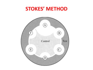
  • 19. STOKES METHOD • The comparative disk test • Uses both a test organism and a control organism on the same plate. • The control organism is of defined sensitivity to the antibiotics being tested. • Allows a direct comparison of the diameter of the zones of inhibition between the test and control organisms.
  • 20. STOKES METHOD • The MHA plate is divided into 3 parts. • Test organism is inoculated on the central one third and control strain on upper and lower thirds of the plate. In modified Stokes disk diffusion method- the test bacterium is inoculated over the upper and lower thirds of the plate and control on central one third. • An Uninoculated gap of 2-3 mm wide should separate, the test and the control area on which the antibiotic disks are applied. • The plates are then incubated at 370C for 16-18 hours.
  • 21. Reporting in Stokes Method • The sensitivity report is prepared by comparing the zones of inhibition of control and test bacterium. • The radius of the inhibition zone from the edge of the disk to the edge of the zone is measured. Result is interpreted as: • Sensitive (S): Zone radius is wider than or equal to, or not >3 mm smaller than the control. • Intermediate ( I): Zone radius is >2 mm but smaller than the control by >3 mm. • Resistant (R): No zone of inhibition or zone radius measures 2 mm or less.
  • 24. BROTH DILUTION METHOD • Procedure • Making serial dilutions (2-fold) of antibiotic in broth Mueller-Hinton, Tryptic Soy Broth Inoculation of fix bacterial inoculum, incubate overnight Controls: • no inoculum • no antibiotic Turbidity visualization  MIC MBC can be obtained by subculturing from each tube to a nutrient agar plate without any antimicrobial agent
  • 26. BROTH DILUTION METHOD 26 Day 1 Add 1 ml of test bacteria (1*106 CFU/ml) to tubes containing 1 ml broth and concentration of antibiotic (mg/l) Controls: C1 = No antibiotic, check viability on agar plates immediately C2 = No test bacteria Bacterial conc.= 5*105 CFU/ml Incubate 35 oC, o/n 128 64 32 16 8 4 2 C1 C2 64 32 16 8 4 2 1 C1 C2
  • 27. 27 BROTH DILUTION METHOD Day 2 Record visual turbidity Subculture non-turbid tubes to agar plates (use 0.01 ml standard loop) MIC = 16 mg/l 64 32 16 8 4 2 1 C1 C2 0.01 ml (spread plate), Incubate 35 oC, o/n 64 32 16 Day 3 Determine CFU on plates: At 16 mg/ = 700 CFU/ml > 0.1% of 5*105 CFU/ml MBC = 32 mg/l
  • 28. Antimicrobial susceptibility testing using micro-broth dilutions • • • • • • • • • • • • • 96 well microtiter plate ug/ml 64 32 16 8 4 2
  • 29. Epsilometer /E test • This is a quantitative method of detecting MIC by using the principles of both dilution and diffusion of antibiotic into the medium • The Etest technique comprises a predefined gradient (serial dilution) of antibiotic concentrations on a plastic absorbent strip. • applied to a lawn inoculum of a bacterium. following incubation of the test organism, an elliptical zone of inhibition is produced surrounding the strip • The antibiotic conc. at which the ellipse edge intersect the strip is taken as MIC value. 29
  • 31. AUTOMATED SYSTEMS • Systems are computer assisted and have sophisticated software to analyse the growth rate and determine the antibiotic susceptibility report. • Detect growth in microvolumes of broth with various dilutions of antimicrobials • Detection via photometric, turbidimetric, or fluorometric methods • Types BD Phoenix system (Becton Dickinson) Vitek 1 and 2 (bioMerieLLx) Micro Scan Walk Away system TREK Sensititre
  • 32. AUTOMATED SYSTEMS • Advantages Increased reproducibility Decreased labor costs Rapid results Software • Detects multi-drug resistances • ESBLs • Correlates bacterial ID with sensitivity • Disadvantages – Cost