SlideShare a Scribd company logo
Systemic mycoses
- DR. ANKUR KUMAR
Systemic mycoses
Disease Causative agent
1. Histoplasmosis Histoplasma capsulatum
2. Blastomycosis Blastomyces dermatitidis
3. Coccidioidomycosis Cocddioides immitis
4. Paracoccidioidomycosis Paracoccidioides brasiliensis
HISTOPLASMOSIS
• Systemic granulomatous disease caused by a
dimorphic fungus, Histoplasma capsulatum.
• Species name is misnomer as it is not
capsulated.
• Also known as Darling's disease (because they
was first reported by Samuel Darling in1905 )
HISTOPLASMOSIS- Pathogenesis
• Transmitted by- inhalation of spores (i.e.
microconidia) which usually circulate in the air
after the contaminated soil is disturbed.
• Spores enter into the lungs engulfed
inside the alveolar macrophages
transform into yeast forms .
• Yeasts survive within the phagolysosome of
the macrophage by producing alkaline
substances (bicarbonate and ammonia).
HISTOPLASMOSIS- Clinical Manifestations
• Pulmonary hlstoplasmosis:
 Most common form.
 Acute form presents with mild flu like illness, pulmonary infiltrates
in chest X-ray with hilar or mediastinal lymphadenopadiy.
 Chronic cavitary histoplasmosis may be seen in smokers with
underlying structural lung disease.
• Mucocutaneous histoplasmosis
 Skin and oral mucosal lesions may develop secondary to
pulmonary infection.
 Oral lesions are particularly seen in Indian patients.
• Disseminated histoplasmosis:
 Develops if CMI is very low (e.g. untreated HIV infected people or
following organ transplantation).
 Common sites affected- bone marrow, spleen, liver, eyes and
adrenal glands
HISTOPLASMOSIS- Laboratory Diagnosis
• Specimens:
Sputum, aspirate from bone marrow & lymph
node, blood and biopsies from skin & mucosa.
• Direct microscopy:
Histopathological staining (PAS, Giemsa or GMS
stain) – Shows tiny oval yeast cells (2-4 µm size)
with narrow based budding within macrophages
with an underlying granulomatous response.
HISTOPLASMOSIS- Laboratory Diagnosis
• Culture:
Gold standard method of diagnosis.
Media- SDA, blood agar and BHI agar
Incubated simultaneously at 25°C and 37°C.
At 25°C:
 Mycelial phase
 Produces white to buff brown colonies, consist of 2 types of
conidia or spores:
1) Tuberculate macroconidia- typical thick walls and finger-like
projections which is a characteristic feature
2) Microconidia are smaller, thin, and smooth-walled.
At 37°C:
 Gets converted into yeast form (creamy white colonies), which is
best developed in special media like Kelley's media.
HISTOPLASMOSIS- Laboratory Diagnosis
• Serology:
Antibodies in serum - detected by CFT and
imununodiffusion test.
Antibodies appear after 1 month of infection; hence
are more useful in chronic stage (often negative in
early course and in disseminated stage).
 False positive result may occur due to past infection
or cross infection with blastomyces.
• Skin test:
 May be done to demonstrate delayed type hypersensitivity
response to histoplasmin antigen, which indicates prior
exposure
Histoplasma capsulatum (schematic diagram)
A. Yeast form;
B. Mycelial form;
C. 2-4 µm yeast cells with narrow based budding (Giemsa stain)
D. Mold form, septate thin hyphae with tuberculate macroconidia (arrows showing)
HISTOPLASMOSIS- TREATMENT
• Liposomal amphotericin B
 Is the antifungal agent of choice in acute pulmonary and
disseminated histoplasmosis.
• ltraconazole
Recommended for chronic cavitary pulmonary
histoplasmosis.
BLASTOMYCOSIS
• Also known as North American blastomycosis
or Gilchrist's disease or Chicago disease
• Is a fungal infection of humans and other
animals also- dogs and cats, caused,
Blastomyces dermatitidis.
BLASTOMYCOSIS- Pathogenesis
• Transmitted by
Inhalation of the conidia of B. dermatitidis.
Spores enter into the lungs, and are engulfed by
alveolar macrophages, where they get converted
into yeast phase.
This yeast expresses a 120-kDa glycoprotein called
BAD-I (B. dermatitidis adhesin-1) which is an
essential virulence factor and also a major inducer
of cellular and humoral immune responses.
BLASTOMYCOSIS- Clinlcal Manifestations
• Acute pulmonary blastomycosis - Most common
form.
• Extrapulmonary manifestations
Skin involvement - Most common extrapulmonary
form; characterized by either verrucous (more
common) or ulcerative type of skin lesions.
Osteomyelitis may develop along with contiguous soft
tissue abscesses and draining sinuses.
Prostate and epididymis involvement in men.
 Central nervous system (CNS) involvement has been
reported in - 40% of AIDS patients.
Brain abscess is the usual presentation, followed by
cranial or spinal epidural abscess and meningitis.
BLASTOMYCOSIS- Laboratory Diagnosis
• Antibody detection: lmmunodiffusion test
specific for B. dermatitidis has been developed
against yeast phase antigens such as antigen-A,
BAD- I and ASWS antigen (alkali soluble water
soluble).
• Antigen detection assay- detect Blastomyces
antigen in urine (more sensitive) and in serum is
commercially available.
• Molecular methods- DNA probe hybridization and
real time PCR
BLASTOMYCOSIS- Laboratory Diagnosis
• Histopathological staining of the tissue biopsy
specimens reveals thick-walled round yeast cells
of 8- 15 µm size with single broad-based budding
(figure of 8 appearance)
• Culture media - SDA, blood agar and BH I.
At 250C- Mycelia form containing hyphae with small
pear-shaped conidia are produced;
At 370C- mold to yeast conversion takes place.
• Skin test: done to demonstrate delayed type
hypersensitivity to blastomycin antigen.
Blastomyces
A. Schematic (Mycelial and yeast form);
B. Histopathological stain (arrow shows) broad based
budding yeast cells (figure o f 8 appearance)
BLASTOMYCOSIS- TREATMENT
• Liposomal amphotericin B
 is the antifungal agent of choice in most of the cases.
• ltraconazole
in immunocompetent patients with mild
pulmonary or non CNS extrapulmonary
blastomycosis
COCCIDIOIDOMYCOSIS
• Also called desert rheumatism or Valley fever
or California fever
• Is a systemic fungal disease caused by a
dimorphic soil dwelling fungus- Coccidioides
(C. immitis and C. posadasii).
COCCIDIOIDOMYCOSIS -Pathogenesis
• Transmitted by inhalation of arthroconidia.
• In lungs, they enlarge, become rounded, and
develop internal septations to form large sac like
structures of size up 10-200 µm called spherules-
encompass numerous endospores.
• Spherules may rupture and release packets of
endospores that can disseminate and develop
into new spherules.
• If spherules returned to artificial media or soil,
they revert back to the mycelial stage.
COCCIDIOIDOMYCOSIS - Clinlcal Manifestations
• Asymptomatic- in most of patients (60%).
• Pulmonary coccidioidomycosis - presents as
pneumonia, cavities, pleural effusion or nodule
formation.
• Skin lesions such as rashes or erythema nodosum and
arthritis with joint pain may appear secondary to
pulmonary infection particularly in women.
• Disseminated form:
Males and persons with low CMI (HIV infected patients
with CD4+ T cell count <250/ µI) are at higher risk.
Common sites for dissemination include skin, bone, joints,
soft tissues, and meninges.
COCCIDIOIDOMYCOSIS - Laboratory Diagnosis
• Histopathological staining
H and E stain, PAS or CMS of sputum or tissue biopsy
specimens demonstrates spherules which are large
sac like structures (20-80 µm size), have thick, double
refractile wall, and are filled with endospores
• Cultures on SDA –
 produces mycelial growth, described as fragmented hyphae
consisting of barrel-shaped arthrospores with alternate cells
distorted (empty cells)
 Coccidioides differs from other dimorphic fungi as it grows as
mold at both 25°C and 37°C in usual culture media (forms
spherules at 37°C in certain special culture media only).
 Cultures are highly infectious: lead to accidental inhalation of
spores in laboratories, require biosafety level-3 precautions.
COCCIDIOIDOMYCOSIS - Laboratory Diagnosis
• Serology: Antibodies are detected by
immunodiffusion test and CFT.
• Skin test: It is done by fungal extaracts
(coccidioidin or spherulin);
 produces at least a 5 mm induration within 48
hours after injection (delayed hypersensitivity
reaction) indicates past infection.
Cocddioides
A. Spherules (schematic);
B. Hyphae with arthroconidia (schematic);
C. Spherules (PAS staining);
D. Hyphae with arthroconidia (LPCB mount)
TREATMENT- Coccidioidomycosis
• Itraconazole -drug of choice
except for diffuse pneumonia with pulmonary
sequelae where amphotericin B is recommended.
PARACOCCIDIOIDOMYCOSIS
• Also known as South American blastomycosis,
Lutz-Splendore-de Almeida disease
• Is a systemic disease caused by the dimorphic
fungus Paracoccidioides brasiliensis.
Pathogenesis and clinical Manifestations
• Transmission- by inhalation of spores,
• Transform into the yeast phase in lungs.
• Occurs as two major forms.
1. Acute form (or juvenile type):
affects young adults under 30 years age.
less common variety, but more severe form, manifests as
disseminated infection involving multiple viscera and is
refractory to t/t.
2. Chronic form (or adult form):
accounts for 90% of cases
predominantly affects older men.
results from reactivation of quiescenct lung lesions.
less severe form, manifested as progressive pulmonary
disease affecting lower lobes, with fibrosis.
Skin, oral mucosal lesions and cervical lymphadenopathy
PARACOCCIDIOIDOMYCOSIS-Laboratory Diagnosis
• Histopathological staining - of pus, tissue biopsies or
sputum
 reveals round thick-walled yeasts, which multiple narrow-
necked buds attached circumferencially giving rise to Mickeiy
mouse or pilot wheel appearance.
• Culture on SDA
 yields mycelial form at 250C
 converts in to yeast phase at 370C when grown in BHI agar
supplemented with blood and glutamine.
• Serology:
 Antibodies are detected by immunodiffusion, and by ELISA,
using gp43 antigen of P. brasiliensis.
• Skin test:
 demonstrates delayed hypersensitivity response against
paracoccidioidin antigen.
Paracoccidioidomycosis
A. Schematic representation of mycelial and yeast forms;
B. Methenamine silver staining shows yeast from (pilot
wheel appearance)
TREATMENT- Paracoccidioidomycosis
• ltraconazole - is the treatment of choice
except for the seriously ill patients where
Amphotericin B is recommended.
Sulfonamides are effective, but the response is
slow with frequent relapses.
THANK
YOU

More Related Content

PPSX
Systemic mycosis
PPTX
Microbial metabolism
PPT
Ascaris lumbricoides
PPTX
Laminar air flow
PPT
Utero vaginal prolapse
PDF
All diseases and syndromes USMLE Step1 and Step2
PDF
Introduction to medical virology
PPTX
CLS Blastomyces dermatitidis.pptx
Systemic mycosis
Microbial metabolism
Ascaris lumbricoides
Laminar air flow
Utero vaginal prolapse
All diseases and syndromes USMLE Step1 and Step2
Introduction to medical virology
CLS Blastomyces dermatitidis.pptx

What's hot (20)

PPTX
Cryptococcosis
PPTX
HISTOPLASMOSIS.pptx
PPTX
Salmonella typhi
PPTX
15. shigella
PDF
PPTX
PENICILLOSIS.pptx
PPTX
CLS Coccidioidomyces.pptx
PPTX
Yersinia pestis .....
PDF
Mycology - all about fungi
PPT
Salmonella
PPTX
Pseudomonas
PPTX
Zygomycosis
PPT
Subcutaneous mycoses
PPTX
Aspergillosis- Dr. Praveen kumar Doddamani
PPTX
Bacillus species
PPTX
Oncogenic viruses
PPTX
Histoplasmosis
Cryptococcosis
HISTOPLASMOSIS.pptx
Salmonella typhi
15. shigella
PENICILLOSIS.pptx
CLS Coccidioidomyces.pptx
Yersinia pestis .....
Mycology - all about fungi
Salmonella
Pseudomonas
Zygomycosis
Subcutaneous mycoses
Aspergillosis- Dr. Praveen kumar Doddamani
Bacillus species
Oncogenic viruses
Histoplasmosis
Ad

Similar to Systemic mycoses (20)

PPTX
Systemic Fungal Infections and its dideases
DOCX
MICROBIOLOGY ASSIGNMENT.docx1HGWUYGYETYDGNBK
PPSX
SYSTEMIC MYCOSES `
PPTX
systemic mycosis.pptx(dimorphic fungi), candidiasis
PDF
Coccidioidomycosis and Paracoccidioidomycosis
PDF
Histoplasmosis and Blastomycosis: Systemic mysosis
PPT
Chapter 5 systemic mycoses.pptx.ppt
PPTX
Chapter 5 systemic mycoses.pptx
PPTX
Subcutaneous,Systemic, Oppurtunistic mycosis
PPTX
Medically Important Histoplasma species .pptx
PPT
Systemic mycose
PPT
Systemic mycoses by Dr. Rakesh Prasad Sah
PPT
11- Chronic inflammations -Tuberculosis.ppt
PPT
09 infectious disease Fungal infections
PDF
Systemic Endemic Mycoses-HBCP.pdf
PPTX
Blasto.pptx
PPTX
HISTOPLASMOSIS /SYSTEMIC MYCOSES/ Histoplasma capsulatum/ Darling's disease.pptx
PPTX
Infectious Diseases Plate Round 9-11-24..pptx
PPTX
Dimorphic Systemic Mycoses.pptx
PPT
Systemic mycosis
Systemic Fungal Infections and its dideases
MICROBIOLOGY ASSIGNMENT.docx1HGWUYGYETYDGNBK
SYSTEMIC MYCOSES `
systemic mycosis.pptx(dimorphic fungi), candidiasis
Coccidioidomycosis and Paracoccidioidomycosis
Histoplasmosis and Blastomycosis: Systemic mysosis
Chapter 5 systemic mycoses.pptx.ppt
Chapter 5 systemic mycoses.pptx
Subcutaneous,Systemic, Oppurtunistic mycosis
Medically Important Histoplasma species .pptx
Systemic mycose
Systemic mycoses by Dr. Rakesh Prasad Sah
11- Chronic inflammations -Tuberculosis.ppt
09 infectious disease Fungal infections
Systemic Endemic Mycoses-HBCP.pdf
Blasto.pptx
HISTOPLASMOSIS /SYSTEMIC MYCOSES/ Histoplasma capsulatum/ Darling's disease.pptx
Infectious Diseases Plate Round 9-11-24..pptx
Dimorphic Systemic Mycoses.pptx
Systemic mycosis
Ad

More from DR. ANKUR KUMAR (20)

PPTX
Viruses 1
PPTX
Streptococcus ldentification
PPT
Air water milk
PPTX
Hypersensitivity
PPTX
Diagnosis &amp; blood stages of malaria .
PPTX
Immune responses
PPTX
Immunization programme
PPTX
Viral diagnosis
PPTX
Bacterial staining
PPTX
Fungal diagnosis
PPTX
Cestodes
PPTX
PPTX
Examination of water
PPTX
Mcqs & case discussion meningitis
PPTX
Leprosy 2
PPT
Air bacteriology
PPTX
Picornavirus
PPTX
Actinomycetes
PPTX
Viruses 1
Streptococcus ldentification
Air water milk
Hypersensitivity
Diagnosis &amp; blood stages of malaria .
Immune responses
Immunization programme
Viral diagnosis
Bacterial staining
Fungal diagnosis
Cestodes
Examination of water
Mcqs & case discussion meningitis
Leprosy 2
Air bacteriology
Picornavirus
Actinomycetes

Recently uploaded (20)

PPTX
surgery guide for USMLE step 2-part 1.pptx
PPTX
Respiratory drugs, drugs acting on the respi system
PDF
NEET PG 2025 | 200 High-Yield Recall Topics Across All Subjects
PPTX
CME 2 Acute Chest Pain preentation for education
PPTX
Pathophysiology And Clinical Features Of Peripheral Nervous System .pptx
PPT
Copy-Histopathology Practical by CMDA ESUTH CHAPTER(0) - Copy.ppt
PPTX
15.MENINGITIS AND ENCEPHALITIS-elias.pptx
PPT
ASRH Presentation for students and teachers 2770633.ppt
PDF
CT Anatomy for Radiotherapy.pdf eryuioooop
DOCX
NEET PG 2025 | Pharmacology Recall: 20 High-Yield Questions Simplified
PPTX
Acid Base Disorders educational power point.pptx
PPTX
History and examination of abdomen, & pelvis .pptx
PPTX
neonatal infection(7392992y282939y5.pptx
PPTX
ca esophagus molecula biology detailaed molecular biology of tumors of esophagus
DOC
Adobe Premiere Pro CC Crack With Serial Key Full Free Download 2025
PDF
Medical Evidence in the Criminal Justice Delivery System in.pdf
PDF
Human Health And Disease hggyutgghg .pdf
PDF
Intl J Gynecology Obste - 2021 - Melamed - FIGO International Federation o...
PPTX
Neuropathic pain.ppt treatment managment
PDF
Deadly Stampede at Yaounde’s Olembe Stadium Forensic.pdf
surgery guide for USMLE step 2-part 1.pptx
Respiratory drugs, drugs acting on the respi system
NEET PG 2025 | 200 High-Yield Recall Topics Across All Subjects
CME 2 Acute Chest Pain preentation for education
Pathophysiology And Clinical Features Of Peripheral Nervous System .pptx
Copy-Histopathology Practical by CMDA ESUTH CHAPTER(0) - Copy.ppt
15.MENINGITIS AND ENCEPHALITIS-elias.pptx
ASRH Presentation for students and teachers 2770633.ppt
CT Anatomy for Radiotherapy.pdf eryuioooop
NEET PG 2025 | Pharmacology Recall: 20 High-Yield Questions Simplified
Acid Base Disorders educational power point.pptx
History and examination of abdomen, & pelvis .pptx
neonatal infection(7392992y282939y5.pptx
ca esophagus molecula biology detailaed molecular biology of tumors of esophagus
Adobe Premiere Pro CC Crack With Serial Key Full Free Download 2025
Medical Evidence in the Criminal Justice Delivery System in.pdf
Human Health And Disease hggyutgghg .pdf
Intl J Gynecology Obste - 2021 - Melamed - FIGO International Federation o...
Neuropathic pain.ppt treatment managment
Deadly Stampede at Yaounde’s Olembe Stadium Forensic.pdf

Systemic mycoses

  • 2. Systemic mycoses Disease Causative agent 1. Histoplasmosis Histoplasma capsulatum 2. Blastomycosis Blastomyces dermatitidis 3. Coccidioidomycosis Cocddioides immitis 4. Paracoccidioidomycosis Paracoccidioides brasiliensis
  • 3. HISTOPLASMOSIS • Systemic granulomatous disease caused by a dimorphic fungus, Histoplasma capsulatum. • Species name is misnomer as it is not capsulated. • Also known as Darling's disease (because they was first reported by Samuel Darling in1905 )
  • 4. HISTOPLASMOSIS- Pathogenesis • Transmitted by- inhalation of spores (i.e. microconidia) which usually circulate in the air after the contaminated soil is disturbed. • Spores enter into the lungs engulfed inside the alveolar macrophages transform into yeast forms . • Yeasts survive within the phagolysosome of the macrophage by producing alkaline substances (bicarbonate and ammonia).
  • 5. HISTOPLASMOSIS- Clinical Manifestations • Pulmonary hlstoplasmosis:  Most common form.  Acute form presents with mild flu like illness, pulmonary infiltrates in chest X-ray with hilar or mediastinal lymphadenopadiy.  Chronic cavitary histoplasmosis may be seen in smokers with underlying structural lung disease. • Mucocutaneous histoplasmosis  Skin and oral mucosal lesions may develop secondary to pulmonary infection.  Oral lesions are particularly seen in Indian patients. • Disseminated histoplasmosis:  Develops if CMI is very low (e.g. untreated HIV infected people or following organ transplantation).  Common sites affected- bone marrow, spleen, liver, eyes and adrenal glands
  • 6. HISTOPLASMOSIS- Laboratory Diagnosis • Specimens: Sputum, aspirate from bone marrow & lymph node, blood and biopsies from skin & mucosa. • Direct microscopy: Histopathological staining (PAS, Giemsa or GMS stain) – Shows tiny oval yeast cells (2-4 µm size) with narrow based budding within macrophages with an underlying granulomatous response.
  • 7. HISTOPLASMOSIS- Laboratory Diagnosis • Culture: Gold standard method of diagnosis. Media- SDA, blood agar and BHI agar Incubated simultaneously at 25°C and 37°C. At 25°C:  Mycelial phase  Produces white to buff brown colonies, consist of 2 types of conidia or spores: 1) Tuberculate macroconidia- typical thick walls and finger-like projections which is a characteristic feature 2) Microconidia are smaller, thin, and smooth-walled. At 37°C:  Gets converted into yeast form (creamy white colonies), which is best developed in special media like Kelley's media.
  • 8. HISTOPLASMOSIS- Laboratory Diagnosis • Serology: Antibodies in serum - detected by CFT and imununodiffusion test. Antibodies appear after 1 month of infection; hence are more useful in chronic stage (often negative in early course and in disseminated stage).  False positive result may occur due to past infection or cross infection with blastomyces. • Skin test:  May be done to demonstrate delayed type hypersensitivity response to histoplasmin antigen, which indicates prior exposure
  • 9. Histoplasma capsulatum (schematic diagram) A. Yeast form; B. Mycelial form; C. 2-4 µm yeast cells with narrow based budding (Giemsa stain) D. Mold form, septate thin hyphae with tuberculate macroconidia (arrows showing)
  • 10. HISTOPLASMOSIS- TREATMENT • Liposomal amphotericin B  Is the antifungal agent of choice in acute pulmonary and disseminated histoplasmosis. • ltraconazole Recommended for chronic cavitary pulmonary histoplasmosis.
  • 11. BLASTOMYCOSIS • Also known as North American blastomycosis or Gilchrist's disease or Chicago disease • Is a fungal infection of humans and other animals also- dogs and cats, caused, Blastomyces dermatitidis.
  • 12. BLASTOMYCOSIS- Pathogenesis • Transmitted by Inhalation of the conidia of B. dermatitidis. Spores enter into the lungs, and are engulfed by alveolar macrophages, where they get converted into yeast phase. This yeast expresses a 120-kDa glycoprotein called BAD-I (B. dermatitidis adhesin-1) which is an essential virulence factor and also a major inducer of cellular and humoral immune responses.
  • 13. BLASTOMYCOSIS- Clinlcal Manifestations • Acute pulmonary blastomycosis - Most common form. • Extrapulmonary manifestations Skin involvement - Most common extrapulmonary form; characterized by either verrucous (more common) or ulcerative type of skin lesions. Osteomyelitis may develop along with contiguous soft tissue abscesses and draining sinuses. Prostate and epididymis involvement in men.  Central nervous system (CNS) involvement has been reported in - 40% of AIDS patients. Brain abscess is the usual presentation, followed by cranial or spinal epidural abscess and meningitis.
  • 14. BLASTOMYCOSIS- Laboratory Diagnosis • Antibody detection: lmmunodiffusion test specific for B. dermatitidis has been developed against yeast phase antigens such as antigen-A, BAD- I and ASWS antigen (alkali soluble water soluble). • Antigen detection assay- detect Blastomyces antigen in urine (more sensitive) and in serum is commercially available. • Molecular methods- DNA probe hybridization and real time PCR
  • 15. BLASTOMYCOSIS- Laboratory Diagnosis • Histopathological staining of the tissue biopsy specimens reveals thick-walled round yeast cells of 8- 15 µm size with single broad-based budding (figure of 8 appearance) • Culture media - SDA, blood agar and BH I. At 250C- Mycelia form containing hyphae with small pear-shaped conidia are produced; At 370C- mold to yeast conversion takes place. • Skin test: done to demonstrate delayed type hypersensitivity to blastomycin antigen.
  • 16. Blastomyces A. Schematic (Mycelial and yeast form); B. Histopathological stain (arrow shows) broad based budding yeast cells (figure o f 8 appearance)
  • 17. BLASTOMYCOSIS- TREATMENT • Liposomal amphotericin B  is the antifungal agent of choice in most of the cases. • ltraconazole in immunocompetent patients with mild pulmonary or non CNS extrapulmonary blastomycosis
  • 18. COCCIDIOIDOMYCOSIS • Also called desert rheumatism or Valley fever or California fever • Is a systemic fungal disease caused by a dimorphic soil dwelling fungus- Coccidioides (C. immitis and C. posadasii).
  • 19. COCCIDIOIDOMYCOSIS -Pathogenesis • Transmitted by inhalation of arthroconidia. • In lungs, they enlarge, become rounded, and develop internal septations to form large sac like structures of size up 10-200 µm called spherules- encompass numerous endospores. • Spherules may rupture and release packets of endospores that can disseminate and develop into new spherules. • If spherules returned to artificial media or soil, they revert back to the mycelial stage.
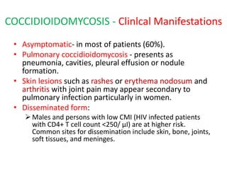
  • 20. COCCIDIOIDOMYCOSIS - Clinlcal Manifestations • Asymptomatic- in most of patients (60%). • Pulmonary coccidioidomycosis - presents as pneumonia, cavities, pleural effusion or nodule formation. • Skin lesions such as rashes or erythema nodosum and arthritis with joint pain may appear secondary to pulmonary infection particularly in women. • Disseminated form: Males and persons with low CMI (HIV infected patients with CD4+ T cell count <250/ µI) are at higher risk. Common sites for dissemination include skin, bone, joints, soft tissues, and meninges.
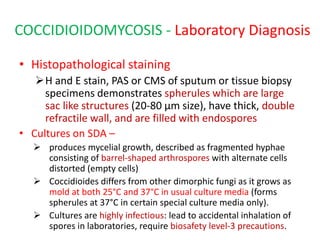
  • 21. COCCIDIOIDOMYCOSIS - Laboratory Diagnosis • Histopathological staining H and E stain, PAS or CMS of sputum or tissue biopsy specimens demonstrates spherules which are large sac like structures (20-80 µm size), have thick, double refractile wall, and are filled with endospores • Cultures on SDA –  produces mycelial growth, described as fragmented hyphae consisting of barrel-shaped arthrospores with alternate cells distorted (empty cells)  Coccidioides differs from other dimorphic fungi as it grows as mold at both 25°C and 37°C in usual culture media (forms spherules at 37°C in certain special culture media only).  Cultures are highly infectious: lead to accidental inhalation of spores in laboratories, require biosafety level-3 precautions.
  • 22. COCCIDIOIDOMYCOSIS - Laboratory Diagnosis • Serology: Antibodies are detected by immunodiffusion test and CFT. • Skin test: It is done by fungal extaracts (coccidioidin or spherulin);  produces at least a 5 mm induration within 48 hours after injection (delayed hypersensitivity reaction) indicates past infection.
  • 23. Cocddioides A. Spherules (schematic); B. Hyphae with arthroconidia (schematic); C. Spherules (PAS staining); D. Hyphae with arthroconidia (LPCB mount)
  • 24. TREATMENT- Coccidioidomycosis • Itraconazole -drug of choice except for diffuse pneumonia with pulmonary sequelae where amphotericin B is recommended.
  • 25. PARACOCCIDIOIDOMYCOSIS • Also known as South American blastomycosis, Lutz-Splendore-de Almeida disease • Is a systemic disease caused by the dimorphic fungus Paracoccidioides brasiliensis.
  • 26. Pathogenesis and clinical Manifestations • Transmission- by inhalation of spores, • Transform into the yeast phase in lungs. • Occurs as two major forms. 1. Acute form (or juvenile type): affects young adults under 30 years age. less common variety, but more severe form, manifests as disseminated infection involving multiple viscera and is refractory to t/t. 2. Chronic form (or adult form): accounts for 90% of cases predominantly affects older men. results from reactivation of quiescenct lung lesions. less severe form, manifested as progressive pulmonary disease affecting lower lobes, with fibrosis. Skin, oral mucosal lesions and cervical lymphadenopathy
  • 27. PARACOCCIDIOIDOMYCOSIS-Laboratory Diagnosis • Histopathological staining - of pus, tissue biopsies or sputum  reveals round thick-walled yeasts, which multiple narrow- necked buds attached circumferencially giving rise to Mickeiy mouse or pilot wheel appearance. • Culture on SDA  yields mycelial form at 250C  converts in to yeast phase at 370C when grown in BHI agar supplemented with blood and glutamine. • Serology:  Antibodies are detected by immunodiffusion, and by ELISA, using gp43 antigen of P. brasiliensis. • Skin test:  demonstrates delayed hypersensitivity response against paracoccidioidin antigen.
  • 28. Paracoccidioidomycosis A. Schematic representation of mycelial and yeast forms; B. Methenamine silver staining shows yeast from (pilot wheel appearance)
  • 29. TREATMENT- Paracoccidioidomycosis • ltraconazole - is the treatment of choice except for the seriously ill patients where Amphotericin B is recommended. Sulfonamides are effective, but the response is slow with frequent relapses.