SlideShare a Scribd company logo
BEDSIDE MONITORING OF
TISSUE PERFUSION AND
   OXYGENATION


                  Dr.Tushar Patil
                       MD
Bedside monitoring of tissue perfusion and oxygenation
Oxygen transport
  involves a series of convective and diffusive processes.
 Convective transport
-bulk movement of oxygen in air or blood
-active, energy consuming processes generating flow
 Diffusive transport
- passive movement of oxygen down its concentration
  gradient across tissue barriers
- across the extracellular matrix
- depends on the oxygen tension gradient and the
  diffusion distance
Bedside monitoring of tissue perfusion and oxygenation
Capillary blood to individual
                 cells
      resting extraction ratio from capillary blood is
      about 25%
     may increase to 70­80% during exercise
     Factors affecting O2 extraction from cappilary
      blood
1.    Rate of O2 delivery to capillary
2.    O2-Hb dissociation relation
3.    Size of capillary to cellular PO2 relation
4.    Diffusion distance to cells
5.    Rate of use of O2 by cells
Bedside monitoring of tissue perfusion and oxygenation
Tolerance to hypoxia of
      various tissues Tissue
    Survival time
1.   Brain<3 min
2.   Kidney and liver15-20 min
3.   Skeletal muscle60-90 min
4.   Vascular smooth muscle24-72 h
5.   Hair and nails -Several days
Tissue Hypoxia in Critically Ill
    disordered regional distribution of blood
   Regional and microcirculatory distribution of
    cardiac output
    endothelial, receptor, neural, metabolic, and
    pharmacological factors
    small resistance arterioles and precapillary
    sphincters
    shunting and tissue hypoxia despite high global
    oxygen delivery and mixed venous saturation.
    reduce regional distribution, particularly to the
    renal and splanchnic capillary beds
Bedside monitoring of tissue perfusion and oxygenation
EFFECTS OF HYPOXIA

   PaO2 level approaches 55mmHg-.short term
    memory loss, euphoria and impaired judgment
    PaO2 30-50mmHg -Progressive loss of
    cognitive and motor functions, increasing
    tachycardia
   PaO2 below 30mmHg-loss of consciousness
Clinical features of tissue
                hypoxia
   Dyspnoea
   Altered mental state
   Tachypnoea or hypoventilation
   Arrhythmias
   Peripheral vasodilatation
   Systemic hypotension
   Coma
   Cyanosis (unreliable)
   Nausea, vomiting, and gastrointestinal disturbance
Monitoring Tissue Perfusion and
              Oxygenation
1.   Clinical Evaluation
2.   Hemodynamic Monitoring
3.   Pulse Oximetry
4.   End Tidal CO2 Monitoring
5.   Monitoring Tissue Hypoxia
6.   Cerebral Oxygenation Monitoring
Clinical Evaluation
 HISTORY
-Dyspnoea
-Cough
-Fever
-Rash
-Discolouration of digits/limbs
-Palpitations
-Altered sensorium
-Convulsions
Clinical Evaluation
 Level of Consciousness
 Evaluation of Peripheral & Central Pulses
 Capillary Refill Time
 Cyanosis
 Respiratory Rate & Pattern
 Blood Pressure
 Systemic Examination
Hemodynamic Monitoring
 Arterial Blood Pressure

-Non Invasive
-Invasive
 Central Venous Pressure Monitoring
 Pulmonary Artery Catheterisation
 Measuring Cardiac Output
Monitoring arterial pressure
    Organ perfusion depends on the organ
    metabolic demand ,perfusion pressure,local
    vasomotor tone and cardiac output
   tissue perfusion is maintained through
    ‘‘autoregulation’’
    Organ perfusion pressure cannot be measured
    directly at the bedside
    As a surrogate for tissue perfusion pressure,
    arterial blood pressure is monitored.
Bedside monitoring of tissue perfusion and oxygenation
Noninvasive measurements of
           arterial pressure
    can be determined either manually or by oscillometric
    method .
    Oscillometric devices, determine MAP and then provide
    readings for systolic and diastolic pressures.
   Oscillometric devices tend to underestimate systolic and
    overestimate diastolic blood pressure
    noninvasive measurements less reliable with marked
    hypovolemia or abnormal cardiac function.
    Oscillometric measurements also limited by cycling
    delay of the device.
Arterial Catheterisation

INDICATIONS
 ABSOLUTE- As a guide to synchronization of intra-aortic
   balloon counter pulsation
 PROBABLE-
1. Guide to management of potent vasodilator drug
   infusions
2. Guide to management of potent vasopressor drug
   infusion
3. As a port for the rapid and repetitive sampling
4. As a monitor of cardiovascular deterioration in patients
Bedside monitoring of tissue perfusion and oxygenation
Arterial Catheterisation
USEFUL APPLICATIONS
 Differentiating cardiac tamponade (pulsus
 paradoxus) from respiration-induced
 swings in systolic BP
 Differentiating hypovolemia from cardiac
 dysfunction as the cause of hemodynamic
Arterial Catherisation
COMPLICATIONS
- temporary occlusion
- hematomas
- Serious ischemic damage
- sepsis
- pseudoaneurysm
Central venous pressure monitoring
Pressure in the large central veins proximal
  to the right atrium relative to atmosphere.
 METHODS
- central venous line / Swan-Ganz catheter
  with distal tip connected to manometer/
  pressure transducer
- noninvasively as jugular venous pressure
Factors affecting measured
                 CVP
1.Central venous blood volume
 Venous return/cardiac output
 Total blood volume
 Regional vascular tone
2.Compliance of central compartment
 Vascular tone
 RV compliance
 Myocardial disease
 Pericardial disease
 Tamponade
3.Tricuspid valve disease
 Stenosis
 Regurgitation
Factors affecting measured
                CVP
4.Cardiac rhythm
 Junctional rhythm
 Atrial fibrillation (AF)
 Atrio ventricular (A-V) dissociation
5.Reference level of transducer
 Positioning of patient
6.Intrathoracic pressure
 Respiration
 IPPV
 Positive end-expiratory pressure (PEEP)
 Tension pneumothorax
Limitations of CVP
 Being  wrongly used as a parameter/ goal
  for replacement of intravascular volume
 The validity as index of RV preload
  nonexistent
 Poor correlation with cardiac index, stroke
  volume, left ventricular end-diastolic
  volume, and right ventricular end-diastolic
  volume
Pulmonary artery
                   catheterization
   Developed in the 1940s and later refined by Swan and Ganz in
    1970

INDICATIONS
 Diagnostic
     – Diagnosis of shock states
     – high- versus low-pressure pulmonary edema
     – primary pulmonary hypertension (PPH)
     – valvular disease, intracardiac shunts, cardiac tamponade, and
       pulmonary embolus (PE)
     – Monitoring complicated AMI
     – hemodynamic instability after cardiac surgery

   Therapeutic
    - Aspiration of air emboli
Bedside monitoring of tissue perfusion and oxygenation
PAC

CONTRAINDICATIONS
 Tricuspid or pulmonary valve mechanical
  prosthesis
 Right heart mass (thrombus and/or tumor)
 Tricuspid or pulmonary valve endocarditis
PAC
MEASURED PARAMETERS
1.  Central Venous Pressure
2.  Pulmonary Capillary Wedge Pressure
3.  Cardiac Index
4.  Stroke Volume Index
5.  LV Stroke Work Index
6.  RVSWI
7.  RV Ejection Fraction
8.  RV End Diastolic Volume
9.  Systemic Vascular Resistance Index
10. Pulmonary Vascular Resistance Index
11. Mixed Venous O2 Saturation
12. O2 delivery
13. O2 uptake
14. O2 exraction Ratio
Complications of PAC
  Venous access complications
 - include arterial puncture
 - hemothorax
 - Pneumothorax
 Arrhythmias
 - PVCs or nonsustained VT
 - Significant VT or ventricular fibrillation
 Right bundle-branch block (RBBB)
 PA rupture
 PAC related infection
 Pulmonary infarction
Measuring Cardiac Output
1. Pulmonary Artery Catheter
2. Pulse Contour Analysis
   -Lithium dilution
    -Transpulmonary Thermodilution
    -Without diluion calibration
3. CO2 Rebreathing
4. Trans thoracic Electrial Bioimpedence
5. Trans Thoracic Echo
6. Esophageal Doppler Monitoring
Pulse Oximetry

PRINCIPLE
 Displayed readings determined primarily by two
   components:
1. The different absorption spectra of oxyhemoglobin and
   deoxyhemoglobin at different wavelengths
2. Pulsatile arterial blood
 Probe (finger, ear, or forehead) contains two light-
   emitting diodes that emit light at 660 nm and 940 nm.
 Photoreceptor receives light, and compares
   absorption two wavelengths,
Bedside monitoring of tissue perfusion and oxygenation
Pulse Oximetry
APPLICATIONS
 indicated in circumstance where hypoxaemia
  May occur.
 should be included in the routine vital signs. .
 continuous monitoring.
 pattern of oxygenation can be recorded.
 can replace arterial blood gas analysis in cases
  where assessment of oxygenation is indication.
 Regulation of oxygen therapy
 Testing adequecy of circulation
Pulse Oximetry
 Improving oximeter signals
•   Warm and rub the skin
•   Apply a topical vasodilator
•   Try a different probe site, especially the
    ear
•   Try a different probe
•   Avoid motion artefact
•   Use a different machine
Bedside monitoring of tissue perfusion and oxygenation
Pulse Oximetry
    PITFALLS
1.   Dyshemoglobinemias
2.   Poor function wiyh poor performance
3.   Difficulty in detecting high oxygen partial
     pressures
4.   Delayed detection of hypoxic events
5.   Erratic performance with irregular rhythms
6.   Nail polish and coverings
7.   Loss of accuracy at low values
8.   Electrical interference
9.   Failure to detect hypoventilation
Monitoring ventilation using end-
          tidal carbon dioxide
    provides information regarding alveolar ventilation.
    PetCO2 - concentration of carbon dioxide at end
    expiration .
    measured in both mechanically ventilated and
    spontaneously breathing patients.
   displayed as either numerical value (capnometry) or as a
    graphic waveform plotted against time (capnography).
    PetCO2 underestimatesPaCO2 by 2 to 5 mm Hg
    because of the influence of dead space ventilation
   relationship between PetCO2 and PaCO2 is unreliable in
    critically ill patients.
Bedside monitoring of tissue perfusion and oxygenation
End Tidal Carbon Dioxide
APPLICATIONS
 Confirming endotracheal tube placement
 detecting endotracheal tube dislodgment
 detecting ventilator malfunction
 assessing the success of cardiopulmonary
  resuscitation
 evaluation of weaning from mechanical
  ventilation
 determining the optimal level of PEEP
End Tidal CO2
     sudden loss of the capnogram waveform
    –ET obstruction
     -extubation
     - ventilator malfunction
     –cardiac arrest
      sudden drop of the waveform
     -partial obstruction of ET
     -an airway leak
     -hypotension
End Tidal CO2
 Capnography    can be used to monitor
  patients in whom hypercarbia may be
  detrimental
 PetCO2 values greater than 40 mm Hg
  correlate with equal or higher value of
  PaCO2
 Elevated PetCo2 indicate sthe need for
  alterations in management
Monitoring Tissue Hypoxia
   Global markers of tissue hypoxia
          - serum lactate
          -central venous oxygen saturation (ScvO2)

   Monitoring regional hypoxia
          -Sublingual Capnometry
          –Gastric Tonometry
          - Orthogonal Polarization Spectroscopy (OPS)
          - Near Infra Red Spectroscopy (NIRS)
          -Trans cutaneous Oxygen Tension
          -Resonance Raman Spectroscopy
Serum Lactate
   byproduct of anaerobic metabolism, resulting from the
    inabilityof pyruvate to enter the Krebs cycle.
   The normal serum value - less than 2 mmol/L.
    lactate levels above 4mmol/L strongly associated with
    worse outcome.
   more important is the time to normalization of
    lactateLevels- ‘‘lactate clearance time.’’
     Prolonged lactate clearance time(>48hrs)-significantly
    higher rates of infection, organ dysfunction, and death
   Better survival correlates with a lactate clearance time
    <24 hrs.
Central venous oxygen saturation
 Mixed venous oxygen saturation (SvO2) - a measure of tissue hypoxia.   o
 Obtained with pulmonary artery catheter.


  FACTORS INFLUENCING SvO2
- arterial oxygen saturation
-   hemoglobin concentration
-   cardiac output
-   tissue oxygen consumption.
-
NORMAL VALUES- 70% to 75%.
- Values below 60% indicate cellular oxidative impairment
- values below50% associated with anaerobic metabolism

pulmonary artery catheters not placed routinely
 ScvO2 - surrogate For SvO2
Bedside monitoring of tissue perfusion and oxygenation
ScvO2
  venous oxygen saturation near the junction of the
  superior vena cava and right atrium.
 obtained from subclavian or internal jugular central
  venous catheter.
 Because ScvO2 neglects venous return from the lower
body, values for ScvO2 typically are 3% to 5% less than
  SvO2
 values < 65% -ongoing oxidative impairment.
 values > 80% - cellular dysfunction with impaired
  oxygen consumption.                            -
       seen in late stages of shock
 To be used in context with other markers of tissue
  perfusion (eg, lactate).
Sublingual capnometry
    studies perfusion of the splanchnic circulation.
    sensor placed under the tongue
    measures partial pressure of carbon dioxide in the
    sublingual tissue (PslCO2).
   Normal values for PslCO2 - 43 to 47 mmHg
    PslCO2 >70 mm Hg - correlates with elevated arterial
    lactate levels
     more important is the ‘‘PslCO2 gap.’’- difference
     between PslCO2 and PaCO2
    A PslCO2 gap of> 25 mm Hg identifies patients at a
    high risk of mortality.
Gastric Tonometry
   Offers an index of aerobic metabolism in gut
    mucosa.
   Based on increase in tissue CO2
   A balloon in stomach,measures intramucosal
    pCO2
   Using this and arterial (HCO3), gastric
    intramucosal PH is calculated
Bedside monitoring of tissue perfusion and oxygenation
Orthogonal polarization
                 spectroscopy
    uses polarized light to visualize the microcirculation directly.
    hemoglobin absorbs polarized light
   real-time images reflected to videomicroscope
    functional capillary density measured.
   sensitive marker of tissue perfusion and an indirect measurement of
    oxygen delivery.
   Tissues evaluated- oral mucosa, sublingual mucosa, rectal
    mucosa,and vaginal mucosa.

   LIMITNG FACTORS-
        -movement artifacts
        -presence of saliva
        -observer related bias
Bedside monitoring of tissue perfusion and oxygenation
Near-infrared spectroscopy
  measures the concentrations of hemoglobin, oxygen saturation,
and cytochrome aa3

    Cytochrome aa3- final receptor in the electron transport chain
                                -                responsible for 90%
    cellular O2 consumption
                  - remains in a reduced state during hypoxia

   used primarily to evaluate the perfusion of skeletal muscles.

PROBLEMS
-signal contamination by light scatter
-variable interpretations of the data
-lack of a reference standard for comparison
Bedside monitoring of tissue perfusion and oxygenation
Transcutaneous oxygen tension
 measure transcutaneous oxygen or carbon dioxide.
   Use heated probes placed on the skin

•markers of regional tissue hypoperfusion
 increased mortality in patients with low transcutaneous oxygen or
high CO2


•LIMITATIONS
-Tissue trauma from probe insertion,
- thermal injury if probes are not moved every4 hours
- lack of established critical values to guide resuscitation .
Bedside monitoring of tissue perfusion and oxygenation
Cerebral Perfusion and
         Oxygenation monitoring

1.   Jugular venous bulb oximetry
2.   Direct brain tissue oxygen tension
3.   Near inrared spectroscopy
4.   Cerebral Microdialysis
5.   Cerebral Blood Flow Monitoring
6.   Oxygen-15 PET
Jugular venous oxygen
               saturation(SjvO2)
   Retrograde placement of jugular venous catheter with oximeter
    cannulate dominant IJV
    catheter tip positioned in jugular bulb
     compatible with MRI.
   SjvO2 - result of the difference between cerebral oxygen delivery
    (supply) and cerebral metabolic rate of oxygen (demand)
    Low SjvO2 (!50% for O10 minutes) -hypoperfusion / increased
               .                                       cerebral
    metabolism.


APPLICATIONS
  - comatose patients (GCS <8)
  -treatment of SAH
- - neurosurgical procedures
Bedside monitoring of tissue perfusion and oxygenation
SjvO2
LIMITATIONS
- changes in arterial oxygen content
-   hemodilution
-   prone position of catheter
-   necessity for frequent calibrations
-   infection
- increase in ICP
-   thrombosis
-   arterial Puncture
- pneumothorax
-   reflects global cerebral oxygenation and does not detect regional
   ischemia in smaller regions ipsilateral or in contralateral
   hemisphere
Brain tissue oxygen
                 pressure(PBO2)
   Small flexible microcatheter inserted into brain parenchyma.
    marker of the balance between regional oxygen supply and use.
   ICP, brain temperature (Licox, Integra Neurosciences) or tissue
    partial pressure of carbon dioxide (PBCO2) and pH (Neurotrend,
    Johnson & Johnson) can be monitored
     Licox device uses polarographic technique by Clark electrode
   Neurotrend uses ‘‘optimal luminescence’’
    catheter should pass through gray matter into white matter
    tunneled after craniotomy or placed through a double
    or triple lumen bolt
     measured tissue volume 17 mm3.
    PBO2 levels highest in dense population of neurons and lower in
    white matter
   PBO2 and amplitude of changes lower with Neurotrend than Licox
    compatible with MRI
Bedside monitoring of tissue perfusion and oxygenation
Near infrared spectroscopy
    monitoring of transmittance across the brain at two or
    more wavelengths
   optical attenuation of the spectra converted into changes
    of cerebral oxygenation
    methods include time-resolved, spatially resolved, and
    phase-resolved spectroscopy
    INVOS system provides a numerical value for oxygen
    saturation using rSO2
   normal range-60-80%
    NIRO oximeters present values for oxygenated and total
    Hb concentration, cytochrome aa3, and a tissue oxyge
    index
Bedside monitoring of tissue perfusion and oxygenation
NIS

APPLICATIONS
 detection of changes during carotid cross-clamping
  during carotid endarterectomy & cardiac surgery
 to detect cerebral vasospasm causing delayed cerebral
  ischemic deficit after SAH
 assessment of perfusion reductions in stroke
 Reconstruction of a three-dimensional image using
  optical tomography
 attractive because applied by attaching pads to the
  forehead or other regions of interest.
Bedside monitoring of tissue perfusion and oxygenation
NIS
LIMITATIONS

  limited and variable penetration of infrared light through
   the skull (2–3 mm, limited to gray matter)
 contamination by extra- and intracranial sources (mixture
   of capillary, venous,and arterial blood), and uniform
   distribution of infrared light in the CSF layer.
 degree of scatter unpredictable
 inconsistent impact of monitoring
of decreased oxygenation on neurologic outcome
Bedside monitoring of tissue perfusion and oxygenation
Cerebral Microdialysis
   bedside monitor to provide on-line analysis of brain tissue
    biochemistry during neurointensive care. The principles and clinical

   double-lumen probe, lined at it tip with dialysis membrane.
   perfused by an inlet tube with fluid isotonic to the tissue interstitium
   perfusate passes along the membrane before exiting collecting
    chamber.
    catheter acts as an artificial blood capillary.
   Measures microdialysate concentrations of glucose, lactate,
    pyruvate, glycerol, and glutamateThe concentration of these
    substances in the microdialysate
   does not correspond to their true extracellular fluid concentration
   proportion of the extracellular fluid concentration the ‘‘relative
    recovery”
Bedside monitoring of tissue perfusion and oxygenation
MD data display


Brain Tissue O2
    monitor

MD Bedside
 Analyser


Jugular Venous
  Saturation
   Monitor
Applications of MD
   Most clinical experience with TBI and SAH
   Severe cerebral hypoxia /ischemia associated with
    marked increases in the lactate-pyruvate ratio
    Ratio greater than 20 to 25 associated with poor
    outcome
   Glycerol is a marker of ischemic cell damage
    Increased MD glycerol concentrations associated with
    poor outcome
   Increased excitatory amino acids and reduced brain
    ECF glucose associated with metabolic catastrophes
    after acute brain injury.
Cerebral Blood Flow Monitoring
 Kety-Schmidt  method
 Radioactive tracer techniques
 Continuous quantitative cerebral blood
  flow monitoring
-Laser Doppler flowmetry
-Thermal diffusion flowmetry
 Double-indicator dilution technique
 Transcranial Doppler ultrasonography
Bedside monitoring of tissue perfusion and oxygenation
Neuroimagong
 [18F]2-deoxy-D-glucose   PET
 oxygen-15 PET
 SPECT
 Xenon-enhanced CT scanning
 perfusion CT
 Perfusion weighted imaging (PWI)
THANKS

More Related Content

PPTX
Neurological monitoring(1)
PDF
ESP block
PPTX
Cardiac output monitoring
PPT
Cardiac output & monitoring
PPTX
Monitoreo hemodinamico avanzado
PPTX
Fractures and dislocations
PPTX
Probiotics
PPTX
Nursing care of the elderly patients
Neurological monitoring(1)
ESP block
Cardiac output monitoring
Cardiac output & monitoring
Monitoreo hemodinamico avanzado
Fractures and dislocations
Probiotics
Nursing care of the elderly patients

What's hot (20)

PPT
Monitoring depth of anaesthesia
PPTX
Interscalene & supraclavicular nerve blocks
PPTX
Bed side pulmonary function tests 7
PPTX
Anaesthesia for renal transplantation
PPTX
O2 cascade flux n odc
PPTX
Anaesthetic management of mitral valvular heart disease
PPT
One lung ventilation
PDF
Audit in anaesthesia
PDF
Fluid responsiveness in critically ill patients
PPTX
Oxygen cascade
PPT
Brachial plexus block
PDF
Management of intraoperative bronchospasm
PPSX
Hemodynamic parameters & fluid therapy Asim
PPT
Simple TCI
PPTX
Newer modes of ventilation
PPTX
Cardio Vascular Monitoring, Anesthesia
PPT
Anaesthesia for cardiac patient undergoing non cardiac surgery
PPTX
Epilepsy and anaesthesia
PPTX
Monitoring depth of anesthesia
PPTX
Anesthesia for coronary artery bypass grafting
Monitoring depth of anaesthesia
Interscalene & supraclavicular nerve blocks
Bed side pulmonary function tests 7
Anaesthesia for renal transplantation
O2 cascade flux n odc
Anaesthetic management of mitral valvular heart disease
One lung ventilation
Audit in anaesthesia
Fluid responsiveness in critically ill patients
Oxygen cascade
Brachial plexus block
Management of intraoperative bronchospasm
Hemodynamic parameters & fluid therapy Asim
Simple TCI
Newer modes of ventilation
Cardio Vascular Monitoring, Anesthesia
Anaesthesia for cardiac patient undergoing non cardiac surgery
Epilepsy and anaesthesia
Monitoring depth of anesthesia
Anesthesia for coronary artery bypass grafting

Similar to Bedside monitoring of tissue perfusion and oxygenation (20)

PPTX
Hemodynamic monitoring in ICU
PPTX
Approach to Shock in Intensive care.pptx
PPT
Dr. Radhey Shyam (presentation)
PPTX
Anesthesia 5th year, 6th & 7th lectures (Dr. Gona)
PPT
Hemodynamics Basic Concepts
PPTX
ARDS- Satya.pptx
PPTX
shock-basicprinciplesinsurgery-221017132732-cbe19616.pptx
PPTX
How to monitor cardiovascular function in critical.pptx
PPTX
5_6280601286203146342 exact science.pptx
PPTX
Pulmonary hypertension
PDF
ASA recommended monitoring
PPTX
Pulmonary artery catheter
PPT
lecture 3 CCNg Respiratory Part One 2023-2024.ppt
PPT
An Introduction To Surgical Icu
PPTX
Respiratory failure
PPTX
Monitoring in mechanical ventilation main
PPTX
2021 Conference hemodynamic monitoring VV ECMO
PPTX
Monitoring Modality in anesthesia
PPTX
Hepatopulmonary syndrome
PPTX
SHOCK- Basic Principles in Surgery.pptx
Hemodynamic monitoring in ICU
Approach to Shock in Intensive care.pptx
Dr. Radhey Shyam (presentation)
Anesthesia 5th year, 6th & 7th lectures (Dr. Gona)
Hemodynamics Basic Concepts
ARDS- Satya.pptx
shock-basicprinciplesinsurgery-221017132732-cbe19616.pptx
How to monitor cardiovascular function in critical.pptx
5_6280601286203146342 exact science.pptx
Pulmonary hypertension
ASA recommended monitoring
Pulmonary artery catheter
lecture 3 CCNg Respiratory Part One 2023-2024.ppt
An Introduction To Surgical Icu
Respiratory failure
Monitoring in mechanical ventilation main
2021 Conference hemodynamic monitoring VV ECMO
Monitoring Modality in anesthesia
Hepatopulmonary syndrome
SHOCK- Basic Principles in Surgery.pptx

Recently uploaded (20)

PPTX
ACID BASE management, base deficit correction
PPTX
History and examination of abdomen, & pelvis .pptx
PPT
Obstructive sleep apnea in orthodontics treatment
PPTX
CEREBROVASCULAR DISORDER.POWERPOINT PRESENTATIONx
DOCX
RUHS II MBBS Microbiology Paper-II with Answer Key | 6th August 2025 (New Sch...
PPTX
Pathophysiology And Clinical Features Of Peripheral Nervous System .pptx
PPTX
1 General Principles of Radiotherapy.pptx
PDF
Handout_ NURS 220 Topic 10-Abnormal Pregnancy.pdf
PDF
NEET PG 2025 | 200 High-Yield Recall Topics Across All Subjects
DOCX
NEET PG 2025 | Pharmacology Recall: 20 High-Yield Questions Simplified
PDF
Khadir.pdf Acacia catechu drug Ayurvedic medicine
PPTX
Uterus anatomy embryology, and clinical aspects
PPTX
POLYCYSTIC OVARIAN SYNDROME.pptx by Dr( med) Charles Amoateng
PPTX
Respiratory drugs, drugs acting on the respi system
PPTX
Note on Abortion.pptx for the student note
PPTX
Chapter-1-The-Human-Body-Orientation-Edited-55-slides.pptx
PPT
1b - INTRODUCTION TO EPIDEMIOLOGY (comm med).ppt
PPTX
Electromyography (EMG) in Physiotherapy: Principles, Procedure & Clinical App...
PPTX
LUNG ABSCESS - respiratory medicine - ppt
PPTX
CME 2 Acute Chest Pain preentation for education
ACID BASE management, base deficit correction
History and examination of abdomen, & pelvis .pptx
Obstructive sleep apnea in orthodontics treatment
CEREBROVASCULAR DISORDER.POWERPOINT PRESENTATIONx
RUHS II MBBS Microbiology Paper-II with Answer Key | 6th August 2025 (New Sch...
Pathophysiology And Clinical Features Of Peripheral Nervous System .pptx
1 General Principles of Radiotherapy.pptx
Handout_ NURS 220 Topic 10-Abnormal Pregnancy.pdf
NEET PG 2025 | 200 High-Yield Recall Topics Across All Subjects
NEET PG 2025 | Pharmacology Recall: 20 High-Yield Questions Simplified
Khadir.pdf Acacia catechu drug Ayurvedic medicine
Uterus anatomy embryology, and clinical aspects
POLYCYSTIC OVARIAN SYNDROME.pptx by Dr( med) Charles Amoateng
Respiratory drugs, drugs acting on the respi system
Note on Abortion.pptx for the student note
Chapter-1-The-Human-Body-Orientation-Edited-55-slides.pptx
1b - INTRODUCTION TO EPIDEMIOLOGY (comm med).ppt
Electromyography (EMG) in Physiotherapy: Principles, Procedure & Clinical App...
LUNG ABSCESS - respiratory medicine - ppt
CME 2 Acute Chest Pain preentation for education

Bedside monitoring of tissue perfusion and oxygenation

  • 1. BEDSIDE MONITORING OF TISSUE PERFUSION AND OXYGENATION Dr.Tushar Patil MD
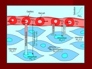
  • 3. Oxygen transport  involves a series of convective and diffusive processes.  Convective transport -bulk movement of oxygen in air or blood -active, energy consuming processes generating flow  Diffusive transport - passive movement of oxygen down its concentration gradient across tissue barriers - across the extracellular matrix - depends on the oxygen tension gradient and the diffusion distance
  • 6. Capillary blood to individual cells  resting extraction ratio from capillary blood is about 25%  may increase to 70­80% during exercise  Factors affecting O2 extraction from cappilary blood 1. Rate of O2 delivery to capillary 2. O2-Hb dissociation relation 3. Size of capillary to cellular PO2 relation 4. Diffusion distance to cells 5. Rate of use of O2 by cells
  • 8. Tolerance to hypoxia of various tissues Tissue  Survival time 1. Brain<3 min 2. Kidney and liver15-20 min 3. Skeletal muscle60-90 min 4. Vascular smooth muscle24-72 h 5. Hair and nails -Several days
  • 9. Tissue Hypoxia in Critically Ill  disordered regional distribution of blood  Regional and microcirculatory distribution of cardiac output  endothelial, receptor, neural, metabolic, and pharmacological factors  small resistance arterioles and precapillary sphincters  shunting and tissue hypoxia despite high global oxygen delivery and mixed venous saturation.  reduce regional distribution, particularly to the renal and splanchnic capillary beds
  • 11. EFFECTS OF HYPOXIA  PaO2 level approaches 55mmHg-.short term memory loss, euphoria and impaired judgment  PaO2 30-50mmHg -Progressive loss of cognitive and motor functions, increasing tachycardia  PaO2 below 30mmHg-loss of consciousness
  • 12. Clinical features of tissue hypoxia  Dyspnoea  Altered mental state  Tachypnoea or hypoventilation  Arrhythmias  Peripheral vasodilatation  Systemic hypotension  Coma  Cyanosis (unreliable)  Nausea, vomiting, and gastrointestinal disturbance
  • 13. Monitoring Tissue Perfusion and Oxygenation 1. Clinical Evaluation 2. Hemodynamic Monitoring 3. Pulse Oximetry 4. End Tidal CO2 Monitoring 5. Monitoring Tissue Hypoxia 6. Cerebral Oxygenation Monitoring
  • 14. Clinical Evaluation  HISTORY -Dyspnoea -Cough -Fever -Rash -Discolouration of digits/limbs -Palpitations -Altered sensorium -Convulsions
  • 15. Clinical Evaluation  Level of Consciousness  Evaluation of Peripheral & Central Pulses  Capillary Refill Time  Cyanosis  Respiratory Rate & Pattern  Blood Pressure  Systemic Examination
  • 16. Hemodynamic Monitoring  Arterial Blood Pressure -Non Invasive -Invasive  Central Venous Pressure Monitoring  Pulmonary Artery Catheterisation  Measuring Cardiac Output
  • 17. Monitoring arterial pressure  Organ perfusion depends on the organ metabolic demand ,perfusion pressure,local vasomotor tone and cardiac output  tissue perfusion is maintained through ‘‘autoregulation’’  Organ perfusion pressure cannot be measured directly at the bedside  As a surrogate for tissue perfusion pressure, arterial blood pressure is monitored.
  • 19. Noninvasive measurements of arterial pressure  can be determined either manually or by oscillometric method .  Oscillometric devices, determine MAP and then provide readings for systolic and diastolic pressures.  Oscillometric devices tend to underestimate systolic and overestimate diastolic blood pressure  noninvasive measurements less reliable with marked hypovolemia or abnormal cardiac function.  Oscillometric measurements also limited by cycling delay of the device.
  • 20. Arterial Catheterisation INDICATIONS  ABSOLUTE- As a guide to synchronization of intra-aortic balloon counter pulsation  PROBABLE- 1. Guide to management of potent vasodilator drug infusions 2. Guide to management of potent vasopressor drug infusion 3. As a port for the rapid and repetitive sampling 4. As a monitor of cardiovascular deterioration in patients
  • 22. Arterial Catheterisation USEFUL APPLICATIONS  Differentiating cardiac tamponade (pulsus paradoxus) from respiration-induced swings in systolic BP  Differentiating hypovolemia from cardiac dysfunction as the cause of hemodynamic
  • 23. Arterial Catherisation COMPLICATIONS - temporary occlusion - hematomas - Serious ischemic damage - sepsis - pseudoaneurysm
  • 24. Central venous pressure monitoring Pressure in the large central veins proximal to the right atrium relative to atmosphere.  METHODS - central venous line / Swan-Ganz catheter with distal tip connected to manometer/ pressure transducer - noninvasively as jugular venous pressure
  • 25. Factors affecting measured CVP 1.Central venous blood volume  Venous return/cardiac output  Total blood volume  Regional vascular tone 2.Compliance of central compartment  Vascular tone  RV compliance  Myocardial disease  Pericardial disease  Tamponade 3.Tricuspid valve disease  Stenosis  Regurgitation
  • 26. Factors affecting measured CVP 4.Cardiac rhythm  Junctional rhythm  Atrial fibrillation (AF)  Atrio ventricular (A-V) dissociation 5.Reference level of transducer  Positioning of patient 6.Intrathoracic pressure  Respiration  IPPV  Positive end-expiratory pressure (PEEP)  Tension pneumothorax
  • 27. Limitations of CVP  Being wrongly used as a parameter/ goal for replacement of intravascular volume  The validity as index of RV preload nonexistent  Poor correlation with cardiac index, stroke volume, left ventricular end-diastolic volume, and right ventricular end-diastolic volume
  • 28. Pulmonary artery catheterization  Developed in the 1940s and later refined by Swan and Ganz in 1970 INDICATIONS  Diagnostic – Diagnosis of shock states – high- versus low-pressure pulmonary edema – primary pulmonary hypertension (PPH) – valvular disease, intracardiac shunts, cardiac tamponade, and pulmonary embolus (PE) – Monitoring complicated AMI – hemodynamic instability after cardiac surgery  Therapeutic - Aspiration of air emboli
  • 30. PAC CONTRAINDICATIONS  Tricuspid or pulmonary valve mechanical prosthesis  Right heart mass (thrombus and/or tumor)  Tricuspid or pulmonary valve endocarditis
  • 31. PAC MEASURED PARAMETERS 1. Central Venous Pressure 2. Pulmonary Capillary Wedge Pressure 3. Cardiac Index 4. Stroke Volume Index 5. LV Stroke Work Index 6. RVSWI 7. RV Ejection Fraction 8. RV End Diastolic Volume 9. Systemic Vascular Resistance Index 10. Pulmonary Vascular Resistance Index 11. Mixed Venous O2 Saturation 12. O2 delivery 13. O2 uptake 14. O2 exraction Ratio
  • 32. Complications of PAC  Venous access complications - include arterial puncture - hemothorax - Pneumothorax  Arrhythmias - PVCs or nonsustained VT - Significant VT or ventricular fibrillation  Right bundle-branch block (RBBB)  PA rupture  PAC related infection  Pulmonary infarction
  • 33. Measuring Cardiac Output 1. Pulmonary Artery Catheter 2. Pulse Contour Analysis -Lithium dilution -Transpulmonary Thermodilution -Without diluion calibration 3. CO2 Rebreathing 4. Trans thoracic Electrial Bioimpedence 5. Trans Thoracic Echo 6. Esophageal Doppler Monitoring
  • 34. Pulse Oximetry PRINCIPLE  Displayed readings determined primarily by two components: 1. The different absorption spectra of oxyhemoglobin and deoxyhemoglobin at different wavelengths 2. Pulsatile arterial blood  Probe (finger, ear, or forehead) contains two light- emitting diodes that emit light at 660 nm and 940 nm.  Photoreceptor receives light, and compares absorption two wavelengths,
  • 36. Pulse Oximetry APPLICATIONS  indicated in circumstance where hypoxaemia May occur.  should be included in the routine vital signs. .  continuous monitoring.  pattern of oxygenation can be recorded.  can replace arterial blood gas analysis in cases where assessment of oxygenation is indication.  Regulation of oxygen therapy  Testing adequecy of circulation
  • 37. Pulse Oximetry  Improving oximeter signals • Warm and rub the skin • Apply a topical vasodilator • Try a different probe site, especially the ear • Try a different probe • Avoid motion artefact • Use a different machine
  • 39. Pulse Oximetry  PITFALLS 1. Dyshemoglobinemias 2. Poor function wiyh poor performance 3. Difficulty in detecting high oxygen partial pressures 4. Delayed detection of hypoxic events 5. Erratic performance with irregular rhythms 6. Nail polish and coverings 7. Loss of accuracy at low values 8. Electrical interference 9. Failure to detect hypoventilation
  • 40. Monitoring ventilation using end- tidal carbon dioxide  provides information regarding alveolar ventilation.  PetCO2 - concentration of carbon dioxide at end expiration .  measured in both mechanically ventilated and spontaneously breathing patients.  displayed as either numerical value (capnometry) or as a graphic waveform plotted against time (capnography).  PetCO2 underestimatesPaCO2 by 2 to 5 mm Hg because of the influence of dead space ventilation  relationship between PetCO2 and PaCO2 is unreliable in critically ill patients.
  • 42. End Tidal Carbon Dioxide APPLICATIONS  Confirming endotracheal tube placement  detecting endotracheal tube dislodgment  detecting ventilator malfunction  assessing the success of cardiopulmonary resuscitation  evaluation of weaning from mechanical ventilation  determining the optimal level of PEEP
  • 43. End Tidal CO2  sudden loss of the capnogram waveform –ET obstruction -extubation - ventilator malfunction –cardiac arrest  sudden drop of the waveform -partial obstruction of ET -an airway leak -hypotension
  • 44. End Tidal CO2  Capnography can be used to monitor patients in whom hypercarbia may be detrimental  PetCO2 values greater than 40 mm Hg correlate with equal or higher value of PaCO2  Elevated PetCo2 indicate sthe need for alterations in management
  • 45. Monitoring Tissue Hypoxia  Global markers of tissue hypoxia - serum lactate -central venous oxygen saturation (ScvO2)  Monitoring regional hypoxia -Sublingual Capnometry –Gastric Tonometry - Orthogonal Polarization Spectroscopy (OPS) - Near Infra Red Spectroscopy (NIRS) -Trans cutaneous Oxygen Tension -Resonance Raman Spectroscopy
  • 46. Serum Lactate  byproduct of anaerobic metabolism, resulting from the inabilityof pyruvate to enter the Krebs cycle.  The normal serum value - less than 2 mmol/L.  lactate levels above 4mmol/L strongly associated with worse outcome.  more important is the time to normalization of lactateLevels- ‘‘lactate clearance time.’’  Prolonged lactate clearance time(>48hrs)-significantly higher rates of infection, organ dysfunction, and death  Better survival correlates with a lactate clearance time <24 hrs.
  • 47. Central venous oxygen saturation  Mixed venous oxygen saturation (SvO2) - a measure of tissue hypoxia. o  Obtained with pulmonary artery catheter. FACTORS INFLUENCING SvO2 - arterial oxygen saturation - hemoglobin concentration - cardiac output - tissue oxygen consumption. - NORMAL VALUES- 70% to 75%. - Values below 60% indicate cellular oxidative impairment - values below50% associated with anaerobic metabolism pulmonary artery catheters not placed routinely ScvO2 - surrogate For SvO2
  • 49. ScvO2  venous oxygen saturation near the junction of the superior vena cava and right atrium.  obtained from subclavian or internal jugular central venous catheter.  Because ScvO2 neglects venous return from the lower body, values for ScvO2 typically are 3% to 5% less than SvO2  values < 65% -ongoing oxidative impairment.  values > 80% - cellular dysfunction with impaired oxygen consumption. - seen in late stages of shock  To be used in context with other markers of tissue perfusion (eg, lactate).
  • 50. Sublingual capnometry  studies perfusion of the splanchnic circulation.  sensor placed under the tongue  measures partial pressure of carbon dioxide in the sublingual tissue (PslCO2).  Normal values for PslCO2 - 43 to 47 mmHg  PslCO2 >70 mm Hg - correlates with elevated arterial lactate levels  more important is the ‘‘PslCO2 gap.’’- difference between PslCO2 and PaCO2  A PslCO2 gap of> 25 mm Hg identifies patients at a high risk of mortality.
  • 51. Gastric Tonometry  Offers an index of aerobic metabolism in gut mucosa.  Based on increase in tissue CO2  A balloon in stomach,measures intramucosal pCO2  Using this and arterial (HCO3), gastric intramucosal PH is calculated
  • 53. Orthogonal polarization spectroscopy  uses polarized light to visualize the microcirculation directly.  hemoglobin absorbs polarized light  real-time images reflected to videomicroscope  functional capillary density measured.  sensitive marker of tissue perfusion and an indirect measurement of oxygen delivery.  Tissues evaluated- oral mucosa, sublingual mucosa, rectal mucosa,and vaginal mucosa.  LIMITNG FACTORS- -movement artifacts -presence of saliva -observer related bias
  • 56. Near-infrared spectroscopy  measures the concentrations of hemoglobin, oxygen saturation, and cytochrome aa3  Cytochrome aa3- final receptor in the electron transport chain - responsible for 90% cellular O2 consumption - remains in a reduced state during hypoxia  used primarily to evaluate the perfusion of skeletal muscles. PROBLEMS -signal contamination by light scatter -variable interpretations of the data -lack of a reference standard for comparison
  • 59. Transcutaneous oxygen tension  measure transcutaneous oxygen or carbon dioxide.  Use heated probes placed on the skin •markers of regional tissue hypoperfusion increased mortality in patients with low transcutaneous oxygen or high CO2 •LIMITATIONS -Tissue trauma from probe insertion, - thermal injury if probes are not moved every4 hours - lack of established critical values to guide resuscitation .
  • 61. Cerebral Perfusion and Oxygenation monitoring 1. Jugular venous bulb oximetry 2. Direct brain tissue oxygen tension 3. Near inrared spectroscopy 4. Cerebral Microdialysis 5. Cerebral Blood Flow Monitoring 6. Oxygen-15 PET
  • 62. Jugular venous oxygen saturation(SjvO2)  Retrograde placement of jugular venous catheter with oximeter  cannulate dominant IJV  catheter tip positioned in jugular bulb  compatible with MRI.  SjvO2 - result of the difference between cerebral oxygen delivery (supply) and cerebral metabolic rate of oxygen (demand)  Low SjvO2 (!50% for O10 minutes) -hypoperfusion / increased . cerebral metabolism. APPLICATIONS - comatose patients (GCS <8) -treatment of SAH - - neurosurgical procedures
  • 64. SjvO2 LIMITATIONS - changes in arterial oxygen content - hemodilution - prone position of catheter - necessity for frequent calibrations - infection - increase in ICP - thrombosis - arterial Puncture - pneumothorax - reflects global cerebral oxygenation and does not detect regional ischemia in smaller regions ipsilateral or in contralateral hemisphere
  • 65. Brain tissue oxygen pressure(PBO2)  Small flexible microcatheter inserted into brain parenchyma.  marker of the balance between regional oxygen supply and use.  ICP, brain temperature (Licox, Integra Neurosciences) or tissue partial pressure of carbon dioxide (PBCO2) and pH (Neurotrend, Johnson & Johnson) can be monitored  Licox device uses polarographic technique by Clark electrode  Neurotrend uses ‘‘optimal luminescence’’  catheter should pass through gray matter into white matter  tunneled after craniotomy or placed through a double or triple lumen bolt  measured tissue volume 17 mm3.  PBO2 levels highest in dense population of neurons and lower in white matter  PBO2 and amplitude of changes lower with Neurotrend than Licox  compatible with MRI
  • 69. Near infrared spectroscopy  monitoring of transmittance across the brain at two or more wavelengths  optical attenuation of the spectra converted into changes of cerebral oxygenation  methods include time-resolved, spatially resolved, and phase-resolved spectroscopy  INVOS system provides a numerical value for oxygen saturation using rSO2  normal range-60-80%  NIRO oximeters present values for oxygenated and total Hb concentration, cytochrome aa3, and a tissue oxyge index
  • 71. NIS APPLICATIONS  detection of changes during carotid cross-clamping during carotid endarterectomy & cardiac surgery  to detect cerebral vasospasm causing delayed cerebral ischemic deficit after SAH  assessment of perfusion reductions in stroke  Reconstruction of a three-dimensional image using optical tomography  attractive because applied by attaching pads to the forehead or other regions of interest.
  • 73. NIS LIMITATIONS  limited and variable penetration of infrared light through the skull (2–3 mm, limited to gray matter)  contamination by extra- and intracranial sources (mixture of capillary, venous,and arterial blood), and uniform distribution of infrared light in the CSF layer.  degree of scatter unpredictable  inconsistent impact of monitoring of decreased oxygenation on neurologic outcome
  • 75. Cerebral Microdialysis  bedside monitor to provide on-line analysis of brain tissue biochemistry during neurointensive care. The principles and clinical  double-lumen probe, lined at it tip with dialysis membrane.  perfused by an inlet tube with fluid isotonic to the tissue interstitium  perfusate passes along the membrane before exiting collecting chamber.  catheter acts as an artificial blood capillary.  Measures microdialysate concentrations of glucose, lactate, pyruvate, glycerol, and glutamateThe concentration of these substances in the microdialysate  does not correspond to their true extracellular fluid concentration  proportion of the extracellular fluid concentration the ‘‘relative recovery”
  • 77. MD data display Brain Tissue O2 monitor MD Bedside Analyser Jugular Venous Saturation Monitor
  • 78. Applications of MD  Most clinical experience with TBI and SAH  Severe cerebral hypoxia /ischemia associated with marked increases in the lactate-pyruvate ratio  Ratio greater than 20 to 25 associated with poor outcome  Glycerol is a marker of ischemic cell damage  Increased MD glycerol concentrations associated with poor outcome  Increased excitatory amino acids and reduced brain ECF glucose associated with metabolic catastrophes after acute brain injury.
  • 79. Cerebral Blood Flow Monitoring  Kety-Schmidt method  Radioactive tracer techniques  Continuous quantitative cerebral blood flow monitoring -Laser Doppler flowmetry -Thermal diffusion flowmetry  Double-indicator dilution technique  Transcranial Doppler ultrasonography
  • 81. Neuroimagong  [18F]2-deoxy-D-glucose PET  oxygen-15 PET  SPECT  Xenon-enhanced CT scanning  perfusion CT  Perfusion weighted imaging (PWI)